吉林大学学报(医学版) ›› 2022, Vol. 48 ›› Issue (1): 104-110.doi: 10.13481/j.1671-587X.20220113

• 基础研究 • 上一篇    下一篇

miRNA-27a对实验性肺结核大鼠免疫功能的调控作用及其机制

韩娜1,刘凡平1,田彦卿1,郑志清1,郎伟明1,王倩1,刘亚涛1,朱建光2()   

  1. 1.河北大学附属医院结核科,河北 保定 071000
    2.中国人民解放军陆军第八十二集团军医院 耳鼻喉科,河北 保定 071000
  • 收稿日期:2021-05-24 出版日期:2022-01-28 发布日期:2022-01-17
  • 通讯作者: 朱建光 E-mail:zxfj6098@163.com
  • 作者简介:韩 娜(1981-),女,河北省保定市人,副主任医师,医学硕士,主要从事肺结核基础和临床方面的研究。
  • 基金资助:
    河北省卫健委医学科学研究计划项目(20190937)

Regulatory effect of miRNA-27a on immune function in experimental pulmonary tuberculosis rats and its mechanism

Na HAN1,Fanping LIU1,Yanqing TIAN1,Zhiqing ZHENG1,Weiming LANG1,Qian WANG1,Yatao LIU1,Jianguang ZHU2()   

  1. 1.Department of Tuberculosis,Affiliated Hospital,Hebei University,Baoding 071000,China
    2.Department of Otolaryngology,No. 82 Military Hospital of PLA,Baoding 071000,China
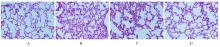
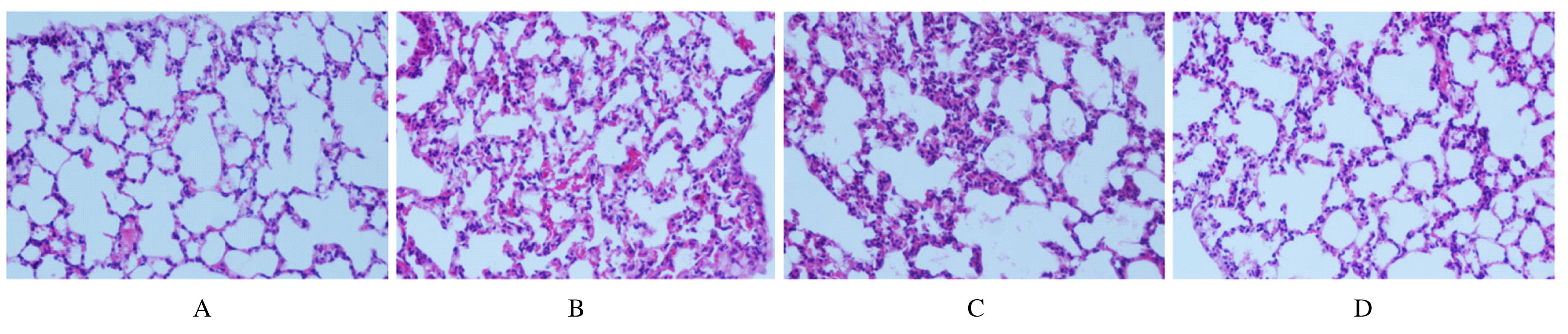
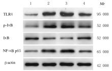
  • Received:2021-05-24 Online:2022-01-28 Published:2022-01-17
  • Contact: Jianguang ZHU E-mail:zxfj6098@163.com

摘要: 目的

探讨miRNA-27a对实验性肺结核模型大鼠免疫功能的调控作用,阐明其可能的作用机制。

方法

经尾静脉注射结核杆菌悬液建立肺结核大鼠模型,将造模成功的大鼠随机分为模型组、miR-27a激动剂对照(agomir-NC)组和miR-27a激动剂(agomir)组,另取正常大鼠设为对照组,每组12只。agomir-NC组和agomir组大鼠经尾静脉注射agomir-NC或miR-27a agomir干预,每天1次,连续5 d。3周后,计算各组大鼠脾脏和胸腺指数,HE染色观察各组大鼠肺组织病理形态表现,流式细胞术检测各组大鼠外周血T淋巴细胞亚群百分率,酶联免疫吸附测定(ELISA)法检测各组大鼠肺组织中白细胞介素1β(IL-1β)、白细胞介素6(IL-6)和肿瘤坏死因子α(TNF-α)水平,实时荧光定量PCR(RT-qPCR)法检测各组大鼠肺组织中miR-27a表达水平,Western blotting法检测各组大鼠肺组织中Toll样受体4(TLR4)、核因子κB (NF-κB)抑制蛋白α(IκBα)、磷酸化IκB(p-IκB)和NF-κB p65蛋白表达水平。

结果

与对照组比较,模型组大鼠肺组织损伤严重,脾脏及胸腺指数、外周血中CD4+T淋巴细胞百分率和肺组织中miR-27a表达水平及IκB蛋白表达水平均明显降低(P<0.05),外周血中CD8+T淋巴细胞百分率及肺组织中IL-1β、IL-6、TNF-α水平和TLR4、p-IκB及NF-κB p65蛋白表达水平均明显升高(P<0.05);与模型组比较,agomir组大鼠肺组织病理形态表现明显改善,脾脏和胸腺指数、外周血中CD4+T淋巴细胞百分率、肺组织中miR-27a表达水平及IκB蛋白表达水平均明显升高(P<0.05),外周血中CD8+T淋巴细胞百分率及肺组织中IL-1β、IL-6、TNF-α水平和TLR4、p-IκB、NF-κB p65蛋白表达水平均明显降低(P<0.05),agomir-NC组大鼠上述各指标差异均无统计学意义(P>0.05)。

结论

过表达miR-27a可能通过抑制TLR4/NF-κB信号通路活化,减少炎症因子释放,调节机体免疫反应,从而改善肺结核大鼠肺损伤。

关键词: 微小RNA, miRNA-27a, 肺结核, Toll样受体4/核因子κB信号通路, 免疫功能, 炎症

Abstract: Objective

To investigate the regulatory effect of miRNA-27a on the immune function of the experimental pulmonary tuberculosis model rats, and to clarify its possible mechanism.

Methods

The rat models of pulmonary tuberculosis were established by injecting Mycobacterium tuberculosis suspension through tail vein, after successful modeling, the model rats were randomly divided into model group, miR-27a agomir control (agomir-NC) group and miR-27a agomir (agomir) group,and another normal rats were used as control group; there were 12 mice in each groups.The rats in agomir-NC group or miR-27a agomir group were injected with agomir-NC or miR-27a agomir via tail vein once a day for 5 consecutive days. Three weeks later,the spleen and thymus indexes of the rats in various groups were calculated, the pathomorphology of lung tissue of the rats in various groups were observed by HE staining; the percentages of T lymphocyte subsets in peripheral blood of the rats in various groups were detected by flow cytometry; the levels of interleukin-1β (IL-1β), interleukin-6 (IL-6) and tumor necrosis factor-α (TNF-α) in lung tissue of the rats in various groups were determined by enzyme linked immunosorbent assay (ELISA); the expression levels of miR-27a in lung tissue of the rats in various groups were detected by real-time fluorescence quantificative PCR (RT-qPCR) method; the expression levels of Toll-like receptor 4 (TLR4), nuclear factor kappa-B (NF-κB) inhibitor α (IκBα), phosporylated-IκB(p-IκB) and NF-κB p65 in lung tissue were detected by Western blotting method.

Results

Compared with control group,the lung injury of rats in model group was serious, the spleen and thymus indexes, the percentages of CD4+T cells in peripheral blood, the expression level of miR-27a and the expression level of IκB protein in lung tissue were significantly decreased (P<0.05); the percentages of CD8+T cells in peripheral blood, the levels of IL-1β, IL-6, TNF-α and the expression levels of TLR4, p-IκB and NF-κB p65 proteins in lung tissue were significantly increased (P<0.05). Compared with model group, the pathomorphology of lung tissue of the rats in agomir group was significantly improved, the spleen and thymus indexes,the percentages of CD4+T cells in peripheral blood, the expression level of miR-27a and the expression level of IκB protein in lung tissue were significantly increased (P<0.05); the percentage of CD8+T cells in peripheral blood, the levels of IL-1β, IL-6, TNF-α and the expression levels of TLR4, p-IκB and NF-κB p65 proteins in lung tissue of the rats were significantly decreased (P<0.05); there were no statistically significant differences in the above indexes of the rats in agomir-NC group (P>0.05).

Conclusion

Overexpression of miR-27a may improve the lung injury in the pulmonary tuberculosis rats by inhibiting activation of TLR4/NF-κB signaling pathway, reducing the release of inflammatory factors and regulating the body's immune response.

Key words: MiRNA-27a, Pulmonary tuberculosis, Toll-like receptor 4/nuclear factor kappa-B signaling pathway, Immune function, Inflammation

中图分类号: 

  • R-332