Journal of Jilin University(Medicine Edition) ›› 2026, Vol. 52 ›› Issue (4): 1151-1160.doi: 10.13481/j.1671-587X.20260428
• Imageology • Previous Articles
Evaluation of CT radiomics combined with clinical imaging features on invasiveness of mixed ground-glass nodule-like pulmonary adenocarcinoma
Hongyu SHI1,Lei XU1( ),Lei ZHAO2
- 1.Department of Respiratory and Critical Care Medicine,Affiliated Hospital,Inner Mongolia Medical University,Hohhot 010050,China
2.Department of Diagnostic Imaging,Affiliated Hospital,Inner Mongolia Medical University,Hohhot 010050,China
-
Received:2025-10-30Accepted:2025-12-23Online:2026-07-28Published:2026-07-27 -
Contact:Lei XU E-mail:lxu81@126.com

CLC Number:
- R734.2
Cite this article
Hongyu SHI,Lei XU,Lei ZHAO. Evaluation of CT radiomics combined with clinical imaging features on invasiveness of mixed ground-glass nodule-like pulmonary adenocarcinoma[J].Journal of Jilin University(Medicine Edition), 2026, 52(4): 1151-1160.
share this article
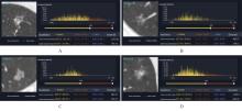
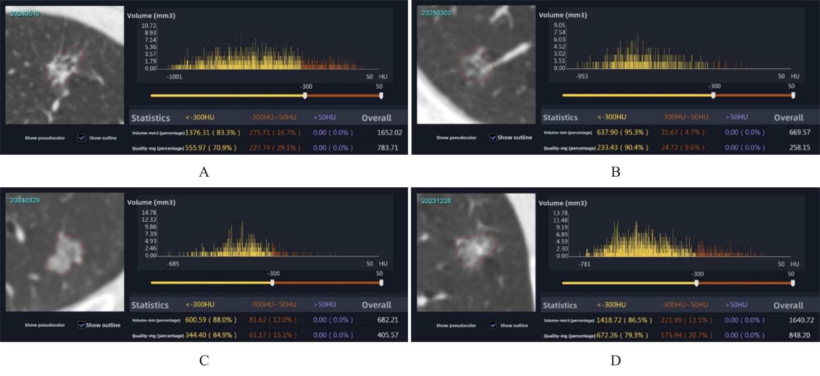
Fig. 1
CT manifestations and radiologic analysis of AAH(A), AIS(B), MIA(C), and IAC(D)"
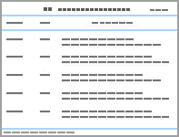
Tab.1
CT morphological characteristics of mGGNs patients"
| Characteristic | Indolent group (n=73) | Invasive group (n=205) | P |
|---|---|---|---|
| Nodule location [n(%)] | 0.480 | ||
| Right upper lobe | 25 (34.25) | 93 (45.37) | |
| Right middle lobe | 5 (6.85) | 12 (5.85) | |
| Right lower lobe | 10 (13.70) | 29 (14.15) | |
| Left upper lobe | 23 (31.51) | 49 (23.90) | |
| Left lower lobe | 10 (13.70) | 22 (10.73) | |
| Spiculation [n(%)] | 0.001 | ||
| Absent | 55 (75.34) | 110 (53.66) | |
| Present | 18 (24.66) | 95 (46.34) | |
| Lobulation [n(%)] | <0.001 | ||
| Absent | 24 (32.88) | 10 (4.88) | |
| Present | 49 (67.12) | 195 (95.12) | |
| Vacuole sign [n(%)] | 0.695 | ||
| Absent | 63 (86.30) | 173 (84.39) | |
| Present | 10 (13.70) | 32 (15.61) | |
| Air bronchogram sign [n(%)] | 0.005 | ||
| Absent | 51 (69.86) | 104 (50.73) | |
| Present | 22 (30.14) | 101 (49.27) | |
| Pleural indentation [n(%)] | <0.001 | ||
| Absent | 39 (53.42) | 64 (31.22) | |
| Present | 34 (46.58) | 141 (68.78) | |
| Spinous process [n(%)] | <0.001 | ||
| Absent | 72 (98.63) | 161 (78.54) | |
| Present | 1 (1.37) | 44 (21.46) | |
| Vascular convergence [n(%)] | 0.222 | ||
| Absent | 69 (94.52) | 184 (89.76) | |
| Present | 4 (5.48) | 21 (10.24) | |
| Vessel penetration [n(%)] | 0.368 | ||
| Absent | 71 (97.26) | 192 (93.66) | |
| Present | 2 (2.74) | 13 (6.34) | |
| Long diameter [M(P25,P75),mm] | 14.00 (11.00, 17.00) | 19.00 (16.00, 23.00) | <0.001 |
| Short diameter [M(P25,P75),mm] | 12.00 (9.00, 13.00) | 14.00 (11.00, 17.00) | <0.001 |
| Mean CT value [M(P25,P75),HU] | -420.50 (-500.30, -356.20) | -327.40 (-420.30, -234.40) | <0.001 |
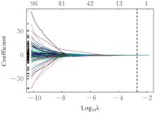
Fig. 2
Radiomics LASSO regression variable selection path plot"
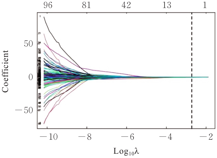
Fig. 3
Radiomics LASSO regression 10-fold cross-validation plot"
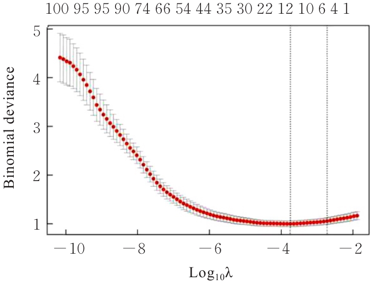
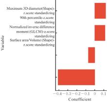
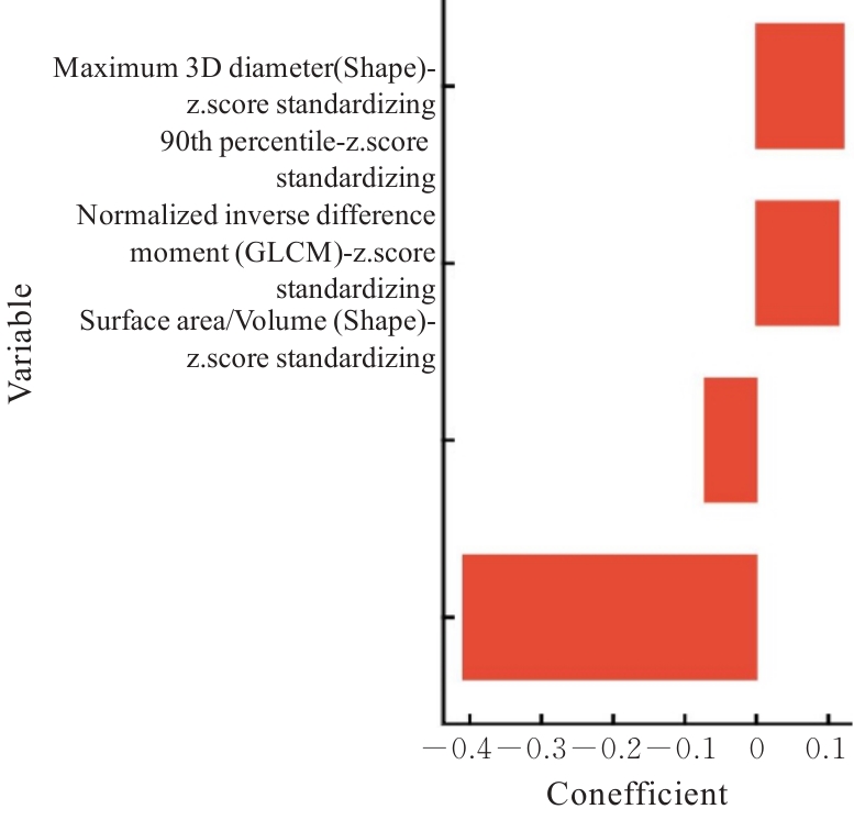
Fig. 4
Radiomics LASSO regression coefficients"
Tab.2
Univariate and multivariate Logistic regression analyses of predictors associated with invasiveness of GGN"
| Characteristic | Univariable | Multivariable | ||||
|---|---|---|---|---|---|---|
| OR | 95% CI | P | OR | 95% CI | P | |
| Nodule location | ||||||
| Right upper lobe | — | — | ||||
| Right middle lobe | 0.645 | 0.208, 2.003 | 0.448 | |||
| Right lower lobe | 0.780 | 0.335, 1.812 | 0.563 | - | - | - |
| Left upper lobe | 0.573 | 0.295, 1.112 | 0.100 | |||
| Left lower lobe | 0.591 | 0.248, 1.409 | 0.236 | |||
| Spiculation | ||||||
| Absent | — | — | — | — | ||
| Present | 2.639 | 1.450, 4.803 | 0.001 | 0.933 | 0.414, 2.102 | 0.867 |
| Lobulation | ||||||
| Absent | — | — | — | — | ||
| Present | 9.551 | 4.285, 21.287 | <0.001 | 3.724 | 1.373, 10.100 | 0.010 |
| Vacuole sign | ||||||
| Absent | — | — | ||||
| Present | 1.165 | 0.542, 2.508 | 0.696 | - | - | - |
| Air bronchogram sign | ||||||
| Absent | — | — | — | — | ||
| Present | 2.251 | 1.273, 3.981 | 0.005 | 0.515 | 0.235, 1.125 | 0.096 |
| Pleural indentation | ||||||
| Absent | — | — | — | — | ||
| Present | 2.527 | 1.463, 4.365 | <0.001 | 0.849 | 0.410, 1.759 | 0.659 |
| Spinous process | ||||||
| Absent | — | — | — | — | ||
| Present | 19.677 | 2.659, 145.604 | 0.004 | 4.406 | 0.533, 36.417 | 0.169 |
| Vascular convergence | ||||||
| Absent | — | — | ||||
| Present | 1.969 | 0.652, 5.941 | 0.229 | - | - | - |
| Vessel penetration | ||||||
| Absent | — | — | - | - | - | |
| Present | 2.404 | 0.529, 10.918 | 0.256 | |||
| Long diameter (mm) | 1.334 | 1.224, 1.454 | <0.001 | 1.318 | 1.152, 1.509 | <0.001 |
| Short diameter (mm) | 1.292 | 1.180, 1.415 | <0.001 | 1.010 | 0.862, 1.184 | 0.900 |
| Mean CT value (HU) | 1.006 | 1.004, 1.009 | <0.001 | 1.006 | 1.003, 1.009 | <0.001 |
Fig. 5
Nomogram of combined model for predicting mGGN pulmonary adenocarcinoma"
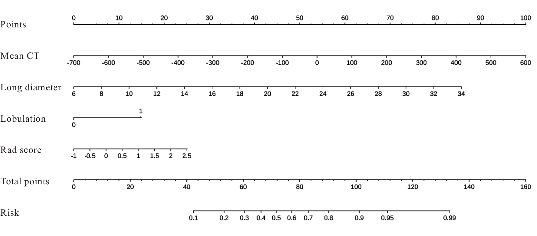
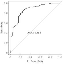
Fig. 6
ROC curves of nomogram"
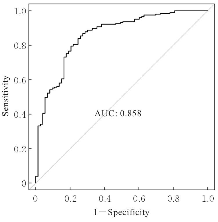
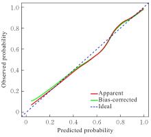
Fig. 7
Calibration curves of nomogram"
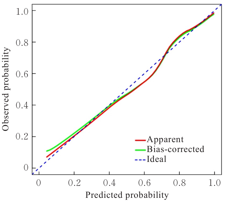
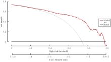
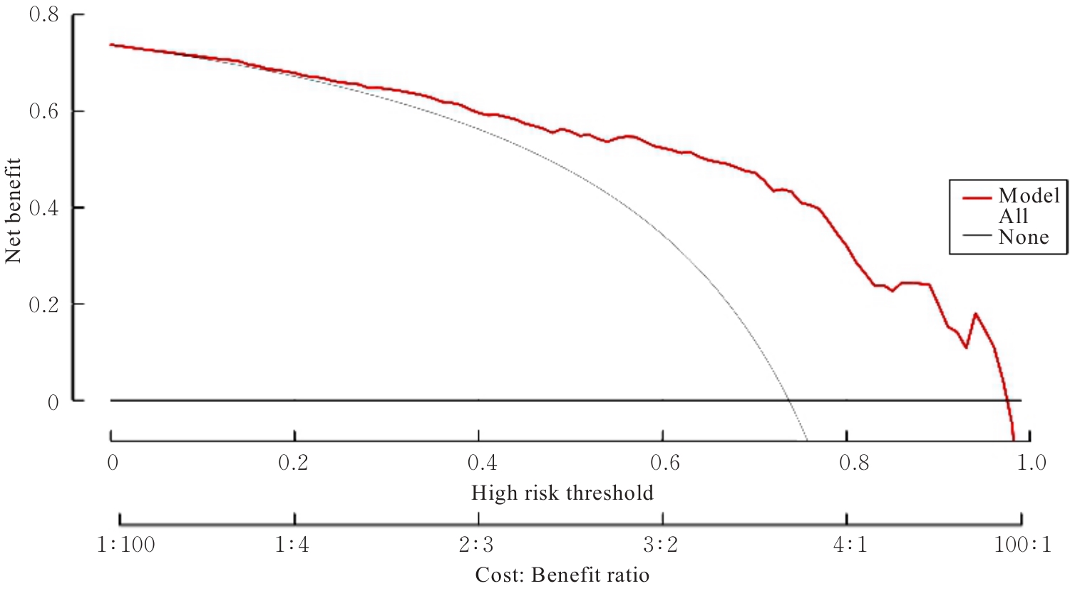
Fig. 8
DCA curves of nomogram"
| [1] | FAN X, LIANG C, MA X Q, et al. Clinical, imaging, and pathological-molecular characteristics associated with stage IA invasive lung adenocarcinoma recurrence after sub-lobar resection[J]. Acad Radiol, 2025, 32(1): 450-459. |
| [2] | OH J, PIAO Z, CHO H J, et al. CT-based three-dimensional invasiveness analysis of adenocarcinoma presenting as pure ground-glass nodules[J]. Transl Cancer Res, 2023, 12(4): 765-773. |
| [3] | 曹立武, 谢召勇, 朱志磊, 等. 磨玻璃结节肺腺癌组织学与CT表现特点对比研究[J]. 中国CT和MRI杂志, 2022, 20(7): 48-51. |
| [4] | MENG Q C, ZHENG C B, GUO L W, et al. Construction and validation of a risk score system for diagnosing invasive adenocarcinoma presenting as pulmonary pure ground-glass nodules: a multi-center cohort study in China[J]. Quant Imaging Med Surg, 2024, 14(7): 4864-4877. |
| [5] | ZHENG H, CHEN W, QI W Y, et al. Enhancing the prediction of the invasiveness of pulmonary adenocarcinomas presenting as pure ground-glass nodules: Integrating intratumor heterogeneity score with clinical-radiological features via machine learning in a multicenter study[J]. Digit Health, 2024, 10: 20552076241289181. |
| [6] | TANG Y H, LI M Z, LIN B K, et al. Deep learning-assisted development and validation of an algorithm for predicting the growth of persistent pure ground-glass nodules[J]. Transl Lung Cancer Res, 2023, 12(12): 2494-2504. |
| [7] | 中华医学会呼吸病学分会,中国肺癌防治联盟专家组.肺结节诊治中国专家共识(2024年版)[J]中华结核和呼吸杂志, 2024,47(8):716-729 |
| [8] | GRADISHAR W J, MORAN M S, ABRAHAM J, et al. Breast Cancer, Version 3.2024, NCCN Clinical Practice Guidelines in Oncology[J]. J Natl Compr Canc Netw, 2024, 22(5): 331-357. |
| [9] | HU X F, GONG J, ZHOU W, et al. Computer-aided diagnosis of ground glass pulmonary nodule by fusing deep learning and radiomics features[J]. Phys Med Biol, 2021, 66(6): 065015. |
| [10] | GAO R J, GAO Y H, ZHANG J, et al. A nomogram for predicting invasiveness of lung adenocarcinoma manifesting as pure ground-glass nodules: incorporating subjective CT signs and histogram parameters based on artificial intelligence[J]. J Cancer Res Clin Oncol, 2023, 149(17): 15323-15333. |
| [11] | LIU J N, QI L L, WANG Y W, et al. Development of a combined radiomics and CT feature-based model for differentiating malignant from benign subcentimeter solid pulmonary nodules[J]. Eur Radiol Exp, 2024, 8(1): 8. |
| [12] | LI Q Q, XIAO T J, LI J D, et al. The diagnosis and management of multiple ground-glass nodules in the lung[J]. Eur J Med Res, 2024, 29(1): 305. |
| [13] | BARTLETT E C, KEMP S V, RAWAL B, et al. Defining growth in small pulmonary nodules using volumetry: results from a “coffee-break” CT study and implications for current nodule management guidelines[J]. Eur Radiol, 2022, 32(3): 1912-1920. |
| [14] | GONG H P, SI H F, XING J L, et al. Value of MSCT multiplanar reconstruction in the differential diagnosis of ground glass nodular lung adenocarcinoma[J] Chin J CT MRI, 2023, 21(2): 52-54. |
| [15] | ZHENG C Q, WANG H F, LIU Q Z, et al. Application effect of low-dose spiral CT on pulmonary nodules and its diagnostic value for benign and malignant nodules[J]. Am J Transl Res, 2023, 15(1): 256-263. |
| [16] | 洪静静, 文 戈, 张 飞, 等. 预测肺腺癌浸润性: 基于临床特征和普通CT平扫影像学特征的联合模型[J]. 分子影像学杂志, 2024, 47(11): 1225-1230. |
| [17] | KWON H J, PARK U H, GOH C J, et al. Enhancing lung cancer classification through integration of liquid biopsy multi-omics data with machine learning techniques[J]. Cancers, 2023, 15(18): 4556. |
| [18] | HU K M, GAO L L, ZHANG R Y, et al. Clinical application of serum seven tumour-associated autoantibodies in patients with pulmonary nodules[J]. Heliyon, 2024, 10(9): e30576. |
| [19] | DONG N, YAN Y M, LI Y X, et al. CT-based habitat radiomics for preoperative differentiation of adenocarcinoma in situ/minimally invasive adenocarcinoma from invasive adenocarcinoma manifesting as ground-glass nodules: a multicenter study[J]. Front Oncol, 2025, 15: 1660071. |
| [20] | WU S J, FAN L F, WU Y M, et al. Deep transfer learning radiomics combined with explainable machine learning for preoperative thymoma risk prediction based on CT[J]. Eur J Radiol, 2025, 190: 112266. |
| [21] | QI S L, ZUO Y F, CHANG R S, et al. Using CT radiomic features based on machine learning models to subtype adrenal adenoma[J]. BMC Cancer, 2023, 23(1): 111. |
| [22] | DONG H, XI Y Z, LIU K, et al. A Radiological-Radiomics model for differentiation between minimally invasive adenocarcinoma and invasive adenocarcinoma less than or equal to 3 cm: a two-center retrospective study[J]. Eur J Radiol, 2024, 176: 111532. |
| [23] | 刘 建, 师 金, 田 国. 基于SEER数据库的年轻肺腺癌晚期患者预后预测及风险分层的模型构建[J]. 解放军医学杂志, 2024, 49(8): 889-896. |
| [24] | NOBEL T B, CARR R A, CASO R, et al. Primary lung cancer in women after previous breast cancer[J]. BJS Open, 2021, 5(6): zrab115. |
| [25] | CHEN P F, GONG H Y, ZHANG L, et al. CT radiomics combined with neural networks predict the malignant degree of pulmonary grinding glass nodules[J]. Front Med, 2025, 12: 1603472. |
| [26] | YANG X L, WANG J Y, WANG P, et al. Deep learning model using CT images for longitudinal prediction of benign and malignant ground-glass nodules[J]. Eur J Radiol, 2025, 190: 112252. |
| [27] | YANG Y T, XU J, WANG W, et al. Meta-analysis of the correlation between CT-based features and invasive properties of pure ground-glass nodules[J]. Asian J Surg, 2023, 46(9): 3405-3416. |
| [28] | HE S Y, CHEN C E, WANG Z G, et al. The use of the mean computed-tomography value to predict the invasiveness of ground-glass nodules: a meta-analysis[J]. Asian J Surg, 2023, 46(2): 677-682. |
| [1] | Feida YAN,Mingdi CHEN,Zhiwei LIANG. Inhibitory effect of TPX2 silencing on immune escape in non-small cell lung cancer A549 cells by regulating Wnt/β-catenin signaling pathway [J]. Journal of Jilin University(Medicine Edition), 2026, 52(4): 1011-1021. |
| [2] | Di ZHANG,Zhiguang GU,Xiangkai ZHAO,Menghan LI,Bin YANG,Xiaoyu HAO,Pengpeng WANG,Zuyun LIU,Ming ZHANG,Dongsheng HU,Wei WANG. Construction of phenotypic age models using different combinations of multiple clinical biomarkers and in fluence factors of phenotypic age acceleration [J]. Journal of Jilin University(Medicine Edition), 2026, 52(4): 1071-1076. |
| [3] | Rui LI,Xiaoyan HAO,Liu YANG,Jiayun LIU,Mu HE. Construction and validation of machine learning-based prediction models for esophageal cancer using routine clinical laboratory data [J]. Journal of Jilin University(Medicine Edition), 2026, 52(4): 1086-1095. |
| [4] | Jiaqi GUO,Lei XU. Comparison of predictive performance of risk assessment models of COMPASS-CAT, Padua, Caprini, and Khorana for venous thromboembolism in lung cancer patients [J]. Journal of Jilin University(Medicine Edition), 2026, 52(4): 1116-1127. |
| [5] | Jing YU,Yinzhuo HUANG,Zhongwei ZHOU,Yuxin XIE,Qian MAO. Analysis on influencing factors in type 2 diabetes mellitus patients complicated with lower limb atherosclerosis and construction of nomogram model [J]. Journal of Jilin University(Medicine Edition), 2026, 52(3): 806-812. |
| [6] | Yuyang LI,Xiang LI,He SHI,Yiwen QIN,Zhaorong XIAO,Sihao CHEN,Cong ZHAO,Weiwei LIU. Analyses of etiology characteristics and risk factors for postoperative infection in patients underwent oral and maxillofacial surgery [J]. Journal of Jilin University(Medicine Edition), 2026, 52(2): 507-512. |
| [7] | Xiao LI,Li WANG,Wen WU,Rui WANG,Aiying ZHANG,Shuo ZHANG,Rujia JIANG,Yaning MENG. Clinical outcomes and risk factors analysis on infection in patients with bullous pemphigoid [J]. Journal of Jilin University(Medicine Edition), 2025, 51(5): 1312-1317. |
| [8] | Xiaoshuang HE,Lina XU,Mei CUI,Yu ZHAO,Bei WANG,Zheng HUANG,Yuchao WANG,Wenyan XIN,Chao WU. Effects of lncRNA DUXAP8 in lung cancer A549 cells-derived exosomes on lung cancer cell growth and its mechnism [J]. Journal of Jilin University(Medicine Edition), 2025, 51(4): 958-967. |
| [9] | Jiahui FENG,Renjie LIU,Xuan CHEN. Analysis on risk factors of development of acute hydrocephalus in patients with aneurysmal subarachnoid hemorrhage [J]. Journal of Jilin University(Medicine Edition), 2025, 51(3): 763-769. |
| [10] | Suzhen YUAN,Yan JIN,Wenwen WANG. Analysis on clinical characteristics of patients with ovarian clear cell carcinoma and ovarian endometriosis [J]. Journal of Jilin University(Medicine Edition), 2024, 50(6): 1677-1682. |
| [11] | Honghong LI,Na YU,Minghao SHI,Ying SUN,Yao LI,Zhongjun SHEN,Xiaoyi LIU,Liyan ZHAO. Predictive value of new thrombotic risk assessment model for venous thromboembolism in patients with malignant tumors [J]. Journal of Jilin University(Medicine Edition), 2024, 50(5): 1390-1399. |
| [12] | Jiayuan YU,Di ZHAO,Xin JIANG,Jing XU,Lili JIANG,Hongyu JIANG. Association analysis on peripheral blood lymphocyte subsets and occurrence of carotid atherosclerosis [J]. Journal of Jilin University(Medicine Edition), 2024, 50(5): 1400-1405. |
| [13] | Peng QI,Xianying MENG,Meihua PIAO,Qiang ZHANG. Network Meta-analysis on risk factors of recurrence of papillary thyroid microcarcinoma [J]. Journal of Jilin University(Medicine Edition), 2023, 49(6): 1504-1512. |
| [14] | Xiunan FENG,Lu CHEN,Yu LONG,Zhenyu JIANG,Ling ZHAO. Analysis on clinical characteristics and risk factors of patients with Sjogren’s syndrome complicated with interstitial lung disease [J]. Journal of Jilin University(Medicine Edition), 2023, 49(6): 1513-1518. |
| [15] | Xue DONG, Jinfeng ZANG, Caifeng XU, Hebing LIU, Zhaohua CHENG. Construction and validation of risk prediction model of enteral nutrition feeding intolerance of patients with severe acute pancreatitis [J]. Journal of Jilin University(Medicine Edition), 2023, 49(6): 1586-1592. |
|
||