Journal of Jilin University(Medicine Edition) ›› 2026, Vol. 52 ›› Issue (4): 932-941.doi: 10.13481/j.1671-587X.20260406
• Research in basic medicine • Previous Articles Next Articles
Inhibitory effect of Codonopsis pilosula polysaccharides on colorectal cancer liver metastasis through TGF-β/Smad signaling pathway
Guiyan TANG1,Dapeng CUI2,Zhongshuai SUN2,Bin ZHANG3( ),Taiyuan MA1()
- 1.Department of Colorectal and Anal Surgery,General Surgery Center,First Hospital,Jilin University,Changchun 130021,China
2.Department of Hepatobiliary Surgery,First Affiliated Hospital,Hebei North University,Zhangjiakou 075000,China
3.Department of General Surgery,Second Hospital,Baoding City,Hebei Province,Baoding 071000,China
-
Received:2025-10-05Accepted:2025-12-11Online:2026-07-28Published:2026-07-27 -
Contact:Bin ZHANG,Taiyuan MA E-mail:15712576000@163.com;mty15804302180@fooxmail.com

CLC Number:
- R735.3
Cite this article
Guiyan TANG,Dapeng CUI,Zhongshuai SUN,Bin ZHANG,Taiyuan MA. Inhibitory effect of Codonopsis pilosula polysaccharides on colorectal cancer liver metastasis through TGF-β/Smad signaling pathway[J].Journal of Jilin University(Medicine Edition), 2026, 52(4): 932-941.
share this article
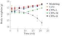
Fig. 1
Body weights of mice in various groups at different time points"
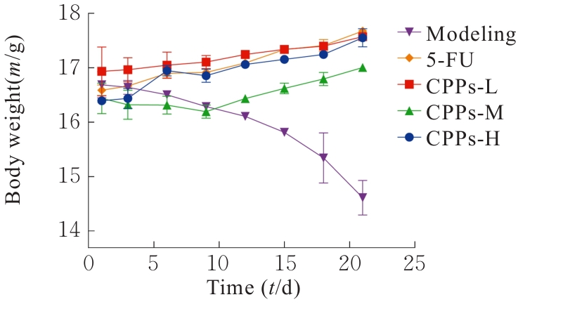

Fig. 2
Patho morphology of liver metastatic focus of mice in various groups after treated with CPPs"

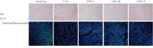
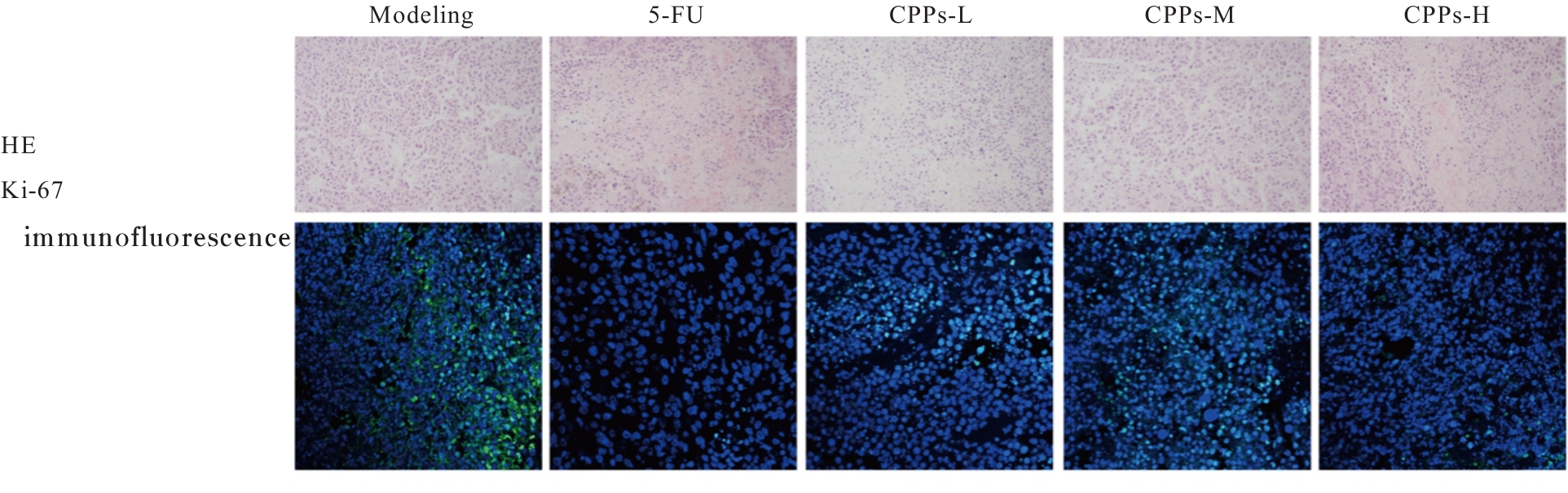
Fig. 3
Proliferation activities of tumor cells in liver metastatic focus of mice in various groups after treated with CPPs (×200)"
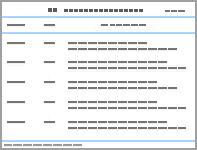
Tab.1
Fluorescen ROI in liver region of mice in various groups"
| Group | ROI | |
|---|---|---|
| Time(t/d) 7 | 21 | |
| Modeling | 89.43±12.18 | 1 243.47±55.77 |
| 5-FU | 90.98±13.06 | 730.03±15.76* |
| CPPs-L | 88.79±12.05 | 1 189.18±179.62*○ |
| CPPs-M | 88.81±21.85 | 899.54±238.00*△○ |
| CPPs-H | 83.72±23.75 | 761.29±181.36*△#○ |
Tab.2
Levels of TNF-α, IL-1β and IL-6 in seurm of mice in various groups [n=3, x±s, ρB/(ng·L-1)]"
| Group | TNF-α | IL-1β | IL-6 |
|---|---|---|---|
| Modeling | 215.14±3.86 | 183.95±8.53 | 200.44±15.49 |
| 5-FU | 91.47±3.24* | 53.00±5.43* | 63.14±9.90* |
| CPPs-L | 196.87±8.91*○ | 147.86±7.95* | 166.74±12.25* |
| CPPs-M | 140.89±17.04*△ | 113.43±16.91*△ | 140.03±11.64*△ |
| CPPs-H | 122.69±11.38*△# | 84.83±12.80*△#○ | 98.81±23.21*△#○ |
Tab.3
Expression levels of E-cadherin, N-cadherin, and Vimentin mRNA in liver tumor tissue of mice in various groups"
| Group | E-Cadherin | N-Cadherin | Vimentin |
|---|---|---|---|
| Modeling | 1.00±0.00 | 1.00±0.00 | 1.00±0.00 |
| 5-FU | 3.16±0.61* | 0.43±0.11* | 0.51±0.10* |
| CPPs-L | 1.10±0.16*○ | 0.81±0.10*○ | 0.71±0.08*○ |
| CPPs-M | 1.86±0. 26*△○ | 0.62±0.10*△○ | 0.59±0.05*○ |
| CPPs-H | 2.76±0.30*△#○ | 0.45±0.12*△# | 0.46±0.07*△# |
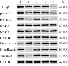
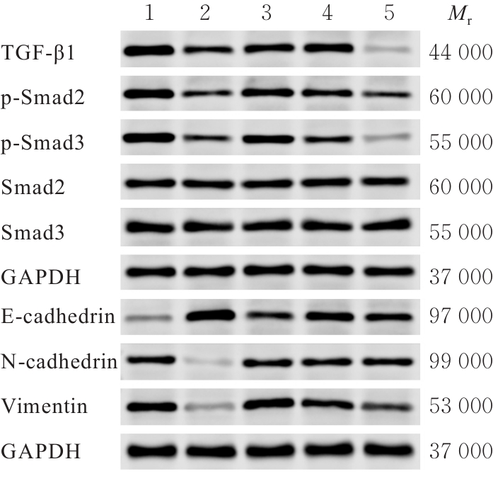
Fig. 4
Electrophoregram of expressions of TGF-β1, Smad2/3, p-Smad2/3, E-cadherin, N-cadherin, and Vimentin proteins in liver tumor tissue of mice in various groups"
| [1] | BRAY F, LAVERSANNE M, SUNG H, et al. Global cancer statistics 2022: GLOBOCAN estimates of incidence and mortality worldwide for 36 cancers in 185 countries[J]. CA Cancer J Clin, 2024, 74(3): 229-263. |
| [2] | SIEGEL R L, WAGLE N S, CERCEK A, et al. Colorectal cancer statistics, 2023[J]. CA Cancer J Clin, 2023, 73(3): 233-254. |
| [3] | HUANG J J, LUCERO-PRISNO D E 3rd, ZHANG L, et al. Updated epidemiology of gastrointestinal cancers in East Asia[J]. Nat Rev Gastroenterol Hepatol, 2023, 20(5): 271-287. |
| [4] | MORRIS V K, KENNEDY E B, BAXTER N N, et al. Treatment of metastatic colorectal cancer: ASCO guideline[J]. J Clin Oncol, 2023, 41(3): 678-700. |
| [5] | SHIN A E, GIANCOTTI F G, RUSTGI A K. Metastatic colorectal cancer: mechanisms and emerging therapeutics[J]. Trends Pharmacol Sci, 2023, 44(4): 222-236. |
| [6] | LIU B L, ZHOU H Y, TAN L C, et al. Exploring treatment options in cancer: Tumor treatment strategies[J]. Sig Transduct Target Ther, 2024, 9(1): 175. |
| [7] | LAWRENCE R, WATTERS M, DAVIES C R, et al. Circulating tumour cells for early detection of clinically relevant cancer[J]. Nat Rev Clin Oncol, 2023, 20(7): 487-500. |
| [8] | LIU A Y, YU C X, QIU C, et al. PRMT5 methylating SMAD4 activates TGF-β signaling and promotes colorectal cancer metastasis[J]. Oncogene, 2023, 42(19): 1572-1584. |
| [9] | SABOUNI E, NEJAD M M, MOJTABAVI S, et al. Unraveling the function of epithelial-mesenchymal transition (EMT) in colorectal cancer: Metastasis, therapy response, and revisiting molecular pathways[J]. Biomedecine Pharmacother, 2023, 160: 114395. |
| [10] | THAPA R, AFZAL O, AFZAL M, et al. From LncRNA to metastasis: The MALAT1-EMT axis in cancer progression[J]. Pathol Res Pract, 2024, 253: 154959. |
| [11] | BARDELČÍKOVÁ A, ŠOLTYS J, MOJŽIŠ J. Oxidative stress, inflammation and colorectal cancer: an overview[J]. Antioxidants (Basel), 2023, 12(4): 901. |
| [12] | NIGAM M, MISHRA A P, DEB V K, et al. Evaluation of the association of chronic inflammation and cancer: Insights and implications[J]. Biomed Pharmacother, 2023, 164: 115015. |
| [13] | CHEN G M, JIANG N, ZHENG J P, et al. Structural characterization and anti-inflammatory activity of polysaccharides from Astragalus membranaceus [J]. Int J Biol Macromol, 2023, 241: 124386. |
| [14] | SHA W J, ZHAO B, WEI H Z, et al. Astragalus polysaccharide ameliorates vascular endothelial dysfunction by stimulating macrophage M2 polarization via potentiating Nrf2/HO-1 signaling pathway[J]. Phytomedicine, 2023, 112: 154667. |
| [15] | YANG Q, MENG D D, ZHANG Q Y, et al. Advances in research on the anti-tumor mechanism of Astragalus polysaccharides[J]. Front Oncol, 2024, 14: 1334915. |
| [16] | YANG C, LI D, KO C N, et al. Active ingredients of traditional Chinese medicine for enhancing the effect of tumor immunotherapy[J]. Front Immunol, 2023, 14: 1133050. |
| [17] | XU Q, CHENG W, WEI J R, et al. Synergist for antitumor therapy: Astragalus polysaccharides acting on immune microenvironment[J]. Discov Oncol, 2023, 14(1): 179. |
| [18] | XIONG H T, HAN X P, CAI L, et al. Natural polysaccharides exert anti-tumor effects as dendritic cell immune enhancers[J]. Front Oncol, 2023, 13: 1274048. |
| [19] | CHEN R Q, LI Y T, ZUO L, et al. Astragalus polysaccharides inhibits tumor proliferation and enhances cisplatin sensitivity in bladder cancer by regulating the PI3K/AKT/FoxO1 axis[J]. Int J Biol Macromol, 2025, 311: 143739. |
| [20] | WANG F H, ZHANG X T, TANG L, et al. The Chinese Society of Clinical Oncology (CSCO): Clinical guidelines for the diagnosis and treatment of gastric cancer, 2023[J]. Cancer Commun, 2024, 44(1): 127-172. |
| [21] | SATHE A, MASON K, GRIMES S M, et al. Colorectal cancer metastases in the liver establish immunosuppressive spatial networking between tumor-associated macrophages and fibroblasts[J]. Clin Cancer Res, 2023, 29(1): 244-260. |
| [22] | BOND M J G, BOLHUIS K, LOOSVELD O J L, et al. First-line systemic treatment strategies in patients with initially unresectable colorectal cancer liver metastases (CAIRO5): an open-label, multicentre, randomised, controlled, phase 3 study from the Dutch Colorectal Cancer Group[J]. Lancet Oncol, 2023, 24(7): 757-771. |
| [23] | LINARES J, SALLENT-ARAGAY A, BADIA-RAMENTOL J, et al. Long-term platinum-based drug accumulation in cancer-associated fibroblasts promotes colorectal cancer progression and resistance to therapy[J]. Nat Commun, 2023, 14(1): 746. |
| [24] | FASANO M, PIROZZI M, MICELI C C, et al. TGF-β modulated pathways in colorectal cancer: new potential therapeutic opportunities[J]. Int J Mol Sci, 2024, 25(13): 7400. |
| [25] | CHEN Z D, LIANG H Y, YAN X X, et al. Astragalus polysaccharide promotes autophagy and alleviates diabetic nephropathy by targeting the lncRNA Gm41268/PRLR pathway[J]. Ren Fail, 2023, 45(2): 2284211. |
| [26] | SUN L I, ZHUO S C, LI X X, et al. Astragalus polysaccharide enhances the therapeutic efficacy of cisplatin in triple-negative breast cancer through multiple mechanisms[J]. Oncol Res, 2025, 33(3): 641-651. |
| [27] | NIAN Q, LIU R X, ZENG J H. Unraveling the pathogenesis of myelosuppression and therapeutic potential of natural products[J]. Phytomedicine, 2024, 132: 155810. |
| [28] | TYLUTKA A, WALAS Ł, ZEMBRON-LACNY A. Level of IL-6, TNF, and IL-1β and age-related diseases: a systematic review and meta-analysis[J]. Front Immunol, 2024, 15: 1330386. |
| [29] | GUO Q, JIN Y Z, CHEN X Y, et al. NF-κB in biology and targeted therapy: new insights and translational implications[J]. Sig Transduct Target Ther, 2024, 9(1): 53. |
| [30] | HU Z Y, SUI Q H, JIN X, et al. IL6-STAT3-C/EBPβ-IL6 positive feedback loop in tumor-associated macrophages promotes the EMT and metastasis of lung adenocarcinoma[J]. J Exp Clin Cancer Res, 2024, 43(1): 63. |
| [31] | LEONE G M, MANGANO K, PETRALIA M C, et al. Past, present and (foreseeable) future of biological anti-TNF alpha therapy[J]. J Clin Med, 2023, 12(4): 1630. |
| [32] | AUYEUNG K K, HAN Q B, KO J K. Astragalus membranaceus: a review of its protection against inflammation and gastrointestinal cancers[J]. Am J Chin Med, 2016, 44(1): 1-22. |
| [33] | WU Q, YANG Z P, NIE Y Z, et al. Multi-drug resistance in cancer chemotherapeutics: Mechanisms and lab approaches[J]. Cancer Lett, 2014, 347(2): 159-166. |
| [34] | DUAN C Y, YU M J, XU J Y, et al. Overcoming Cancer Multi-drug Resistance (MDR): Reasons, mechanisms, nanotherapeutic solutions, and challenges[J]. Biomed Pharmacother, 2023, 162: 114643. |
| [1] | Qiaotong REN,Weihua WU,Xingxiang WANG,Shichao WANG,Yipeng CHENG,Chun LI. Effect of interleukin-17 recepter B gene silencing on proliferation, invasion and apoptosis of lung adenocarcinoma A549 cells and its mechanism [J]. Journal of Jilin University(Medicine Edition), 2026, 52(3): 738-745. |
| [2] | Hong LI,Panpan YU,Zouyu ZHAO,Chongfeng SUN,Hui QIAO,Ping YANG. Expression of GSTO1 in ovarian cancer tissue and effect of N-glycosylation site mutations on biological behaviors of epithelial ovarian cancer cells [J]. Journal of Jilin University(Medicine Edition), 2026, 52(2): 469-482. |
| [3] | Pingsheng ZHU,Sitang GE,Lugen ZUO,Deli CHEN,Yangyang ZHANG. Effect of miR-325-3p targeting PRELID1 gene in regulation of EMT pathway on invasion and migration of colon cancer cells and their mechanisms [J]. Journal of Jilin University(Medicine Edition), 2025, 51(5): 1185-1193. |
| [4] | Zhongjun SHEN,Yao ZHAO,Mingbo JIA,Liyan ZHAO. Research progress in effects of hypoxia-inducible factors on cell migration and invasion during epithelial-mesenchymal transition in glioma cells [J]. Journal of Jilin University(Medicine Edition), 2025, 51(4): 1145-1154. |
| [5] | Hongli LI,Mengyao WANG,Yangyang LIU,Hui ZHANG,Li LI. Effect of KHSRP on biological behavior of colorectal cancer cells through activation of JAK/STAT signaling pathway [J]. Journal of Jilin University(Medicine Edition), 2025, 51(4): 996-1006. |
| [6] | Xingxiang WANG,Ying ZHAO,Qiaotong REN,Hefei WANG,Gang PU,Chun LI. Promotive effect of M2 macrophages on epithelial-mesenchymal transition and cisplatin resistance in non-small cell lung cancer A549 cells by regulating NF-κB signaling pathway [J]. Journal of Jilin University(Medicine Edition), 2025, 51(3): 642-652. |
| [7] | Changyu SHI,Yong LI,Jing DENG,Chunmei PIAO,Ming JIN. Bioinformatics analysis on adjustment effect of colorectal liver metastases model in mice based on complement alternative pathway and its experimental verification [J]. Journal of Jilin University(Medicine Edition), 2025, 51(3): 703-715. |
| [8] | Fan WANG,Xin WEN,Yixuan WANG,Yuan WANG. Effect of gap junction β2 on prognosis of patients with lung adenocarcinoma and biological behavior of lung adenocarcinoma A549 cells [J]. Journal of Jilin University(Medicine Edition), 2025, 51(3): 716-726. |
| [9] | Yan WANG,Zouyu ZHAO,Panpan YU,Ping YANG. Expression of I kappa B kinase-interacting protein in cervical cancer tissue and its effect on proliferation, migration and invasion of cervical cancer cells [J]. Journal of Jilin University(Medicine Edition), 2025, 51(2): 341-351. |
| [10] | Feina WANG,Xuguang MI,Xiuying LIN,Jianhua FU,Lei LIU,Xinyue YU,Huanhuan ZANG,Linjun LIU,Shiling CHEN,Yanqiu FANG. Effect of Wnt/β-catenin signaling pathway inhibitor MSAB on fibrogenic responses of human endometrial stromal cells [J]. Journal of Jilin University(Medicine Edition), 2024, 50(5): 1266-1274. |
| [11] | Bingbing HU,Kangning LUO,Su PENG,Yuzhong ZHOU,Maoliang CHEN,Changhua LIU. Improvement effect of down-regulation of miR-208a on 5-FU resistance in colorectal cancer cells through targeting SFRP1 for mediating Wnt signaling pathway [J]. Journal of Jilin University(Medicine Edition), 2024, 50(4): 947-955. |
| [12] | Shilei GAO,Jiaqiang WANG,Weitao YAO,Zhichao TIAN,Chao LI,Xiaoxiao LIANG,Xin WANG. Effect of miR-761 on epithelial-mesenchymal transition in osteosarcoma MG63 cells by regulating tumor-associated macrophage polarization [J]. Journal of Jilin University(Medicine Edition), 2024, 50(4): 978-988. |
| [13] | Jing TANG,Huan LI,Shuo ZHANG,Ligang JING. Analysis on association between serum homocysteine and inflammatory response and oxidative stress in patients with acute ischemic stroke [J]. Journal of Jilin University(Medicine Edition), 2024, 50(3): 786-790. |
| [14] | Haifeng WEI,Zhiqiang NI,Yanhong WEI,Qilai WANG,Shouqing LI,Yinfu MA,Yan TAN,Yanqiu FANG. Effects of miR-126 over-expression and ADAM9 gene silencing on biological behavior of gastric cancer SGC-7901 cells and their mechanisms [J]. Journal of Jilin University(Medicine Edition), 2024, 50(2): 310-319. |
| [15] | Buqi NA,Chunji QUAN,Fang ZHAO,Fan YANG,Ru XIAO,Xuemei JIN,Zhenling LI. Effect of histone deacetylase inhibitor CUDC-101 on DNA damage, migration, and epithelial-mesenchymal transition of prostate cancer DU145 cells [J]. Journal of Jilin University(Medicine Edition), 2024, 50(2): 400-410. |
|
||