Journal of Jilin University(Medicine Edition) ›› 2026, Vol. 52 ›› Issue (4): 977-986.doi: 10.13481/j.1671-587X.20260410
• Research in basic medicine • Previous Articles Next Articles
Protective effect of triptolide on hippocampal neuron injury in neonatal rats with hypoxic-ischemic encephalopathy by regulating PTEN/PI3K/AKT signaling pathway
Yunxia MA,Dan LIU,Ying ZHANG,Shifeng MA( )
)
- Department of Pediatrics,General Hospital,Tianjin Medical University Tianjin 300052,China
-
Received:2025-09-04Accepted:2025-11-10Online:2026-07-28Published:2026-07-27 -
Contact:Shifeng MA E-mail:2020@tmu.edu.cn

CLC Number:
- R743.3
Cite this article
Yunxia MA,Dan LIU,Ying ZHANG,Shifeng MA. Protective effect of triptolide on hippocampal neuron injury in neonatal rats with hypoxic-ischemic encephalopathy by regulating PTEN/PI3K/AKT signaling pathway[J].Journal of Jilin University(Medicine Edition), 2026, 52(4): 977-986.
share this article
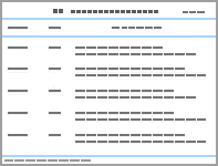
Tab.1
Neurological function scores of neonatal rats in various groups"
| Group | Neurological function score |
|---|---|
| Sham operation | 0.00±0.00 |
| HIE | 2.80±0.19* |
| L-TP | 2.21±0.15△ |
| H-TP | 1.35±0.16△# |
| H-TP+Ad-PTEN | 2.16±0.18*○ |
| F | 603.875 |
| P | <0.001 |
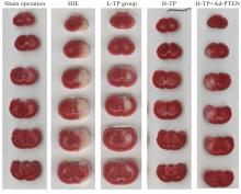
Fig. 1
Brain infarction status of rats in various groups"
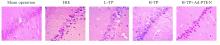
Fig. 2
Pathomorphology of hippocampal CA1 region of rats in various groups (HE,×200)"
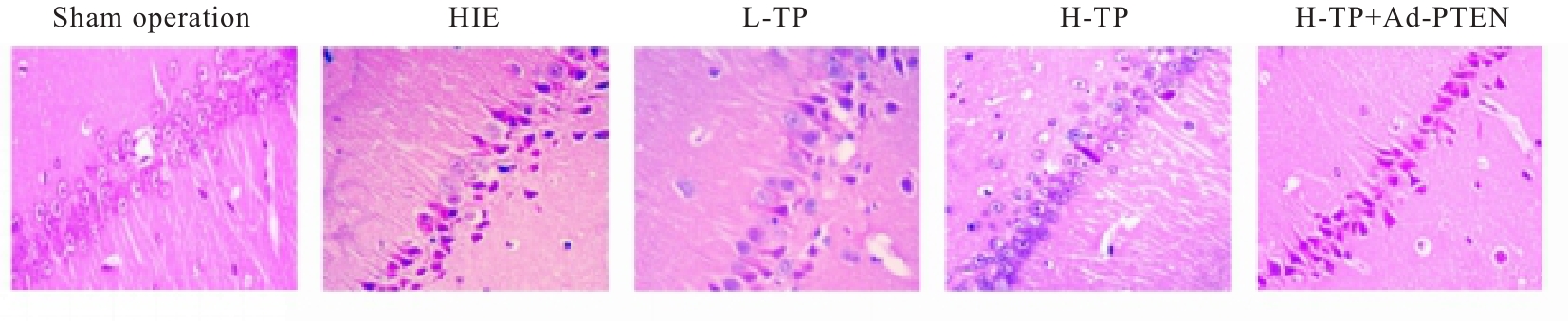
Fig. 3
Levels of IL-6, IL-8 and TNF-α in serum of rats in various groups"
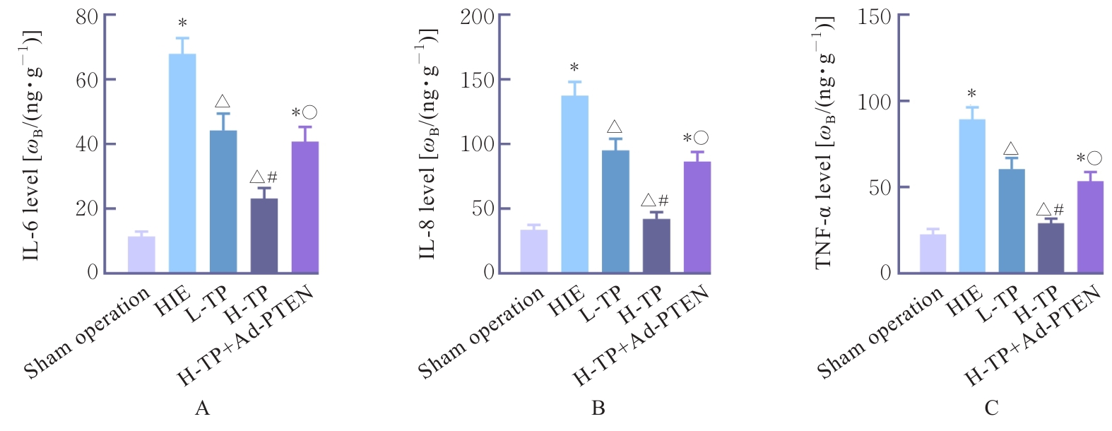
Fig. 4
SOD(A), CAT(B) activities and GSH(C) levels in brain tissue of rats in various groups"
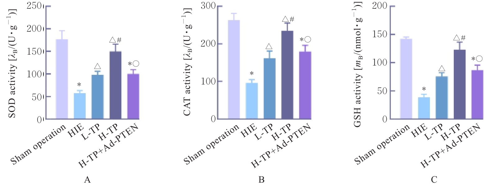
Fig. 5
TUNEL of staining hippocampal neurons(A-E) and number of positive cells(F) in rats in various groups"
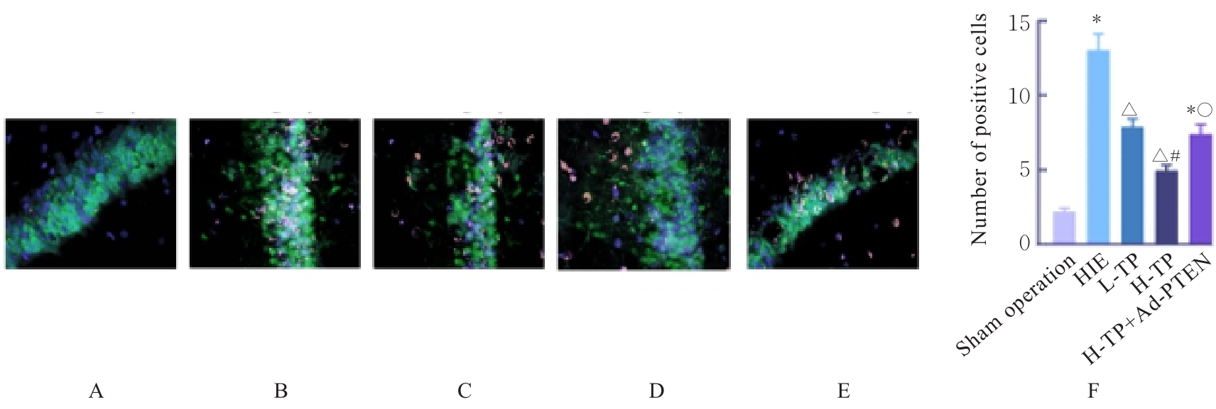
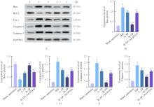
Fig. 6
Electrophoregram and histograms of expressions of apoptosis-related proteins in brain tissue of rats in various groups"
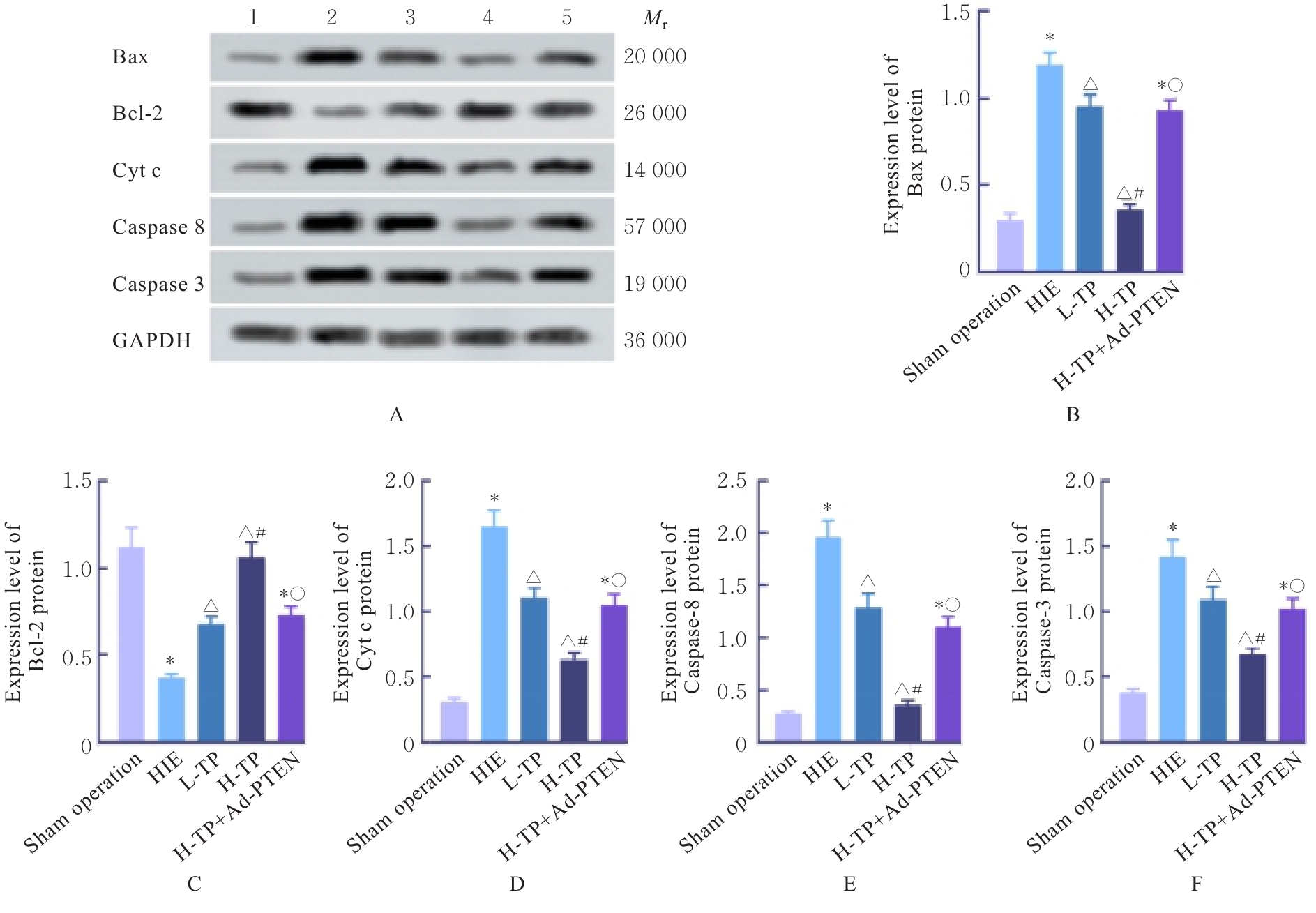
Fig. 7
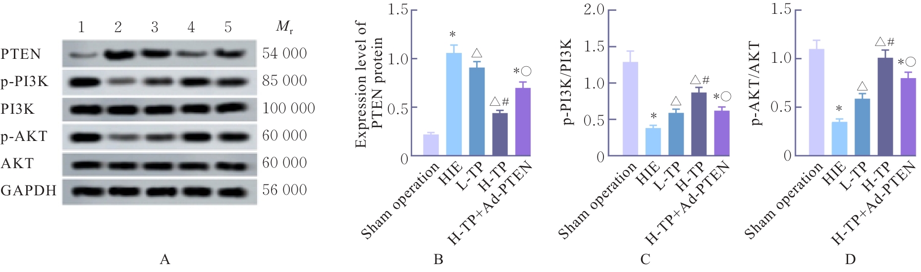
Electrophoregram and histograms of expressions of PTEN, p-PI3K, PI3K, p-AKT and AKT proteins in brain tissue of rats in various groups"
| [1] | HUANG Q Y, TANG J H, XIANG Y, et al. 4-Benzyl-2-methyl-1, 2, 4-thiadiazolidine-3, 5-dione rescues oligodendrocytes ferroptosis leading to myelin loss and ameliorates neuronal injury facilitating memory in neonatal hypoxic-ischemic brain damage[J]. Exp Neurol, 2025, 390: 115262. |
| [2] | HTUN Y, NAKAMURA S, KUSAKA T. Hydrogen and therapeutic gases for neonatal hypoxic-ischemic encephalopathy: potential neuroprotective adjuncts in translational research[J]. Pediatr Res, 2021, 89(4): 753-759. |
| [3] | OORSCHOT D E, SIZEMORE R J, AMER A R. Treatment of neonatal hypoxic-ischemic encephalopathy with erythropoietin alone, and erythropoietin combined with hypothermia: history, current status, and future research[J]. Int J Mol Sci, 2020, 21(4): 1487. |
| [4] | SONG J, HE G N, DAI L. A comprehensive review on celastrol, triptolide and triptonide: Insights on their pharmacological activity, toxicity, combination therapy, new dosage form and novel drug delivery routes[J]. Biomedecine Pharmacother, 2023, 162: 114705. |
| [5] | CUI Y R, JIANG X J, FENG J. The therapeutic potential of triptolide and celastrol in neurological diseases[J]. Front Pharmacol, 2022, 13: 1024955. |
| [6] | ZHU J J, YU B Y, HUANG X K, et al. Neferine protects against hypoxic-ischemic brain damage in neonatal rats by suppressing NLRP3-mediated inflammasome activation[J]. Oxid Med Cell Longev, 2021, 2021: 6654954. |
| [7] | 李春雨, 刘思思, 李 辰, 等. 补肾益髓方含药骨髓间充质干细胞外泌体调控miR-223介导PTEN/PI3K/Akt通路抑制CPZ小鼠神经元凋亡的机制研究[J]. 环球中医药, 2025, 18(3): 457-467. |
| [8] | 姬 斌, 刘丽娟. 西红花苷调节PTEN-PI3K/Akt信号通路保护脑缺血再灌注大鼠神经功能[J]. 解剖科学进展, 2025, 31(2): 189-191, 196. |
| [9] | HUANG Y, BA X, WANG H, et al. Triptolide alleviates collagen-induced arthritis in mice by modulating Treg/Th17 imbalance through the JAK/PTEN-STAT3 pathway[J]. Basic Clin Pharmacol Toxicol, 2023, 133(1): 43-58. |
| [10] | 张艺森, 张 玉, 张 新. 基于Nrf2/NF-κB信号通路研究连翘苷对缺氧缺血性脑病新生大鼠神经元凋亡的影响[J]. 中草药, 2025, 56(10): 3577-3584. |
| [11] | 扎西吉, 张 赟, 马凤美, 等. 基于PKC/ERK通路探讨败酱总黄酮对缺氧缺血性脑病新生大鼠神经元凋亡的影响[J]. 南京医科大学学报(自然科学版), 2023, 43(6): 786-794. |
| [12] | 李向斌, 侯芳芳. 雷公藤甲素通过mTOR/ULK1途径抑制自噬功能减轻脑缺血卒中造成的神经功能损伤[J]. 中国药师, 2022, 25(1): 1-5, 17. |
| [13] | ZHOU J J, YE W, CHEN L, et al. Triptolide alleviates cerebral ischemia/reperfusion injury via regulating the Fractalkine/CX3CR1 signaling pathway[J]. Brain Res Bull, 2024, 211: 110939. |
| [14] | 余 恒, 宋光捷. 去甲斑蝥素通过Nrf2通路保护新生大鼠缺氧缺血性脑损伤的机制[J]. 西北药学杂志, 2022, 37(4): 82-86. |
| [15] | PAN W Y, XU Z M. Triptolide mediates Wnt/β- catenin signalling pathway to reduce cerebral ischemia-reperfusion injury in rats[J]. Folia Neuropathol, 2020, 58(4): 324-333. |
| [16] | ZHENG Y H, LI L Y, CHEN B W, et al. Chlorogenic acid exerts neuroprotective effect against hypoxia-ischemia brain injury in neonatal rats by activating Sirt1 to regulate the Nrf2-NF-κB signaling pathway[J]. Cell Commun Signal, 2022, 20(1): 84. |
| [17] | LU L Y, PANG M D, CHEN T T, et al. Protopine exerts neuroprotective effects on neonatal hypoxic-ischemic brain damage in rats via activation of the AMPK/PGC1α pathway[J]. Drug Des Devel Ther, 2024, 18: 4975-4992. |
| [18] | SHAW P, KUMAR N, SAHUN M, et al. Modulating the antioxidant response for better oxidative stress-inducing therapies: how to take advantage of two sides of the same medal?[J]. Biomedicines, 2022, 10(4): 823. |
| [19] | CHEN Q, LEI Y Q, LIU J F, et al. Triptolide improves neurobehavioral functions, inflammation, and oxidative stress in rats under deep hypothermic circulatory arrest[J]. Aging, 2021, 13(2): 3031-3044. |
| [20] | DIETZ R M, DINGMAN A L, HERSON P S. Cerebral ischemia in the developing brain[J]. J Cereb Blood Flow Metab, 2022, 42(10): 1777-1796. |
| [21] | AYCAN N, ÇAY DEMIR D, YÜREKTÜRK E, et al. Oxidative and antioxidative biomarker profiles in neonatal hypoxic-ischemic encephalopathy: insights for pathophysiology and treatment strategies[J]. Med Sci Monit, 2024, 30: e945045. |
| [22] | SAMAIYA P K, KRISHNAMURTHY S, KUMAR A. Mitochondrial dysfunction in perinatal asphyxia: role in pathogenesis and potential therapeutic interventions[J]. Mol Cell Biochem, 2021, 476(12): 4421-4434. |
| [23] | FANG Y, ZHENG Y H, GAO Q Q, et al. Activation of the Nrf2/Keap1 signaling pathway mediates the neuroprotective effect of Perillyl alcohol against cerebral hypoxic-ischemic damage in neonatal rats[J]. Redox Rep, 2024, 29(1): 2394714. |
| [24] | 张亚茹, 庄 耀, 陶 柱, 等. 基于CREB/BDNF/TrkB信号通路探究雷公藤甲素改善皮质酮诱发小鼠抑郁的作用及机制[J]. 中国药理学通报, 2025, 41(4): 677-685. |
| [25] | 赵彩霞, 刘建芳, 刘 宏, 等. 雷公藤甲素对心力衰竭大鼠心肌损伤及AMPK/mTOR通路的影响[J]. 中南医学科学杂志, 2025, 53(1): 23-27, 69. |
| [26] | RAN Y Y, YE L, DING Z T, et al. Melatonin protects against ischemic brain injury by modulating PI3K/AKT signaling pathway via suppression of PTEN activity[J]. ASN Neuro, 2021, 13: 17590914211022888. |
| [27] | ZHAO M M, GAO J L, ZHANG Y N, et al. Elevated miR-29a contributes to axonal outgrowth and neurological recovery after intracerebral hemorrhage via targeting PTEN/PI3K/Akt pathway[J]. Cell Mol Neurobiol, 2021, 41(8): 1759-1772. |
| [28] | WU H, PEI H Y, LIU J Z, et al. Protective effect of total saponins of ginseng stems and leaves (GSLS) on chlorpyrifos-induced brain toxicity in mice through the PTEN/PI3K/AKT axis[J]. Aging, 2022, 14(22): 8982-8999. |
| [29] | GAO Y, GUO Z A, LIU Y Y. Analysis of the potential molecular biology of triptolide in the treatment of diabetic nephropathy: a narrative review[J]. Medicine, 2022, 101(48): e31941. |
| [30] | 王 磊, 吕成银, 刘强晖, 等. 放射学阴性中轴型脊柱关节炎的诊疗进展[J]. 中国实用内科杂志, 2026, 46(3): 245-249. |
| [1] | Hui KE,Lizheng YAN,Yu ZHANG,Jilong MAO,Yufei YAO. Regulation effect of resveratrol on ovarian granulosa cell function in females and mechanism of SIRT1 in regulation process [J]. Journal of Jilin University(Medicine Edition), 2026, 52(4): 1043-1050. |
| [2] | Xin LIU,Yuqing LIU,Hui LI. Research progress in effect of chronic periodontitis on occurrence and development of metabolic fatty liver disease [J]. Journal of Jilin University(Medicine Edition), 2026, 52(4): 1187-1194. |
| [3] | Xiaowei HUANG,Pengyu ZHENG,Xin LI,Chuanbin HONG,Mengmeng LI,Guangfu LYU,Yan XU,Zhe LIN,Jiaming XU. Promotion effect of velvet antler polypeptide on myocardial injury in diabetic cardiomyopathy mice by regulating Keap1/Nrf2/HO-1 signaling pathway [J]. Journal of Jilin University(Medicine Edition), 2026, 52(4): 942-952. |
| [4] | Junqi LI,Jiayun JIANG,Zhongfu ZUO,Hongdan YU. Bioinformatics analysis and in vitro and in vivo experimental verification on protective effect of salidroside on retinal ganglion cells in diabetic rats [J]. Journal of Jilin University(Medicine Edition), 2026, 52(4): 963-976. |
| [5] | Yang LI,Shiyang LI,Hongmei ZHANG,Qianqian LIU,Yubing CHEN. Effect of NGFR gene expression differences on promotion of apoptosis of squamous cell carcinoma and adenocarcinoma cells by PD-1 inhibitor-activated T lymphocytes [J]. Journal of Jilin University(Medicine Edition), 2026, 52(3): 680-693. |
| [6] | Lingyu WANG,Ruili LI,Yu ZHANG,Meitong GUO,Yalong LIANG,Xu HAN,Xiaowei HUANG,Yang ZHANG,Xiuhua YU. Improvement effect of Renshen Jianzhi Tablets on learning and memory ability in cognitive impairment model rats and its protective effect on hippocampal neurons and its mechanism [J]. Journal of Jilin University(Medicine Edition), 2026, 52(3): 719-729. |
| [7] | Qiaotong REN,Weihua WU,Xingxiang WANG,Shichao WANG,Yipeng CHENG,Chun LI. Effect of interleukin-17 recepter B gene silencing on proliferation, invasion and apoptosis of lung adenocarcinoma A549 cells and its mechanism [J]. Journal of Jilin University(Medicine Edition), 2026, 52(3): 738-745. |
| [8] | Qiuping SU,Lei XING,Wei ZHANG,Shan JIANG,Yangyang ZHAO,Fangfang CHEN. Research progress in anti-tumor mechanism of berberine and application of its novel nano-delivery system in tumor therapy [J]. Journal of Jilin University(Medicine Edition), 2026, 52(3): 863-871. |
| [9] | Zheng TANG,Wei LIU,Hongmin HU,Qi LAI,Yan SHI,Shengquan LIU,Jun YANG,Chun CHU. Improvement effect of Qishen Yiqi dropping pills on myocardium injury in rats induced by sorafenib and its mechanism [J]. Journal of Jilin University(Medicine Edition), 2026, 52(2): 308-317. |
| [10] | Ning MA,Shuang LIU,Chengyao WANG,Jiajun LU,Linyu CHEN,Ziyi WANG,Weiqin ZHAO,Weitong WANG,Pengcheng CHE,Hong SUN. Improvement effect of topical application of exosome loaded hydrogel DCC on peritoneal adhesion in mice and its mechanism [J]. Journal of Jilin University(Medicine Edition), 2026, 52(2): 318-329. |
| [11] | Jie YU,Zongkang WANG,Xun LIU,Yanan YANG,Jin TAN. Effect of miR-153-3p/PGC-1α axis on apoptosis of oral squamous cell carcinoma cells by modulation of mitochondrial apoptosis pathway [J]. Journal of Jilin University(Medicine Edition), 2026, 52(2): 348-358. |
| [12] | Jingyuan WANG,Fang CHEN,Yancun LIU,Shixin LI,Songtao SHOU. Protective effect of silencing coagulation factor V gene on septic rats by inhibiting JNK1/2 and p38 MAPK signaling pathways [J]. Journal of Jilin University(Medicine Edition), 2026, 52(2): 418-428. |
| [13] | Liyang BAI,Di MA,Shuo HUANG,Chuan WANG,Jiaxin LIANG,Tian WANG,Liuping CUI,Lingmin ZHAO,Meizhen XIE,Mengyue YAO,Ying CHEN,Lijuan WANG. Effect of carotid plaque composition and HDL-C levels on symptomatic carotid artery stenosis [J]. Journal of Jilin University(Medicine Edition), 2026, 52(2): 491-498. |
| [14] | Huiyan ZHU,Min CHEN,Jinxian LI,Chunli LI. Effect of enriched environment on neurofunctional damage in rats with ischemic stroke via transcription factor EB-mediated autophagy [J]. Journal of Jilin University(Medicine Edition), 2026, 52(1): 116-124. |
| [15] | Yaping LI,Chunyan TAN,Lujie ZHAO,Jiayi ZHAO,Ting LI,Xiao YANG,Xiaoyun YANG. Inductive effect of lysophosphatidic acid combined with 6-hydroxydopamine on apoptosis of SH-SY5Y cells and its mechanism [J]. Journal of Jilin University(Medicine Edition), 2026, 52(1): 152-161. |
|
||