吉林大学学报(医学版) ›› 2026, Vol. 52 ›› Issue (5): 1322-1333.doi: 10.13481/j.1671-587X.20260514

• 基础研究 • 上一篇    

三七总皂苷通过PI3K/Akt信号通路调节有氧糖酵解对大鼠缺血性脑卒中神经损伤的改善作用及其机制

刘刚1,刘雪娟2,黄廷1,杨学3(),庄丽华4   

  1. 1.武汉科技大学附属老年病医院神经内科 武汉科技大学公共卫生学院,湖北 武汉 430205
    2.武汉亚心总医院血液透析室,湖北 武汉 430015
    3.武汉科技大学附属老年病医院神经外科,湖北 武汉 430205
    4.湖北中医药大学临床医学院,湖北 武汉 430065
  • 收稿日期:2025-11-13 接受日期:2026-01-14 出版日期:2026-09-28 发布日期:2026-10-15
  • 通讯作者: 杨学 E-mail:19291047082@163.com
  • 作者简介:刘 刚(1986-),男,湖南省衡阳市人,主治医师,主要从事神经内科基础和临床方面的研究。
  • 基金资助:
    湖北省教育厅科研项目(Q20201106);武汉市卫健委医学科研项目(WX23Z39)

Improvement effect of Panax notoginseng saponins on ischemic stroke-induced neural injury in rats by regulating aerobic glycolysis through PI3K/Akt pathway and its mechanism

Gang LIU1,Xuejuan LIU2,Ting HUANG1,Xue YANG3(),Lihua ZHUANG4   

  1. 1.Department of Neurology,Geriatric Hospital,School of Public Health,Wuhan University of Science and Technology,Wuhan 430205,China
    2.Hemodialysis Room,Wuhan Yaxin General Hospital,Wuhan 430015,China
    3.Department of Neurosurgery,Geriatric Hospital,Wuhan University of Science and Technology,Wuhan 430205,China
    4.Clinical Medical College,Hubei University of Chinese Medicine,Wuhan 430065,China
  • Received:2025-11-13 Accepted:2026-01-14 Online:2026-09-28 Published:2026-10-15
  • Contact: Xue YANG E-mail:19291047082@163.com

摘要:

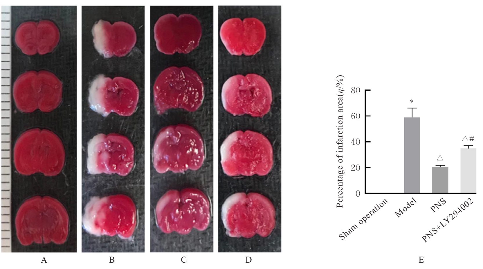
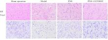
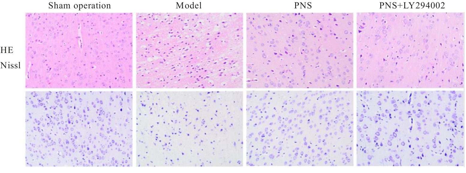
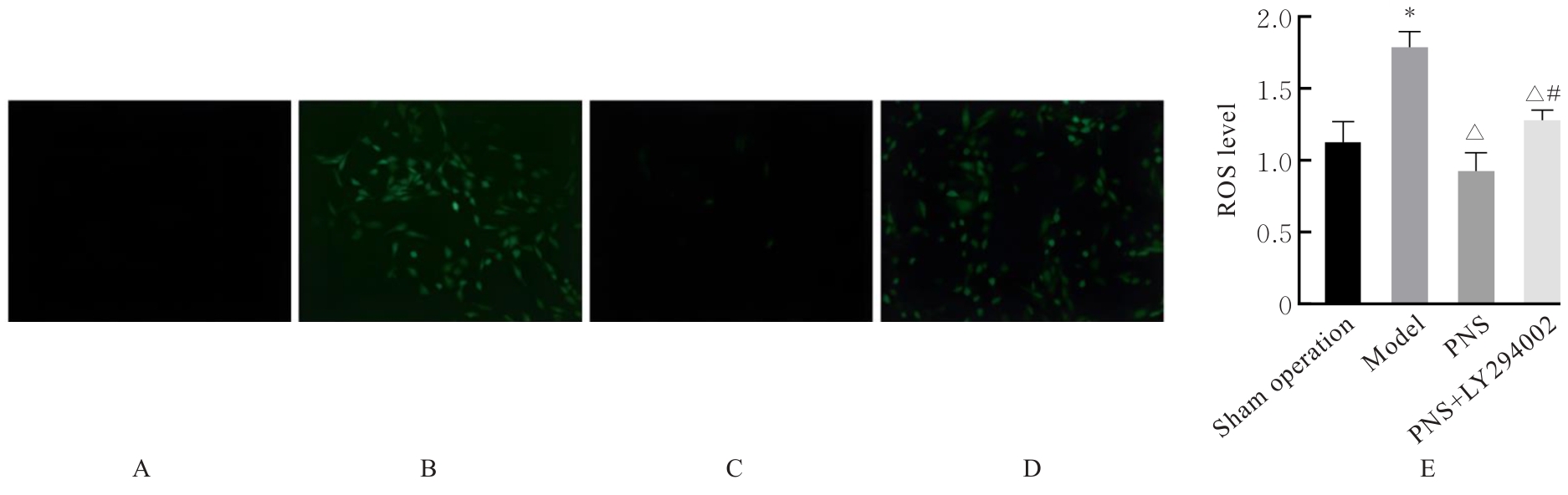
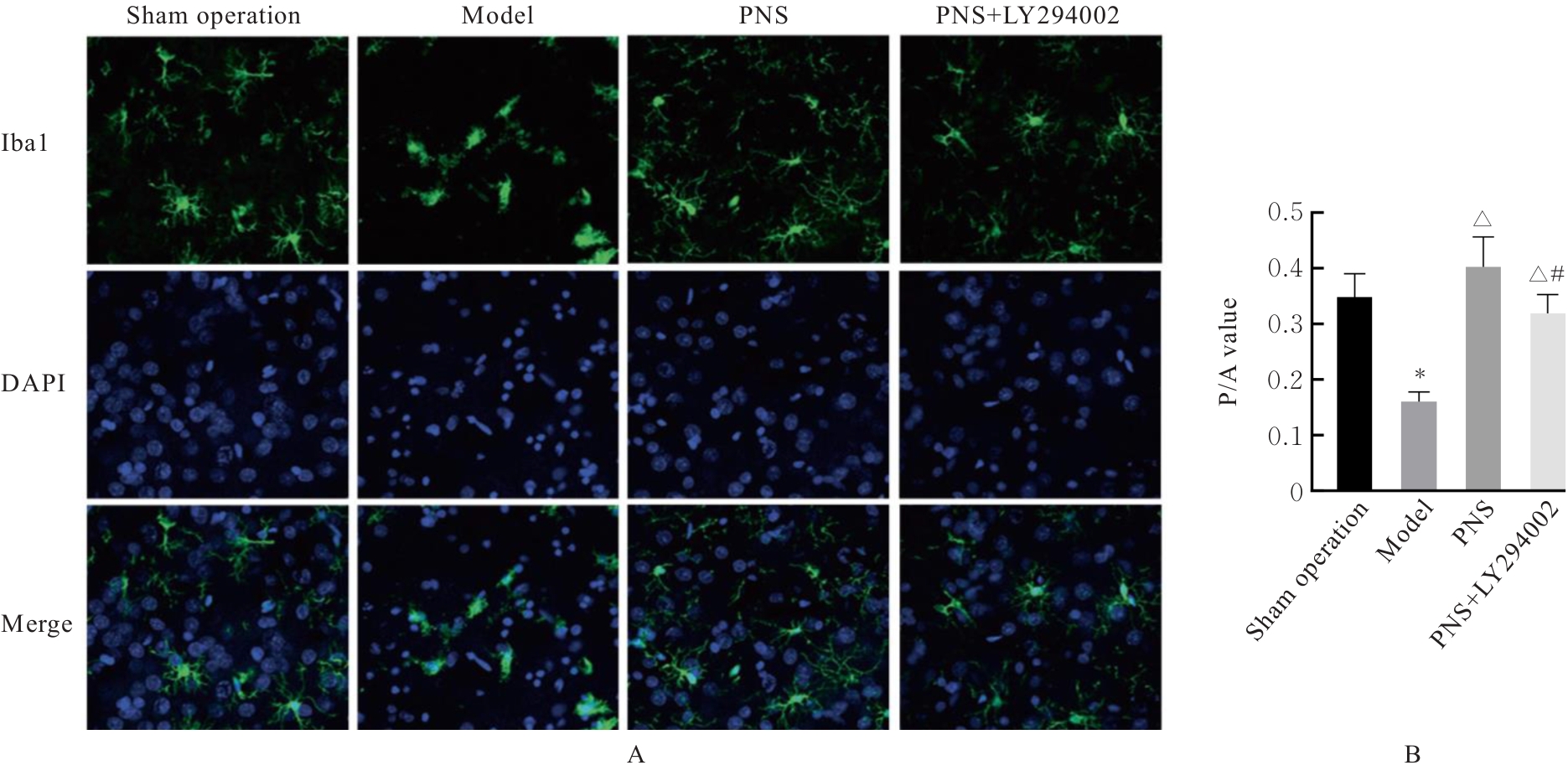
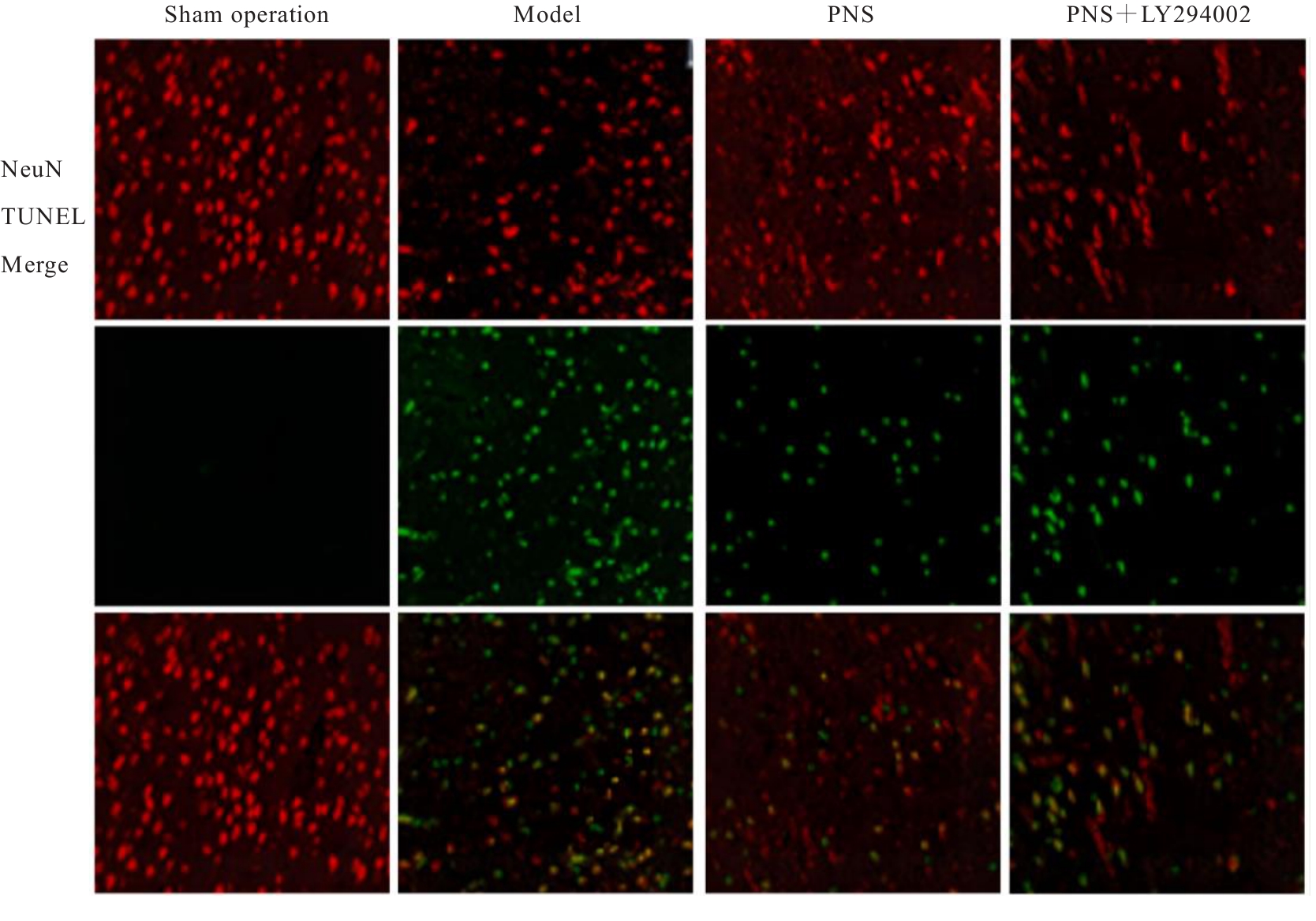
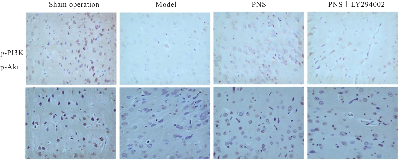
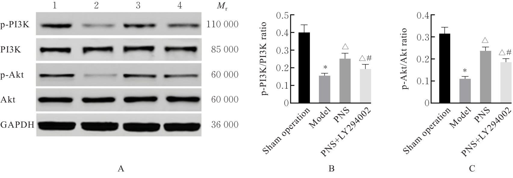
目的 探讨三七总皂苷(PNS)通过调节有氧糖酵解对大鼠缺血性脑卒中神经损伤的改善作用,并阐明其作用机制。 方法 将48只大鼠随机分为假手术组、模型组、PNS组(40 mg·kg-1)和PNS+磷脂酰肌醇3-激酶(PI3K)抑制剂LY294002组(5 mg·kg-1)(PNS+LY294002组),每组12只。除假手术组外,其余组大鼠采用大脑中动脉闭塞/再灌注(MCAO/R)法建立大鼠缺血性脑卒中模型。采用Longa评分法评估各组大鼠神经功能缺损情况,氯化三苯基四氮唑(TTC)染色法检测各组大鼠脑梗死面积百分比,HE染色和尼氏(Nissl)染色观察各组大鼠脑组织病理及神经元形态表现,酶联免疫吸附试验(ELISA)法检测各组大鼠脑组织中乳酸脱氢酶A(LDHA)、白细胞介素(IL)-1β和IL-18水平,实时荧光定量PCR(RT-qPCR)法检测各组大鼠脑组织中己糖激酶(HK)1、HK2、磷酸果糖激酶(PFK)1、PFK2和M2型丙酮酸激酶同工酶(PKM2)mRNA表达水平,免疫荧光染色法检测各组大鼠脑组织细胞中活性氧(ROS)水平和离子化钙结合衔接分子1(Iba1)阳性小胶质细胞周长/面积比(P/A),末端脱氧核苷酸转移酶介导dUTP缺口末端标记(TUNEL)法检测各组大鼠脑组织神经元凋亡率,免疫组织化学染色法和Western blotting法检测各组大鼠脑组织中PI3K/蛋白激酶B(Akt)信号通路相关蛋白表达情况及磷酸化PI3K(p-PI3K)/PI3K和磷酸化Akt(p-Akt)/Akt比值。 结果 与假手术组比较,模型组大鼠神经功能缺损评分明显升高(P<0.05),脑梗死面积百分比明显升高(P<0.05),脑组织病理损伤严重。与模型组比较,PNS组和PNS+LY294002组大鼠神经功能缺损评分均明显降低(P<0.05),脑梗死面积百分比均明显降低(P<0.05),脑组织病理损伤减轻;与PNS组比较,PNS+LY294002组大鼠神经功能缺损评分明显升高(P<0.05),脑梗死面积百分比明显升高(P<0.05),脑组织病理损伤加重。ELISA法,与假手术组比较,模型组大鼠脑组织中LDHA、IL-1β和IL-18水平均明显升高(P<0.05);与模型组比较,PNS组和PNS+LY294002组大鼠脑组织中LDHA、IL-1β及IL-18水平均明显降低(P<0.05);与PNS组比较,PNS+LY294002组大鼠脑组织中LDHA、IL-1β和IL-18水平均明显升高(P<0.05)。RT-qPCR法,与假手术组比较,模型组大鼠脑组织中HK1、HK2、PFK1、PFK2和PKM2 mRNA表达水平均明显降低(P<0.05);与模型组比较,PNS组和PNS+LY294002组大鼠脑组织中HK1、HK2、PFK1、PFK2及PKM2 mRNA表达水平均明显升高(P<0.05);与PNS组比较,PNS+LY294002组大鼠脑组织中HK1、HK2、PFK1、PFK2和PKM2 mRNA表达水平均明显降低(P<0.05)。免疫荧光染色法,与假手术组比较,模型组大鼠脑组织细胞中ROS水平明显升高(P<0.05),Iba1阳性小胶质细胞P/A比值明显降低(P<0.05);与模型组比较,PNS组和PNS+LY294002组大鼠脑组织细胞中ROS水平均明显降低(P<0.05),Iba1阳性小胶质细胞P/A比值均明显升高(P<0.05);与PNS组比较,PNS+LY294002组大鼠脑组织细胞中ROS水平明显升高(P<0.05),Iba1阳性小胶质细胞P/A比值明显降低(P<0.05)。TUNEL法,与假手术组比较,模型组大鼠脑组织中神经元凋亡率明显升高(P<0.05);与模型组比较,PNS组和PNS+LY294002组大鼠脑组织中神经元凋亡率均明显降低(P<0.05);与PNS组比较,PNS+LY294002组大鼠脑组织中神经元凋亡率明显升高(P<0.05)。与假手术组比较,模型组大鼠脑组织中p-PI3K/PI3K和p-Akt/Akt比值均明显降低(P<0.05);与模型组比较,PNS组和PNS+LY294002组大鼠脑组织中p-PI3K/PI3K及p-Akt/Akt比值均明显升高(P<0.05);与PNS组比较,PNS+LY294002组大鼠脑组织中p-PI3K/PI3K和p-Akt/Akt比值均明显降低(P<0.05)。 结论 PNS可有效改善MCAO/R大鼠神经功能缺损,降低脑组织中ROS水平,其作用机制可能与激活PI3K/Akt信号通路、提高糖酵解能力,从而促进小胶质细胞的代谢重编程有关。

关键词: 三七总皂苷, 脑缺血/再灌注损伤, 有氧糖酵解, 细胞凋亡, 缺血性脑卒中

Abstract:

Objective To investigate the improvement effect of Panax notoginseng saponins (PNS) on neurological injury in the rats with ischemic stroke by regulating aerobic glycolysis, and to elucidate its mechanism. Methods Forty-eight rats were randomly divided into sham operation group, model group, PNS group (40 mg·kg-1), and PNS+phosphatidylinositol 3-kinase (PI3K) inhibitor LY294002 group (5 mg·kg-1) PNS+LY294002 group, with 12 rats in each group. Except for sham operation group, the rats in other groups were subjected to middle cerebral artery occlusion/reperfusion (MCAO/R) method to establish ischemic stroke models. Longa scoring method was used to evaluate the neurological deficit of the rats in various groups. Triphenyltetrazolium chloride (TTC) staining method was used to detect the percentages of cerebral infarction area of the rats in various groups. HE staining and Nissl staining methods were used to observe the pathological changes and neuronal morphology in the brain tissue of the rats in various groups. Enzyme-linked immunosorbent assay (ELISA) method was used to detect the levels of lactate dehydrogenase A (LDHA), interleukin (IL)-1β, and IL-18 in the brain tissue of the rats in various groups. Real-time fluorescence quantitative PCR (RT-qPCR) method was used to detect the expression levels of hexokinase (HK)1, HK2, phosphofructokinase(PFK)1, PFK2, and pyruvate kinase M2 (PKM2) mRNA in the brain tissue of the rats in various groups. Immunofluorescence staining method was used to detect the levels of reactive oxygen species (ROS) in the cells and the perimeter/area (P/A) ratio of ionized calcium-binding adapter molecule 1 (Iba1)-positive microglia in the brain tissue of the rats in various groups. Terminal deoxynucleotidyl transferase dUTP nick end labeling(TUNEL) method was used to detect the neuronal apoptosis rates in the brain tissue of the rats in various groups. Immunohistochemistry method and Western blotting method were used to detect the expression levels of PI3K/protein kinase B (Akt) pathway-related proteins and the ratios of phosphorylated PI3K (p-PI3K)/PI3K and phosphorylated Akt (p-Akt)/Akt in the brain tissue of the rats in various groups. Results Compared with sham operation group, the neurological deficit score of the rats in model group was significantly increased (P<0.05), the percentage of cerebral infarction area was significantly increased (P<0.05), and severe pathological damage was observed in the brain tissue of the rats; compared with model group, the neurological deficit scores of the rats in PNS group and PNS+LY294002 group were significantly decreased (P<0.05), the percentages of cerebral infarction area were significantly decreased(P<0.05), and the pathological damages in the brain tissue of the rats were alleviated; compared with PNS group, the neurological deficit score of the rats in PNS+LY294002 group was significantly increased (P<0.05), the percentage of cerebral infarction area was significantly increased (P<0.05), and the pathological damage in brain tissue was aggravated. The ELISA method results showed that compared with sham operation group, the levels of LDHA, IL-1β, and IL-18 in the brain tissue of the rats in model group were significantly increased (P<0.05). Compared with model group, the levels of LDHA, IL-1β, and IL-18 in the brain tissue of the rats in PNS group and PNS+LY294002 group were significantly decreased (P<0.05); compared with PNS group, the levels of LDHA, IL-1β, and IL-18 in the brain tissue of the rats in PNS+LY294002 group were significantly increased (P<0.05). The results of RT-qPCR method showed that compared with sham operation group, the expression levels of HK1, HK2, PFK1, PFK2, and PKM2 mRNA in the brain tissue of the rats in model group were significantly decreased (P<0.05). Compared with model group, the expression levels of the HK1, HK2, PFK1, PFK2, and PKM2 mRNA in the brain tissue of the rats in PNS group and PNS+LY294002 group were significantly increased (P<0.05); compared with PNS group, the expression levels of the HK1, HK2, PFK1, PFK2, and PKM2 mRNA in the brain tissue of the rats in PNS+LY294002 group were significantly decreased (P<0.05). The immunofluorescence staining results showed that compared with sham operation group, the level of ROS in cells in the brain tissue of the rats in model group was significantly increased (P<0.05), and the P/A ratio of Iba1-positive microglia in the brain tissue of the rats was significantly decreased(P<0.05); compared with model group, the levels of ROS in the cells in the brain tissue of the rats in PNS group and PNS+LY294002 group were significantly decreased (P<0.05), and the P/A ratios of Iba1-positive microglia in the brain tissue of the rats were significantly increased (P<0.05); compared with PNS group, the level of ROS in the cells in the brain tissue of the rats in PNS+LY294002 group was significantly increased (P<0.05), and the P/A ratio of Iba1-positive microglia in the brain tissue of the rats was significantly decreased (P<0.05). The TUNEL method results showed that compared with sham operation group, the neuronal apoptosis rate in the brain tissue of the rats in model group was significantly increased (P<0.05); Compared with model group, the neuronal apoptosis rates in the brain tissue of the rats in PNS group and PNS+LY294002 group were significantly decreased (P<0.05); compared with PNS group, the neuronal apoptosis rate in the brain tissue of the rats in PNS+LY294002 group was significantly increased (P<0.05). The Western blotting method results showed that compared with sham operation group, the ratios of p-PI3K/PI3K and p-Akt/Akt in the brain tissue of the rats in model group were significantly decreased (P<0.05); compared with model group, the ratios of p-PI3K/PI3K and p-Akt/Akt in the brain tissue of the rats in PNS group and PNS+LY294002 group were significantly increased (P<0.05); compared with PNS group, the ratios of p-PI3K/PI3K and p-Akt/Akt in the brain tissue of the rats in PNS+LY294002 group were significantly decreased (P<0.05). Conclusion PNS can effectively improve neurological deficit in the MCAO/R rats and reduce the level of ROS in brain tissue of the rats. Its mechanism may be related to activating the PI3K/Akt pathway, enhancing glycolytic capacity, and thereby promoting metabolic reprogramming of microglia.

Key words: Panax notoginseng saponins, Cerebral ischemia/reperfusion injury, Aerobic glycolysis, Apoptosis, Ischemia stroke

中图分类号: 

  • R743