吉林大学学报(医学版) ›› 2026, Vol. 52 ›› Issue (5): 1195-1205.doi: 10.13481/j.1671-587X.20260501

• 基础研究 •    

负载CeO₂的树枝状介孔二氧化硅纳米颗粒对小鼠颌骨骨髓间充质干细胞成骨分化的促进作用及其机制

胡雅雯1,付长慧2,杨燕美3,孟宪伟2,李朋礼4,刘泓琦1,张娜3,顾斌3()   

  1. 1.解放军总医院研究生院,北京 100853
    2.中国科学院理化技术研究所低温重点实验室 纳米材料可控制备与应用实验室,北京 100190
    3.解放军总医院第一医学中心口腔科,北京 100853
    4.空军军医大学口腔医院第二医疗中心,陕西 西安 710032
  • 收稿日期:2025-11-26 接受日期:2026-01-05 出版日期:2026-09-28 发布日期:2026-10-15
  • 通讯作者: 顾斌 E-mail:gubin000@sina.com
  • 作者简介:胡雅雯(1999-),女,山东省东营市人,在读硕士研究生,主要从事干细胞成骨分化方面的研究。
  • 基金资助:
    国家自然科学基金面上项目(62471492)

Promotive effect of osteogenic differentiation of mouse jaw bone marrow mesenchymal stem cells by CeO₂-loaded dendritic mesoporous silica nanoparticles and its mechanism

Yawen HU1,Changhui FU2,Yanmei YANG3,Xianwei MENG2,Pengli LI4,Hongqi LIU1,Na ZHANG3,Bin GU3()   

  1. 1.Graduate School,Chinese PLA General Hospital,Beijing 100853,China
    2.Laboratory of Controllable Preparation and Application of Nanomaterials,Key Laboratory of Cryogenics,Technical Institute of Physics and Chemistry,Chinese Academy of Sciences,Beijing 100190,China
    3.Department of Stomatology,First Medical Center,Chinese PLA General Hospital,Beijing 100853,China
    4.Second Medical Center,Stomatological Hospital,Air Force Military Medical University,Xi’an 710032,China
  • Received:2025-11-26 Accepted:2026-01-05 Online:2026-09-28 Published:2026-10-15
  • Contact: Bin GU E-mail:gubin000@sina.com

摘要:

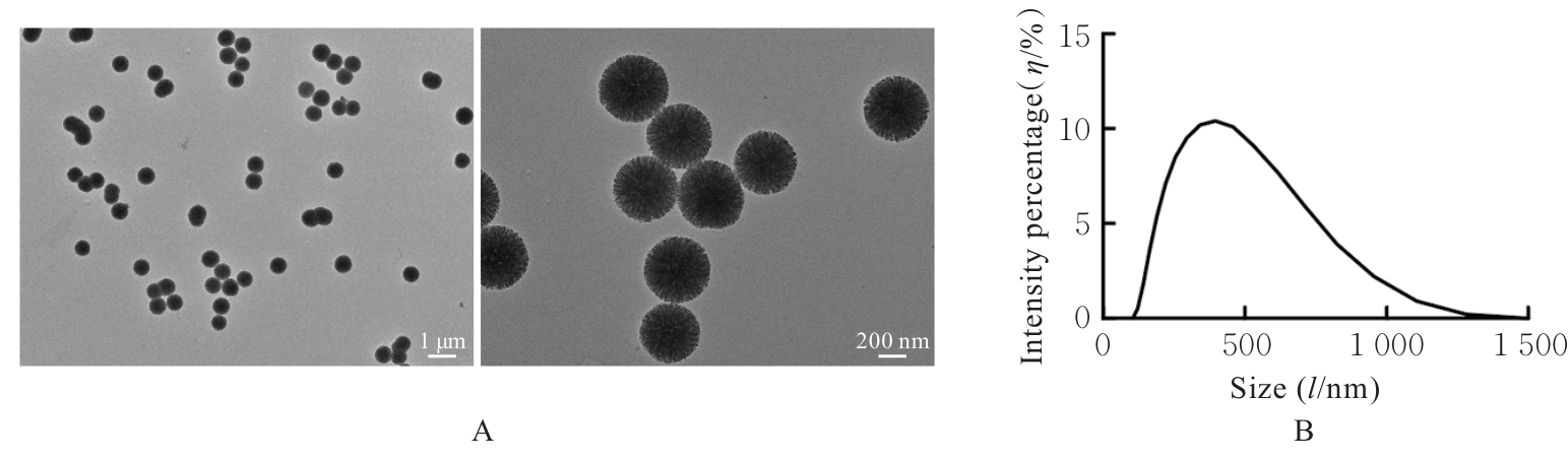
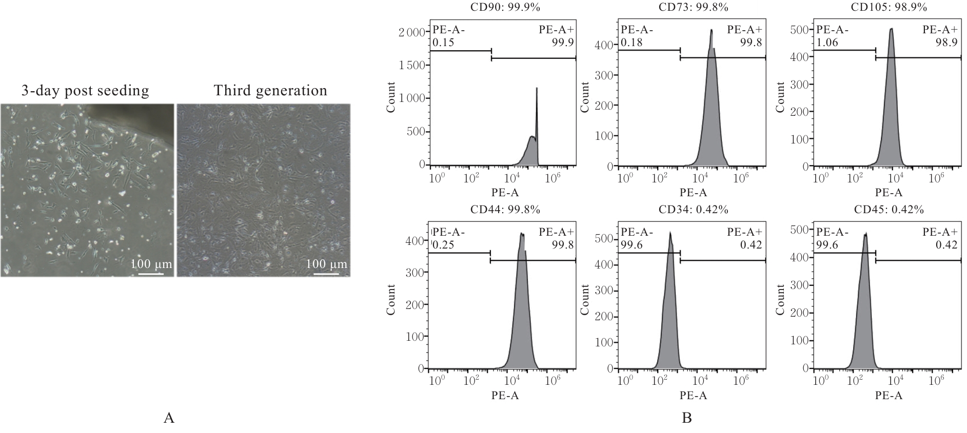
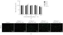
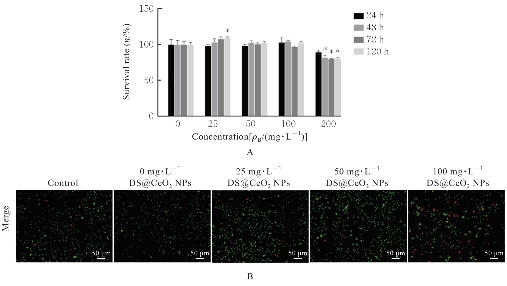
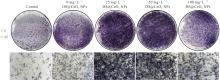
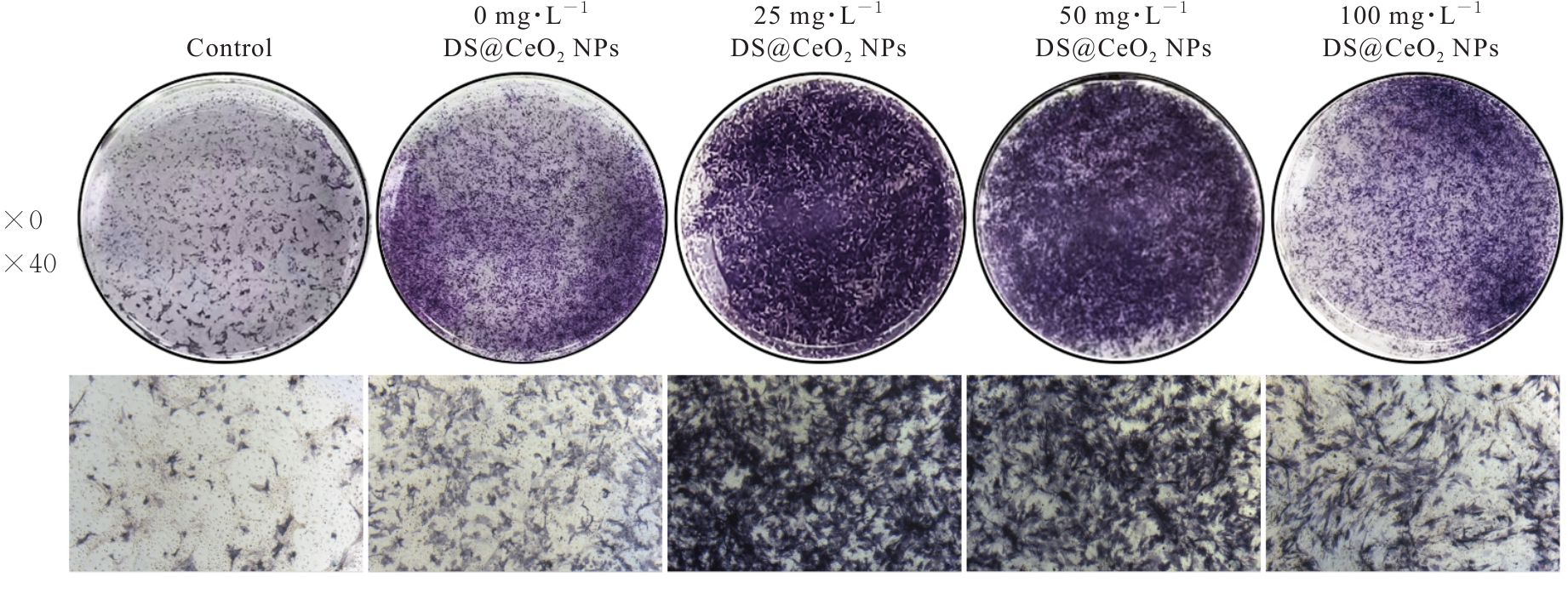
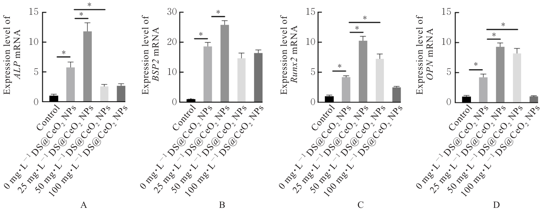
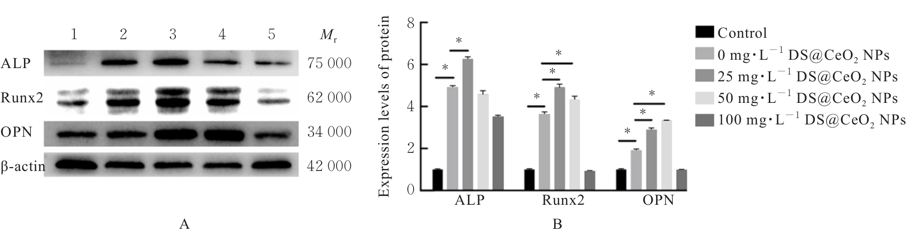
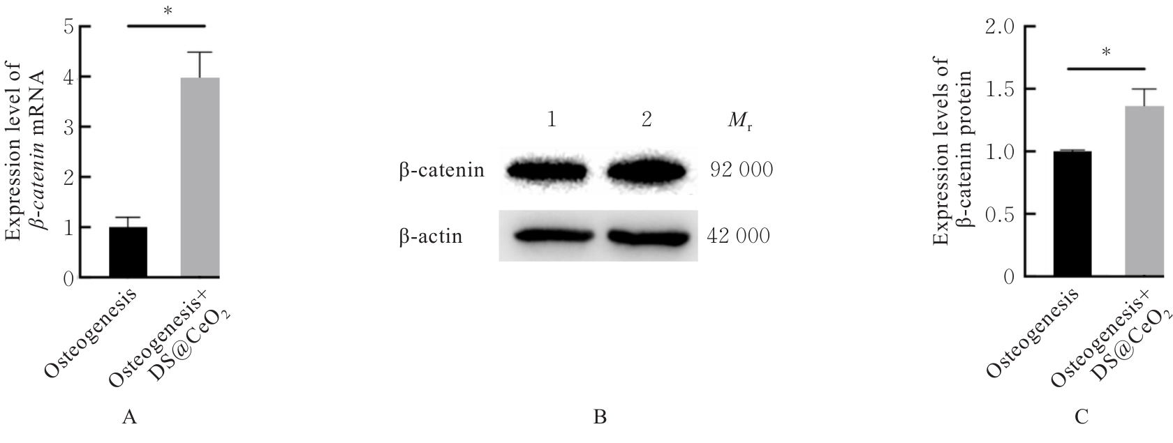
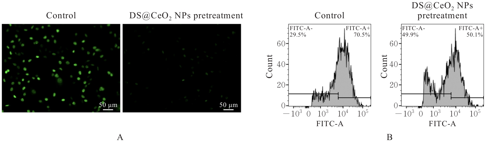
目的 探讨负载二氧化铈(CeO2)的树枝状介孔二氧化硅纳米颗粒(DS@CeO? NPs)对小鼠颌骨骨髓间充质干细胞(JBMMSCs)成骨分化的影响,并阐明其可能的作用机制。 方法 合成DS@CeO2 NPs,采用透射电子显微镜(TEM)和动态光散射法(DLS)检测DS@CeO? NPs的形貌和粒径。采用酶消法提取小鼠JBMMSCs,流式细胞术鉴定细胞表面标志物。将细胞分为空白对照组和不同浓度(25、50、100和200 mg·L-1)DS@CeO2 NPs组,采用细胞计数试剂盒8(CCK-8)法和Calcein/碘化丙啶(PI)染色法检测各组细胞增殖及存活情况。将细胞分为对照组和不同浓度(0、25、50和100 mg·L-1)DS@CeO? NPs成骨诱导组,成骨诱导7 d后,采用碱性磷酸酶(ALP)染色法检测各组细胞中ALP活性,实时荧光定量PCR(RT-qPCR)法检测各组细胞中ALP、Runt相关转录因子2(Runx2)、骨涎蛋白2(BSP2)和骨桥蛋白(OPN)mRNA表达水平,Western blotting法检测各组细胞中ALP、Runx2和OPN蛋白表达水平;另设单纯成骨组和成骨+DS@CeO2 NPs组,采用RT-qPCR法和Western blotting法检测2组细胞中β-连环蛋白(β-catenin)mRNA及蛋白表达水平。在过氧化氢(H2O2)刺激下,将细胞分为对照组和DS@CeO2 NPs预处理组,采用二氯二氢荧光素二乙酯(DCFH-DA)荧光染色法和流式细胞术检测2组细胞中活性氧(ROS)水平。 结果 TEM和DLS,DS@CeO? NPs呈近球形,平均水合粒径为378.2 nm。流式细胞术,细胞中CD73、CD90、CD105和CD44表达率均高于95%,CD34和CD45几乎不表达,可鉴定为JBMMSCs。CCK-8法,与对照组比较,25、50和100 mg·L-1 DS@CeO? NPs组细胞存活率差异无统计学意义(P>0.05),200 mg·L-1 DS@CeO? NPs组细胞存活率明显降低(P<0.05)。ALP染色法,与对照组和0 mg·L-1 DS@CeO2 NPs组比较,25和50 mg·L-1 DS@CeO2 NPs组细胞ALP染色较深且阳性染色面积较大,细胞ALP活性升高。RT-qPCR法和Western blotting法,与0 mg·L-1 DS@CeO? NPs组比较,25 mg·L-1 DS@CeO? NPs组细胞中ALP、BSP2、Runx2和OPN mRNA表达水平以及ALP、Runx2和OPN蛋白表达水平均明显升高(P<0.05)。与单纯成骨组比较,成骨+DS@CeO? NPs组细胞中β-catenin mRNA和蛋白表达水平均明显升高(P<0.05)。DCFH-DA染色法和流式细胞术,与对照组比较,DS@CeO? NPs预处理组细胞中ROS水平降低。 结论 DS@CeO? NPs具有良好的生物相容性,可促进JBMMSCs成骨分化,其作用机制可能与激活Wnt/β-catenin信号通路和抗氧化作用有关。

关键词: 二氧化铈, 树枝状介孔二氧化硅, 颌骨骨髓间充质干细胞, 成骨分化, 无翅型MMTV整合位点家族/β-连环蛋白信号通路, 活性氧

Abstract:

Objective To discuss the effects of CeO2-loaded dendritic mesoporous silica nanoparticles (DS@CeO2 NPs) on the osteogenic differentiation of mouse jaw bone marrow mesenchymal stem cells (JBMMSCs), and to clarify its possible mechanism. Methods DS@CeO2 NPs were synthesized. Transmission electron microscopy (TEM) and dynamic light scattering (DLS) method were used to detect the morphology and particle size of DS@CeO2 NPs. Enzyme digestion method was used to isolate mouse JBMMSCs. Flow cytometry method was used to identify the cell surface markers on JBMMSCs. The cells were divided into blank control group and different concentrations of DS@CeO2 NPs (25, 50, 100, and 200 mg·L-1) groups. Cell counting kit-8 (CCK-8) method and Calcein/propidium iodide (PI) staining method were used to detect the proliferation and survival of the cells in various groups. The cells were further divided into control group and different concentrations of DS@CeO2 NPs osteogenic induction groups (0, 25, 50, and 100 mg·L-1). After 7 d of osteogenic induction, alkaline phosphatase (ALP) staining method was used to detect the ALP activities in the cells in various groups. Real-time fluorescence quantitative PCR (RT-qPCR) method was used to detect the expression levels of ALP, Runt-related transcription factor 2 (Runx2), bone sialoprotein 2 (BSP2), and osteopontin (OPN) mRNA in the cells in various groups. Western blotting method was used to detect the expression levels of ALP, Runx2, and OPN proteins in the cells in various groups. In addition, osteogenesis group and osteogenesis+DS@CeO2 NPs group were established. RT-qPCR method and Western blotting method were used to detect the expression levels of β-catenin mRNA and protein in the cells in two groups. Under hydrogen peroxide (H2O2) stimulation, the cells were divided into control group and DS@CeO2 NPs pretreatment group. 2',7'-dichlorodihydrofluorescein diacetate (DCFH-DA) fluorescence staining method and flow cytometry method were used to detect the levels of reactive oxygen species (ROS) in the cells in two groups. Results The TEM and DLS results showed that DS@CeO2 NPs exhibited a nearly spherical morphology with an average hydrated particle size of 378.2 nm. The flow cytometry results showed that the expression rates of CD73, CD90, CD105, and CD44 in the cells were all higher than 95%, while CD34 and CD45 were almost not expressed, so the cells could be identified as JBMMSCs. The CCK-8 method results showed that compared with control group, there was no statistically significant difference in the survival rates of the cells in 25, 50, and 100 mg·L-1 DS@CeO2 NPs groups (P>0.05), while the survival rate of the cells in 200 mg·L-1 DS@CeO2 NPs group was significantly decreased (P<0.05). The ALP staining results showed that compared with control group and 0 mg·L-1 DS@CeO2 NPs group, the cells in 25 and 50 mg·L-1 DS@CeO2 NPs groups exhibited darker ALP staining and larger positive staining areas, and the ALP activities in the cells were increased. The RT-qPCR and Western blotting results showed that compared with 0 mg·L-1 DS@CeO2 NPs group, the expression levels of ALP, BSP2, Runx2, and OPN mRNA as well as the expression levels of ALP, Runx2, and OPN proteins in the cells in 25 mg·L-1 DS@CeO2 NPs group were significantly increased (P<0.05). Compared with pure osteogenic group, the expression levels of β-catenin mRNA and protein in the cells in osteogenic+DS@CeO2 NPs group were significantly increased (P<0.05). The DCFH-DA staining and flow cytometry results showed that compared with control group, the ROS level in the cells in DS@CeO2 NPs pretreatment group was decreased. Conclusion DS@CeO2 NPs show good biocompatibility and can promote the osteogenic differentiation of JBMMSCs; the mechanism may be related to activation of Wnt/β-catenin signaling pathway and antioxidant effect.

Key words: Cerium oxide, Dendritic mesoporous silica, Jaw bone marrow mesenchymal stem cells, Osteogenic differentiation, Wingless-type MMTV integration site family/β-catenin signaling pathway, Reactive oxygen species

中图分类号: 

  • R584.2