Journal of Jilin University(Medicine Edition) ›› 2026, Vol. 52 ›› Issue (3): 590-601.doi: 10.13481/j.1671-587X.20260302
• Research in basic medicine • Previous Articles Next Articles
Ameliorative effect of Pueraria lobata-derived exosome-like nanovesicles on D-galactose-induced senescence in L929 mouse fibroblasts and its mechanism
Chunrong XIANG,Yong HE,Nuo JIANG,Shouqi HUANG,Li HONG( )
)
- Department of Obstetrics and Gynecology,People’s Hospital,Wuhan University,Wuhan 430060,China
-
Received:2025-09-30Accepted:2025-11-02Online:2026-05-28Published:2026-06-08 -
Contact:Li HONG E-mail:dr_hongli@whu.edu.cn

CLC Number:
- R711.4
Cite this article
Chunrong XIANG,Yong HE,Nuo JIANG,Shouqi HUANG,Li HONG. Ameliorative effect of Pueraria lobata-derived exosome-like nanovesicles on D-galactose-induced senescence in L929 mouse fibroblasts and its mechanism[J].Journal of Jilin University(Medicine Edition), 2026, 52(3): 590-601.
share this article
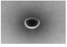
Fig. 1
Morphology of PL-ELNs observed by TEM (×100 000)"
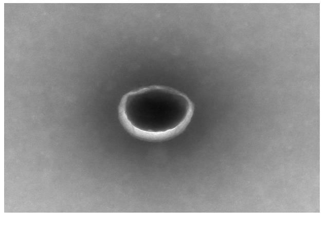
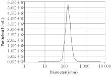
Fig. 2
Particle sizes of PL-ELNs detected by NTA method"
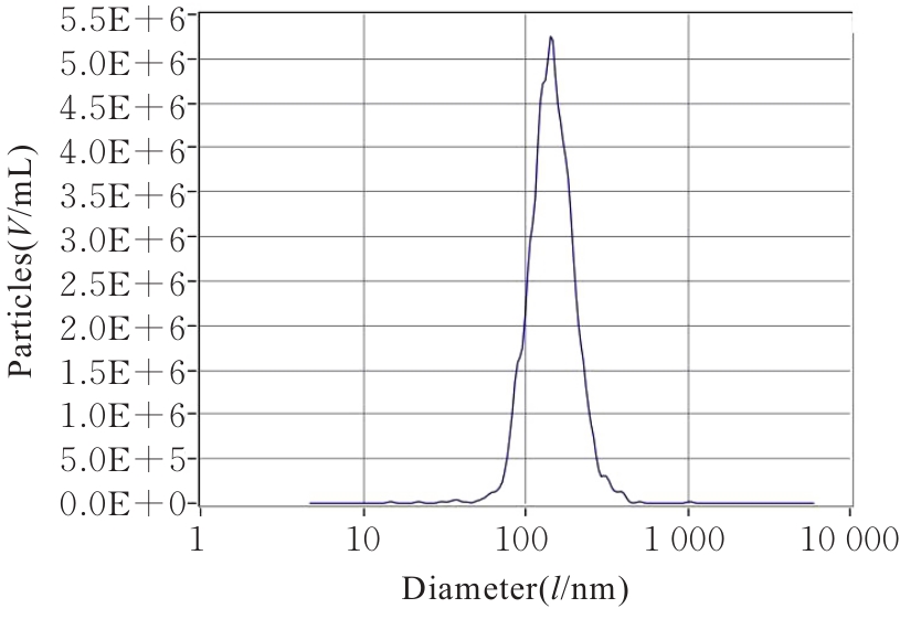
Fig. 3
Uptake of PL-ELNs in L929 cells observed under confocal fluorescence microscope(PKH26 fluorescence dye tracer, ×400)"
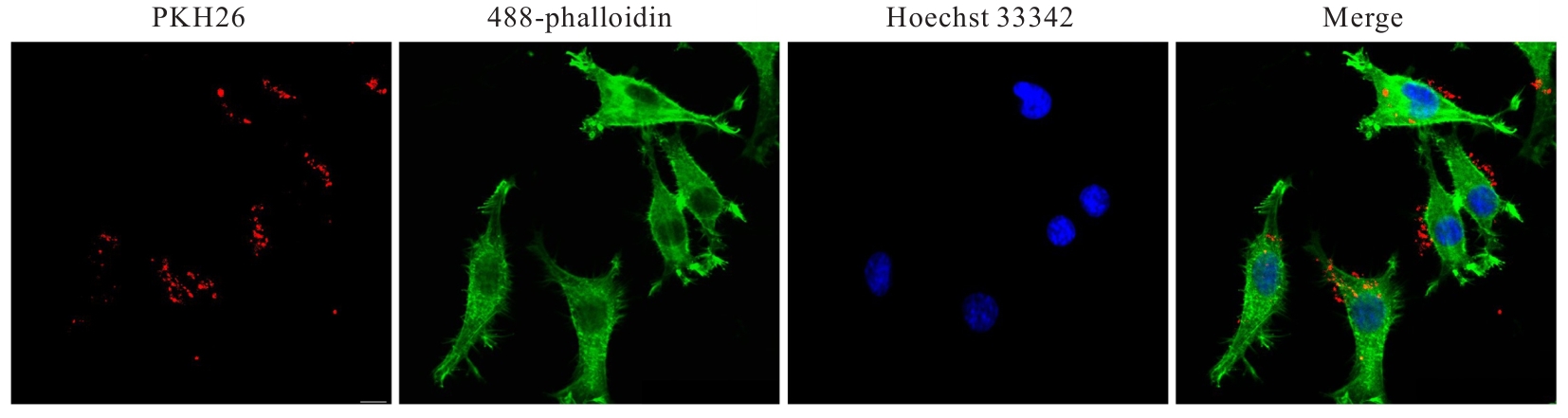
Fig. 4
Activities of L929 cells in various groups after treated with different concentrations of D-gal for 48 h"
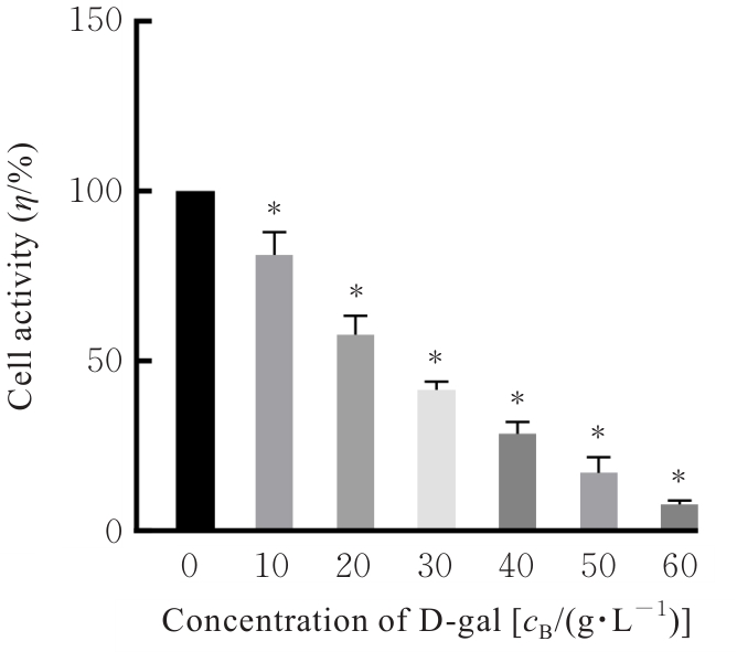
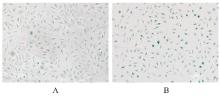
Fig. 5
Morphology of L929 cells in two groups observed by SA-β-gal staining (×200)"
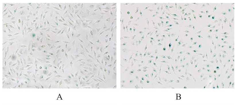
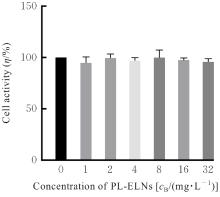
Fig. 6
Activities of L929 cells in various groups after treated with different concentrations of PL-ELNs for 48 h"
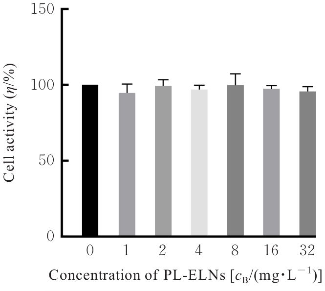
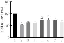
Fig. 7
Activities of L929 cells in various groups"
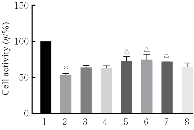

Fig. 8
SA-β-gal staining of L929 cells in various groups(×200)"

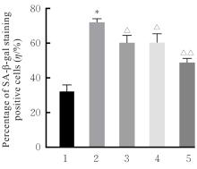
Fig. 9
Percentages of SA-β-gal staining positive L929 cells in various groups"
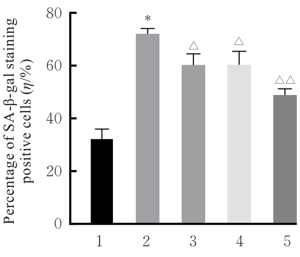
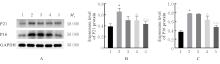
Fig. 10
Electrophoregram(A) and histograms(B,C) of expressions of P21 and P16 proteins in L929 cells in various groups"
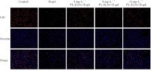
Fig. 11
Proliferation of L929 cells in various groups (EdU staining,×200)"
Fig. 12
Edu positive rates of L929 cells in various groups"
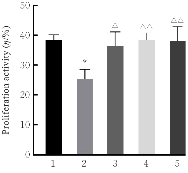
Fig. 13
Flow cytometry diagram(A) and histogram(B) of ROS levels in L929 cells in various groups"
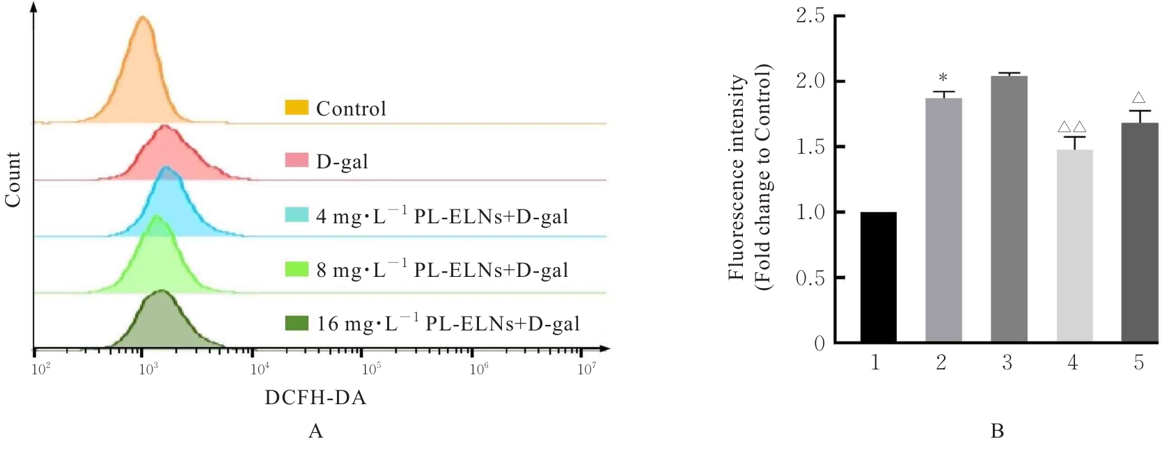
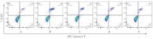
Fig. 14
Apoptotic rates of L929 cells in various groups detected by flow cytometry"
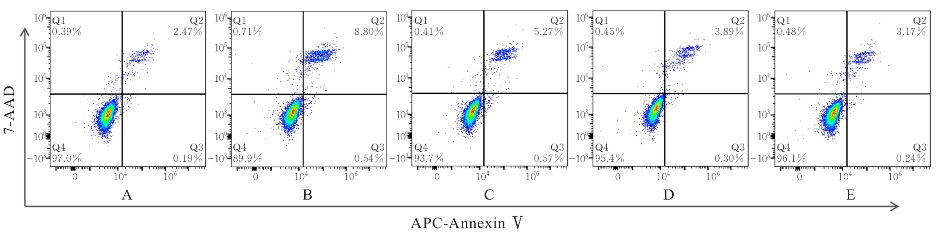
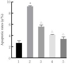
Fig. 15
Apoptotic rates of L929 cells in various groups"
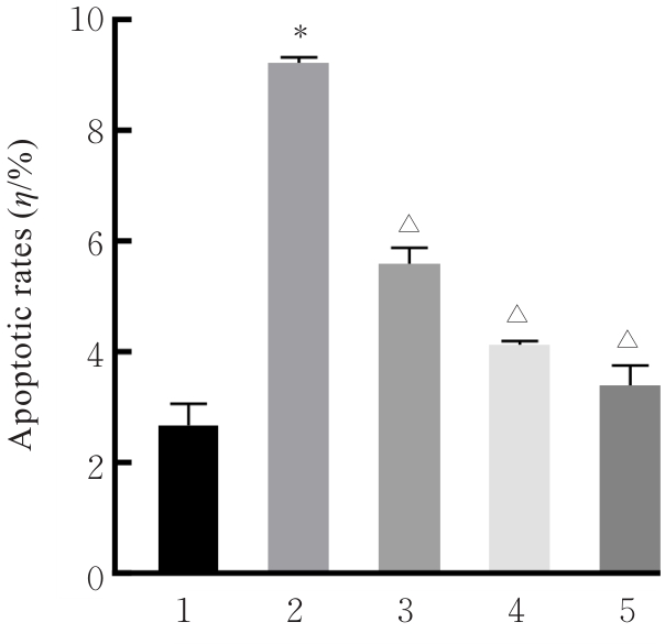
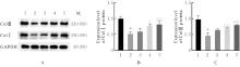
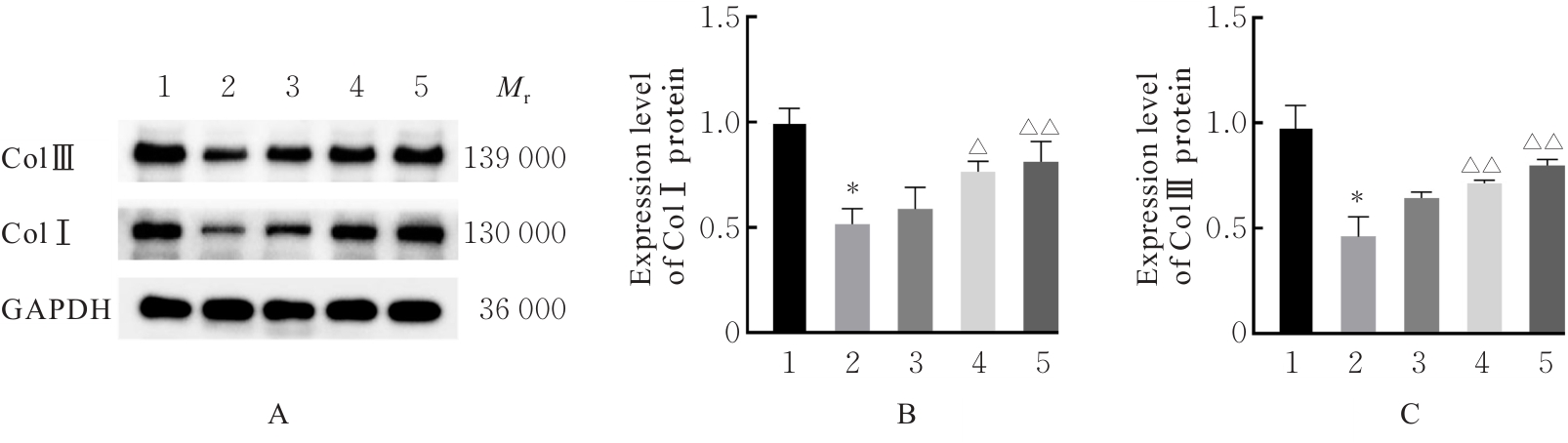
Fig. 16
Electrophoregram(A) and histograms(B, C) of expressions of ColⅠ and ColⅢ proteins in L929 cells in various groups"
| [1] | PEINADO-MOLINA R A, HERNÁNDEZ-MARTÍNEZ A, MARTÍNEZ-VÁZQUEZ S, et al. Pelvic floor dysfunction: prevalence and associated factors[J]. BMC Public Health, 2023, 23(1): 2005. |
| [2] | MANNELLA P, PALLA G, BELLINI M, et al. The female pelvic floor through midlife and aging[J]. Maturitas, 2013, 76(3): 230-234. |
| [3] | DOUMOUCHTSIS S K, CHRYSANTHOPOULOU E L. Urogenital consequences in ageing women[J]. Best Pract Res Clin Obstet Gynaecol, 2013, 27(5): 699-714. |
| [4] | MENACHEM-ZIDON OBEN, REUBINOFF B, SHVEIKY D. Transplantation of mesenchymal stem cells derived from old rats improves healing and biomechanical properties of vaginal tissue following surgical incision in aged rats[J]. Int J Mol Sci, 2024, 25(11): 5714. |
| [5] | PETROS P E. Re: Alterations in connective tissue metabolism in stress incontinence and prolapse[J]. J Urol, 2012, 187(6): 2281. |
| [6] | FANG C Y, ZENG Z T, YE J S, et al. Progress of mesenchymal stem cells affecting extracellular matrix metabolism in the treatment of female stress urinary incontinence[J]. Stem Cell Res Ther, 2025, 16(1): 95. |
| [7] | ZHAO B, LIN H J, JIANG X C, et al. Exosome-like nanoparticles derived from fruits, vegetables, and herbs: innovative strategies of therapeutic and drug delivery[J]. Theranostics, 2024, 14(12): 4598-4621. |
| [8] | CAO M, DIAO N N, CAI X L, et al. Plant exosome nanovesicles (PENs): green delivery platforms[J]. Mater Horiz, 2023, 10(10): 3879-3894. |
| [9] | FENG H, YUE Y, ZHANG Y, et al. Plant-derived exosome-like nanoparticles: emerging nanosystems for enhanced tissue engineering[J]. Int J Nanomedicine, 2024, 19: 1189-1204. |
| [10] | KALLURI R, LEBLEU V S. The biology, function, and biomedical applications of exosomes[J]. Science, 2020, 367(6478): eaau6977. |
| [11] | SUN Z X, ZHENG Y Z, WANG T R, et al. Aloe vera gel and rind-derived nanoparticles mitigate skin photoaging via activation of Nrf2/ARE pathway[J]. Int J Nanomedicine, 2025, 20: 4051-4067. |
| [12] | CHOI W, CHO J H, PARK S H, et al. Ginseng root-derived exosome-like nanoparticles protect skin from UV irradiation and oxidative stress by suppressing activator protein-1 signaling and limiting the generation of reactive oxygen species[J]. J Ginseng Res, 2024, 48(2): 211-219. |
| [13] | SRIWASTVA M K, DENG Z B, WANG B M, et al. Exosome-like nanoparticles from Mulberry bark prevent DSS-induced colitis via the AhR/COPS8 pathway[J]. EMBO Rep, 2022, 23(3): e53365. |
| [14] | HSU P, KAMIJYO Y, KOIKE E, et al. Exosome-like nanovesicles derived from kale juice enhance collagen production by downregulating Smad7 in human skin fibroblasts[J]. Front Nutr, 2025, 12: 1486572. |
| [15] | JEON Y D, LEE J H, LEE Y M, et al. Puerarin inhibits inflammation and oxidative stress in dextran sulfate sodium-induced colitis mice model[J]. Biomedecine Pharmacother, 2020, 124: 109847. |
| [16] | TIYASATKULKOVIT W, MALAIVIJITNOND S, CHAROENPHANDHU N, et al. Pueraria mirifica extract and puerarin enhance proliferation and expression of alkaline phosphatase and type Ⅰ collagen in primary baboon osteoblasts[J]. Phytomedicine, 2014, 21(12): 1498-1503. |
| [17] | KIM M, KIM H S, OH J, et al. Ameliorative effects of Kyung-Ok-Ko and its mixture with Pueraria lobata Ohwi on postmenopausal osteoporosis by promoting phytoestrogenic activity in rats[J]. Front Nutr, 2023, 10: 1171346. |
| [18] | KAMIYA Y, ODAMA M, MIZUGUTI A, et al. Puerarin blocks the aging phenotype in human dermal fibroblasts[J]. PLoS One, 2021, 16(4): e0249367. |
| [19] | LIU S, HU H J, ZHANG M, et al. Puerarin delays mammary gland aging by regulating gut microbiota and inhibiting the p38MAPK signaling pathway[J]. J Agric Food Chem, 2024, 72(19): 10879-10896. |
| [20] | LI Y, HONG L, LIU C, et al. Effect of puerarin on collagen metabolism of fibroblasts in pelvic tissue of women with pelvic organ prolapse[J]. Mol Med Rep, 2018, 17(2): 2705-2711. |
| [21] | LU Y, XU J, TANG R, et al. Edible pueraria Lobata-derived exosome-like nanovesicles ameliorate dextran sulfate sodium-induced colitis associated lung inflammation through modulating macrophage polarization[J]. Biomedecine Pharmacother, 2024, 170: 116098. |
| [22] | ZHAN W Q, DENG M Z, HUANG X Q, et al. Pueraria lobata-derived exosome-like nanovesicles alleviate osteoporosis by enhacning autophagy[J]. J Control Release, 2023, 364: 644-653. |
| [23] | ZHANG J, YU H Y, MAN M Q, et al. Aging in the dermis: Fibroblast senescence and its significance[J]. Aging Cell, 2024, 23(2): e14054. |
| [24] | WANG B M, ZHUANG X Y, DENG Z B, et al. Targeted drug delivery to intestinal macrophages by bioactive nanovesicles released from grapefruit[J]. Mol Ther, 2014, 22(3): 522-534. |
| [25] | PERUT F, RONCUZZI L, AVNET S, et al. Strawberry-derived exosome-like nanoparticles prevent oxidative stress in human mesenchymal stromal cells[J]. Biomolecules, 2021, 11(1): 87. |
| [26] | CHEN Q B, ZU M H, GONG H L, et al. Tea leaf-derived exosome-like nanotherapeutics retard breast tumor growth by pro-apoptosis and microbiota modulation[J]. J Nanobiotechnology, 2023, 21(1): 6. |
| [27] | HUANG Y, LIU H B, SUN X L, et al. Honeysuckle-derived microRNA2911 directly inhibits varicella-zoster virus replication by targeting IE62 gene[J]. J Neurovirol, 2019, 25(4): 457-463. |
| [28] | YAN J Y, CHEN S Y, YI Z M, et al. The role of p21 in cellular senescence and aging-related diseases[J]. Mol Cells, 2024, 47(11): 100113. |
| [29] | STOJANOVIC B, JOVANOVIC I, DIMITRIJEVIC STOJANOVIC M, et al. Oxidative stress-driven cellular senescence: mechanistic crosstalk and therapeutic horizons[J]. Antioxidants (Basel), 2025, 14(8): 987. |
| [30] | VON ZGLINICKI T. Oxidative stress and cell senescence as drivers of ageing: Chicken and egg[J]. Ageing Res Rev, 2024, 102: 102558. |
| [1] | Lina XU,Xiaoshuang HE,Wenyan XIN,Chao WU. Improvement effect of bone marrow mesenchymal stem cell-derived exosomes on airway resistance and lung tissue lesion in mice of asthmatic models and its mechanism [J]. Journal of Jilin University(Medicine Edition), 2026, 52(3): 602-611. |
| [2] | Nuo JIANG,Shufei ZHANG,Li HONG. Effect of selenoprotein N1 gene on senescence of L929 cells and its mechanism [J]. Journal of Jilin University(Medicine Edition), 2026, 52(3): 612-620. |
| [3] | Lingyu WANG,Ruili LI,Yu ZHANG,Meitong GUO,Yalong LIANG,Xu HAN,Xiaowei HUANG,Yang ZHANG,Xiuhua YU. Improvement effect of Renshen Jianzhi Tablets on learning and memory ability in cognitive impairment model rats and its protective effect on hippocampal neurons and its mechanism [J]. Journal of Jilin University(Medicine Edition), 2026, 52(3): 719-729. |
| [4] | Heqiang JIA,Lu PAN,Zhenjiang WANG,Jiayu LU,Dan WANG,Lifeng LIU,Ying DONG,Chunyan YU. Improving effect of Panaxatriol saponins on D-galactose-induced skeletal muscle aging of mice and its mechanism [J]. Journal of Jilin University(Medicine Edition), 2026, 52(3): 730-737. |
| [5] | Ning MA,Shuang LIU,Chengyao WANG,Jiajun LU,Linyu CHEN,Ziyi WANG,Weiqin ZHAO,Weitong WANG,Pengcheng CHE,Hong SUN. Improvement effect of topical application of exosome loaded hydrogel DCC on peritoneal adhesion in mice and its mechanism [J]. Journal of Jilin University(Medicine Edition), 2026, 52(2): 318-329. |
| [6] | Di LI,Yang SU,Yong ZHANG,Jiaoqi WANG,Xiaofei ZHANG,Xuebing ZHAO. Expression levels of miR-328-3p and miR-671-5p in serum exosomes of Kawasaki disease model mice and their significance [J]. Journal of Jilin University(Medicine Edition), 2026, 52(2): 359-365. |
| [7] | Tian LI,Jiafeng WANG,Zhimin ZHANG. Research progress in application of exosomes in diagnosis and treatment of oral diseases [J]. Journal of Jilin University(Medicine Edition), 2026, 52(2): 543-550. |
| [8] | Huiying YANG,Weihong LIANG,Xinru XU,Haodan SUN,Xin JIAO,Haiping WANG. Research progress in effect of bone marrow mesenchymal stem cells on differentiation induction of cardiomyocyte-like cells, protection of cardiomyocytes, and myocardial-related diseases [J]. Journal of Jilin University(Medicine Edition), 2026, 52(1): 281-289. |
| [9] | Xiaoqian TANG,Shengcong WEN,Zhenya DONG,Jingyi CHEN,Yu CAO,Yunhua ZHANG. Improvement effect of engineered exosomes delivering ANGPTL6 mRNA on liver fibrosis in mice [J]. Journal of Jilin University(Medicine Edition), 2025, 51(6): 1452-1463. |
| [10] | Han SUN,Weilun SUN,Huifeng WANG,Wenli MA,Huali XU,Wenwen FU. Optimization of preparation method for D-galactose-induced mouse model of aging-related cognitive dysfunction [J]. Journal of Jilin University(Medicine Edition), 2025, 51(6): 1464-1474. |
| [11] | Jing LIU,Yan WANG,Xu HUANG. Promoting effect of overexpressed circRNAs-modified dental pulp stem cell-derived exosomes on angiogenesis of human umbilical vein endothelial cells [J]. Journal of Jilin University(Medicine Edition), 2025, 51(6): 1561-1570. |
| [12] | Meng XIANG,Bing XU,Peisha WANG,Siqi LIU,Shaohua ZHANG. Effect of exosomes loaded with miR-520a-5p on pregnancy outcomes in fetal mice with intrauterine growth restriction and its mechanism [J]. Journal of Jilin University(Medicine Edition), 2025, 51(5): 1230-1239. |
| [13] | Xiaoshuang HE,Lina XU,Mei CUI,Yu ZHAO,Bei WANG,Zheng HUANG,Yuchao WANG,Wenyan XIN,Chao WU. Effects of lncRNA DUXAP8 in lung cancer A549 cells-derived exosomes on lung cancer cell growth and its mechnism [J]. Journal of Jilin University(Medicine Edition), 2025, 51(4): 958-967. |
| [14] | DILIXIATI·Dilidaer,Lin JIA. Improvement effect of exosomes derived from human adipose-derived stem cells and human dermal fibroblasts on ultraviolet-induced photoaging skin wrinkles in nude mice [J]. Journal of Jilin University(Medicine Edition), 2025, 51(3): 621-631. |
| [15] | Junping WEI,Dajia FU,Qingwen MENG,Daofei LIN,Yanzai LIN. Effect of bone marrow mesenchymal stem cell-derived exosomes on myocardial fibrosis in rats induced by isoproterenol and its mechanism [J]. Journal of Jilin University(Medicine Edition), 2024, 50(5): 1348-1357. |