Journal of Jilin University(Medicine Edition) ›› 2026, Vol. 52 ›› Issue (5): 1263-1272.doi: 10.13481/j.1671-587X.20260508
• Research in basic medicine • Previous Articles
Promotion effect of TXNDC9 gene knockdown on apoptosis and autophagy of colorectal cancer cells and its influence in xenograft tumor in nude mice
Yu ZHANG,Shuguang LI( ),Mingfu CAO,Di HUANG
- Department of Gastrointestinal Tumor Surgery,First Affiliated Hospital,Hebei North University,Zhangjiakou 075000,China
-
Received:2025-10-27Accepted:2026-01-01Online:2026-09-28Published:2026-10-15 -
Contact:Shuguang LI E-mail:lsg3451@163.com

CLC Number:
- R735.3
Cite this article
Yu ZHANG,Shuguang LI,Mingfu CAO,Di HUANG. Promotion effect of TXNDC9 gene knockdown on apoptosis and autophagy of colorectal cancer cells and its influence in xenograft tumor in nude mice[J].Journal of Jilin University(Medicine Edition), 2026, 52(5): 1263-1272.
share this article
Fig. 1
Electrophoregram(A) and histogram(B) of expressions of TXNDC9 protein in cells in various groups"
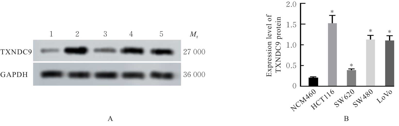
Fig. 2
Clone formation(Crystal violet) and invasion (×40) of HCT116 cells in various groups"
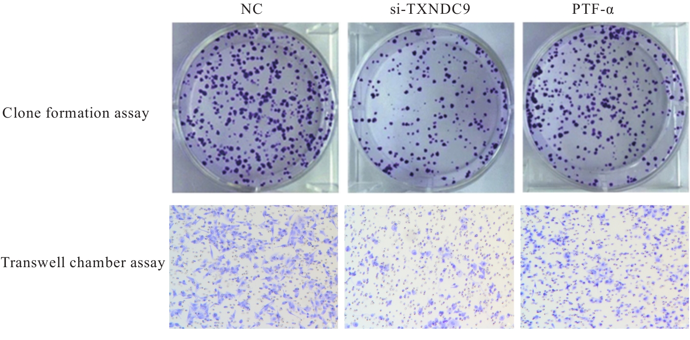
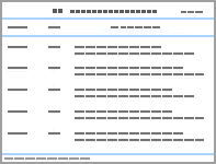
Tab.1
Numbers of clone formation and invasion cells of HCT116 cells in various groups"
| Group | Number of clone formation | Number of invasion cells |
|---|---|---|
| NC | 121.96±15.08 | 244.81±21.65 |
| Si-TXNDC9 | 53.23±3.95* | 67.49±5.71* |
| PTF-α | 95.17±8.89△ | 210.35±23.82△ |
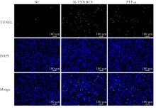
Fig. 3
Apoptosis of HCT116 cells in various groups ( TUNEL)"
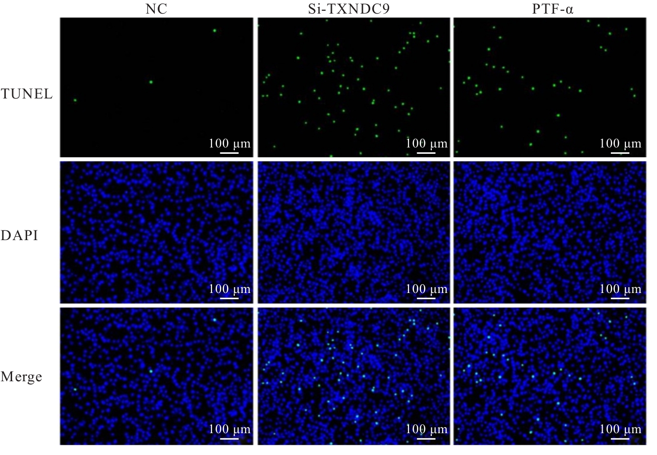
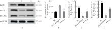
Fig. 4
Electrophoregram(A) and histograms(B-D) of expressions of apoptosis-related proteins in cells in various groups"
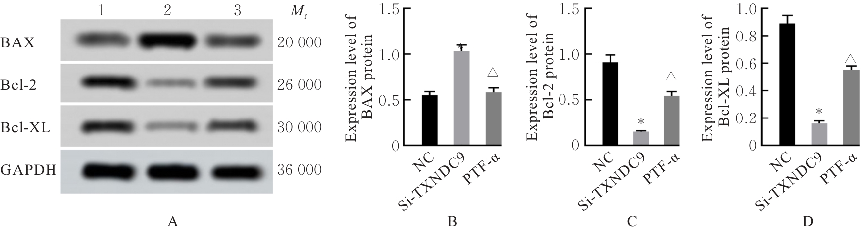
Fig. 5
Distribution of autophagosomes in HCT116 cells in various groups(TEM,×10 000)"
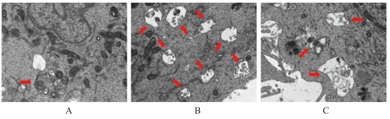
Fig. 6
Electrophoregram(A) and histograms(B-E) of expressions of autophagy-related proteins in HCT116 cells in various groups"
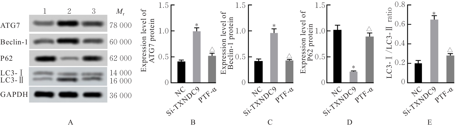
Fig. 7
Electrophoregram(A) and histograms(B-C) of expressions of p53 pathway-related proteins in cells in various groups"
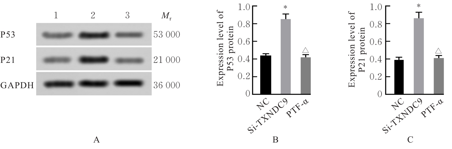
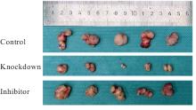
Fig. 8
General morphology of subcutaneous xenograft tumor of nude mice in various groups"
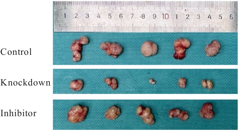
Tab.2
Weights and volumes of transplanted tumors in nude mice in various groups"
| Group | Tumor weight(m/mg) | Tumor volume(V/mm3) | |||
|---|---|---|---|---|---|
| (t/d) 7 | 14 | 21 | 28 | ||
| Control | 765.56±55.49 | 152.01±10.16 | 373.12±25.19 | 586.29±46.47 | 722.29±51.45 |
| Knockdown | 401.19±35.64* | 92.31±8.49* | 161.05±18.62* | 263.49±23.64* | 389.16±35.47* |
| Inhibitor | 605.06±51.19△ | 139.94±14.03△ | 295.16±19.06△ | 432.19±42.31△ | 587.46±49.75△ |
| F | 71.770 | 40.162 | 128.367 | 86.730 | 66.015 |
| P | <0.001 | <0.001 | <0.001 | <0.001 | <0.001 |
| [1] | WU Y J, HE S Y, CAO M D, et al. Comparative analysis of cancer statistics in China and the United States in 2024[J]. Chin Med J, 2024, 137(24): 3093-3100. |
| [2] | 赵英楠, 高君妍, 贺子轩, 等. 中国结直肠癌早筛早诊早治高质量发展的思考[J]. 中国实用内科杂志, 2025, 45(5): 373-380. |
| [3] | HE D, CHEN S L, WANG X Y, et al. Icaritin represses autophagy to promote colorectal cancer cell apoptosis and sensitized low-temperature photothermal therapy via targeting HSP90-TXNDC9 interactions[J]. Adv Sci, 2025, 12(20): 2412953. |
| [4] | SHAO B. Autophagy-dependent apoptosis induction by oridonin are mediated by ROS-dependent AMPK-mTOR-ULK1 pathway in colon cancer[J]. Am J Cancer Res, 2025, 15(4): 1902-1918. |
| [5] | ALAM M, ALAM S, SHAMSI A, et al. Bax/Bcl-2 cascade is regulated by the EGFR pathway: therapeutic targeting of non-small cell lung cancer[J]. Front Oncol, 2022, 12: 869672. |
| [6] | TAUCHER E, MYKOLIUK I, FEDIUK M, et al. Autophagy, oxidative stress and cancer development[J]. Cancers, 2022, 14(7): 1637. |
| [7] | EL-KHOULY D, THABET N A, SAYED-AHMED M, et al. Promotion of autophagy and apoptosis in colorectal cancer exposed to imatinib and thymoquinone[J]. J Biochem Mol Toxicol, 2025, 39(4): e70238. |
| [8] | ZHOU W J, FANG C, ZHANG L W, et al. Thioredoxin domain-containing protein 9 (TXNDC9) contributes to oxaliplatin resistance through regulation of autophagy-apoptosis in colorectal adenocarcinoma[J]. Biochem Biophys Res Commun, 2020, 524(3): 582-588. |
| [9] | 张仙宏, 李 乐. p53调控肿瘤代谢的研究进展[J]. 中国科学: 生命科学, 2023, 53(4): 431-448. |
| [10] | ZHENG T T, CHEN K K, ZHANG X, et al. Knockdown of TXNDC9 induces apoptosis and autophagy in glioma and mediates cell differentiation by p53 activation[J]. Aging, 2020, 12(18): 18649-18659. |
| [11] | JIANG Z S, SUN X G, LI Y, et al. Anlotinib induced ferroptosis through the p53/xCT/GPX4 pathway in non-small cell lung cancer[J]. Transl Oncol, 2025, 53: 102289. |
| [12] | 王洛伟, 杜奕奇, 柏 愚, 等. 中国消化道肿瘤的早诊早治现状及挑战[J]. 中国实用内科杂志, 2025, 45(5): 353-356. |
| [13] | XU W L, TANG Y J, YANG Y, et al. Depletion of CPNE7 sensitizes colorectal cancer to 5-fluorouracil by downregulating ATG9B expression[J]. J Cell Mol Med, 2024, 28(8): e18261. |
| [14] | WANG J, PAN X T, LI J, et al. TXNDC9 knockdown inhibits lung adenocarcinoma progression by targeting YWHAG[J]. Mol Med Rep, 2022, 25(6): 203. |
| [15] | YANG Q C, HAO N, LI R H, et al. High level of TXNDC9 predicts poor prognosis and contributes to the NF-κB-regulated metastatic potential in gastric cancer[J]. Neoplasma, 2022, 69(1): 103-112. |
| [16] | LU A G, WANGPU X, HAN D P, et al. TXNDC9 expression in colorectal cancer cells and its influence on colorectal cancer prognosis[J]. Cancer Investig, 2012, 30(10): 721-726. |
| [17] | OPYDO M, MLYCZYŃSKA A, MLYCZYŃSKA E, et al. Synergistic action of MCL-1 inhibitor with BCL-2/BCL-XL or MAPK pathway inhibitors enhances acute myeloid leukemia cell apoptosis and differentiation[J]. Int J Mol Sci, 2023, 24(8): 7180. |
| [18] | PALABIYIK A. The role of Bcl-2 in controlling the transition between autophagy and apoptosis (Review)[J]. Mol Med Rep, 2025, 32(1): 1-9. |
| [19] | ASHRAFIZADEH M, AHMADI Z, MOHAMMADINEJAD R, et al. Monoterpenes modulating autophagy: a review study[J]. Basic Clin Pharmacol Toxicol, 2020, 126(1): 9-20. |
| [20] | XIE Q Q, LIU Y, LI X F. The interaction mechanism between autophagy and apoptosis in colon cancer[J]. Transl Oncol, 2020, 13(12): 100871. |
| [21] | WU Y X, YE H P, PENG B, et al. MiR-643 functions as a potential tumor suppressor in gastric cancer by inhibiting cell proliferation and invasion via targeting TXNDC9[J]. Ann Clin Lab Sci, 2021, 51(4): 494-502. |
| [22] | WANG Y M, XU X, TANG J, et al. Apatinib induces endoplasmic reticulum stress-mediated apoptosis and autophagy and potentiates cell sensitivity to paclitaxel via the IRE-1α-AKT-mTOR pathway in esophageal squamous cell carcinoma[J]. Cell Biosci, 2021, 11(1): 124. |
| [23] | 王蓓蕾, 于 洋, 韩桂玲, 等. 肺痿冲剂对肺纤维化小鼠肺纤维化、自噬与上皮间质转化的影响[J]. 郑州大学学报(医学版), 2025, 60(5): 613-617. |
| [24] | DEVIS-JAUREGUI L, ERITJA N, DAVIS M L, et al. Autophagy in the physiological endometrium and cancer[J]. Autophagy, 2021, 17(5): 1077-1095. |
| [25] | VOLIK P I, ZAMARAEV A V, EGORSHINA A Y, et al. Ally or traitor: the dual role of p62 in Caspase-2 regulation[J]. Cell Death Dis, 2024, 15: 827. |
| [26] | SONG C F, XU W, WU H K, et al. Photodynamic therapy induces autophagy-mediated cell death in human colorectal cancer cells via activation of the ROS/JNK signaling pathway[J]. Cell Death Dis, 2020, 11: 938. |
| [27] | PANATTA E, ZAMPIERI C, MELINO G, et al. Understanding p53 tumour suppressor network[J]. Biol Direct, 2021, 16(1): 14. |
| [28] | WEN J, WANG D L. Deciphering the PTM codes of the tumor suppressor p53[J]. J Mol Cell Biol, 2022, 13(11): 774-785. |
| [29] | VADDAVALLI P L, SCHUMACHER B. The p53 network: cellular and systemic DNA damage responses in cancer and aging[J]. Trends Genet, 2022, 38(6): 598-612. |
| [30] | YOO H S, WON S B, KWON Y H. Luteolin induces apoptosis and autophagy in HCT116 colon cancer cells via p53-dependent pathway[J]. Nutr Cancer, 2022, 74(2): 677-686. |
| [31] | CHATTERJEE T, GUHA D, DHAR J, et al. Adenosine dialdehyde, a methyltransferase inhibitor, induces colorectal cancer cells apoptosis by regulating PIMT: p53 interaction[J]. Biochem Biophys Res Commun, 2023, 684: 149134. |
| [32] | WANG H L, GUO M, WEI H D, et al. Targeting p53 pathways: mechanisms, structures and advances in therapy[J]. Signal Transduct Target Ther, 2023, 8: 92. |
| [1] | Hui KE,Lizheng YAN,Yu ZHANG,Jilong MAO,Yufei YAO. Regulation effect of resveratrol on ovarian granulosa cell function in females and mechanism of SIRT1 in regulation process [J]. Journal of Jilin University(Medicine Edition), 2026, 52(4): 1043-1050. |
| [2] | Xiaowei HUANG,Pengyu ZHENG,Xin LI,Chuanbin HONG,Mengmeng LI,Guangfu LYU,Yan XU,Zhe LIN,Jiaming XU. Promotion effect of velvet antler polypeptide on myocardial injury in diabetic cardiomyopathy mice by regulating Keap1/Nrf2/HO-1 signaling pathway [J]. Journal of Jilin University(Medicine Edition), 2026, 52(4): 942-952. |
| [3] | Yunxia MA,Dan LIU,Ying ZHANG,Shifeng MA. Protective effect of triptolide on hippocampal neuron injury in neonatal rats with hypoxic-ischemic encephalopathy by regulating PTEN/PI3K/AKT signaling pathway [J]. Journal of Jilin University(Medicine Edition), 2026, 52(4): 977-986. |
| [4] | Jingxin WANG,Yuejuan GAO,Jingnan ZHANG,Jiawen NIU,Chunhua JIN. Effect of sesamin on learning and memory abilities of rats with Alzheimer’s disease model through regulating AMPK/SIRT1 autophagy pathway [J]. Journal of Jilin University(Medicine Edition), 2026, 52(3): 663-671. |
| [5] | Yang LI,Shiyang LI,Hongmei ZHANG,Qianqian LIU,Yubing CHEN. Effect of NGFR gene expression differences on promotion of apoptosis of squamous cell carcinoma and adenocarcinoma cells by PD-1 inhibitor-activated T lymphocytes [J]. Journal of Jilin University(Medicine Edition), 2026, 52(3): 680-693. |
| [6] | Qiaotong REN,Weihua WU,Xingxiang WANG,Shichao WANG,Yipeng CHENG,Chun LI. Effect of interleukin-17 recepter B gene silencing on proliferation, invasion and apoptosis of lung adenocarcinoma A549 cells and its mechanism [J]. Journal of Jilin University(Medicine Edition), 2026, 52(3): 738-745. |
| [7] | Qiuping SU,Lei XING,Wei ZHANG,Shan JIANG,Yangyang ZHAO,Fangfang CHEN. Research progress in anti-tumor mechanism of berberine and application of its novel nano-delivery system in tumor therapy [J]. Journal of Jilin University(Medicine Edition), 2026, 52(3): 863-871. |
| [8] | Ling DENG,Shouqing LI,Chunxue NIU,Xiuying LIN,Xuguang MI,Jianhua FU. Effect of bisphenol AF on senescence of human endometrial stromal cells and inhibitory role of rapamycin [J]. Journal of Jilin University(Medicine Edition), 2026, 52(2): 340-347. |
| [9] | Jie YU,Zongkang WANG,Xun LIU,Yanan YANG,Jin TAN. Effect of miR-153-3p/PGC-1α axis on apoptosis of oral squamous cell carcinoma cells by modulation of mitochondrial apoptosis pathway [J]. Journal of Jilin University(Medicine Edition), 2026, 52(2): 348-358. |
| [10] | Yao ZHENG,Mingxia FU,Weichen WANG,Weiwei CHEN,Yuchen HAN,Yu BAI,Jiajia AN. Effect of sodium selenite on biological behaviors of breast cancer doxorubicin-resistant MCF-7/ADR cells [J]. Journal of Jilin University(Medicine Edition), 2026, 52(2): 410-417. |
| [11] | Yu ZHAO,Xiaoshuang HE,Xiaoyin DONG,Fengyi GAO,Jiageng HE. Regulatory effect of lncRNA urothelial carcinoembryonic antigen 1 on anoikis in human gastrointestinal stromal tumor cells and its mechanism [J]. Journal of Jilin University(Medicine Edition), 2026, 52(2): 429-439. |
| [12] | Huiyan ZHU,Min CHEN,Jinxian LI,Chunli LI. Effect of enriched environment on neurofunctional damage in rats with ischemic stroke via transcription factor EB-mediated autophagy [J]. Journal of Jilin University(Medicine Edition), 2026, 52(1): 116-124. |
| [13] | Yaping LI,Chunyan TAN,Lujie ZHAO,Jiayi ZHAO,Ting LI,Xiao YANG,Xiaoyun YANG. Inductive effect of lysophosphatidic acid combined with 6-hydroxydopamine on apoptosis of SH-SY5Y cells and its mechanism [J]. Journal of Jilin University(Medicine Edition), 2026, 52(1): 152-161. |
| [14] | Jiaxin WANG,Junwen MAO,Xuan ZHANG. Expression of natural autoantibodies against apoptosis inhibitory genes in plasma of patients with hepatocellular carcinoma and its clinical significance [J]. Journal of Jilin University(Medicine Edition), 2026, 52(1): 228-235. |
| [15] | Huaimin LIANG,Jiacheng JIN,Wenhua CHEN,Yuyao LI,Hangyu WANG,Ke ZHANG,Jinhui WANG. UPLC-Q-TOF/MS and network pharmacology analysis and experimental verification based on potential active ingredients and mechanisms of medicinal Mulberry Leaves in anti-acute kidney injury [J]. Journal of Jilin University(Medicine Edition), 2026, 52(1): 56-69. |
|
||