Journal of Jilin University(Medicine Edition) ›› 2026, Vol. 52 ›› Issue (4): 963-976.doi: 10.13481/j.1671-587X.20260409
• Research in basic medicine • Previous Articles Next Articles
Bioinformatics analysis and in vitro and in vivo experimental verification on protective effect of salidroside on retinal ganglion cells in diabetic rats
Junqi LI1,2,Jiayun JIANG3,4,Zhongfu ZUO1,2,Hongdan YU1,2( )
)
- 1.Department of Anatomy,School of Basic Medical Sciences,Jinzhou Medical University,Jinzhou 121001,China
2.Key Laboratory of Diabetes Perception Dysfunction of Liaoning Province,Jinzhou 121001,China
3.Department of Neurology,First Clinical Medical School,Jinzhou Medical University,Jinzhou 121001,China
4.Department of Neurology,Second People’s Hospital,Fuxin City,Liaoning Province,Fuxin 123000,China
-
Received:2025-08-11Accepted:2025-12-25Online:2026-07-28Published:2026-07-27 -
Contact:Hongdan YU E-mail:yuhongdan1116@126.com

CLC Number:
- R774.1
Cite this article
Junqi LI,Jiayun JIANG,Zhongfu ZUO,Hongdan YU. Bioinformatics analysis and in vitro and in vivo experimental verification on protective effect of salidroside on retinal ganglion cells in diabetic rats[J].Journal of Jilin University(Medicine Edition), 2026, 52(4): 963-976.
share this article
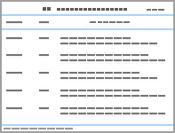
Tab.1
RT-qPCR primers"
| Gene | Sense primer | Anti-sense primer |
|---|---|---|
| GAPDH | GTGGCAAAGTGGAGATTGTTG | CGTTGAATTTGCCGTGAGTG |
| HIF1A | TCTCAGAATGAAGTGTACCCT | GTGCAGTGCAATACCTTCC |
| CASP3 | AACTCTTCATCATTCAGGCCT | CCATATCATCGTCAGTTCCAC |
| NFE2L2 | GCATTTCGCTGAACACAA | CTCTTCCATTTCCGAGTCA |
| CASP8 | GTGAAAAACTGTGTTTCCT | CAGTCAGGATGGTGAGAAT |
| HSPA5 | GAGGAGGACAAGAAGGAGG | CTTGAATACACCGACGCAG |
| MTOR | ATGGCAGGGGACACTTT | CACGGAGAACGAGGACA |
| NLRP3 | AGACATGGGACTCAAGCTC | CTGCAGTTGTCTAACTCCAG |
| NOS3 | GACATTGAGATCAAAGGACTGC | GTAGAGATGGTCCAGTTGGG |
| STRT1 | ACACAAAATCCAGCAACTCAGC | ATTGCTTTCCTTCCACTGCAC |
| TLR4 | CAGTACAACCAGATCCGCT | CCAGGTACAGCTCTTCCAG |
Fig. 1
Identification of primary RG cells"
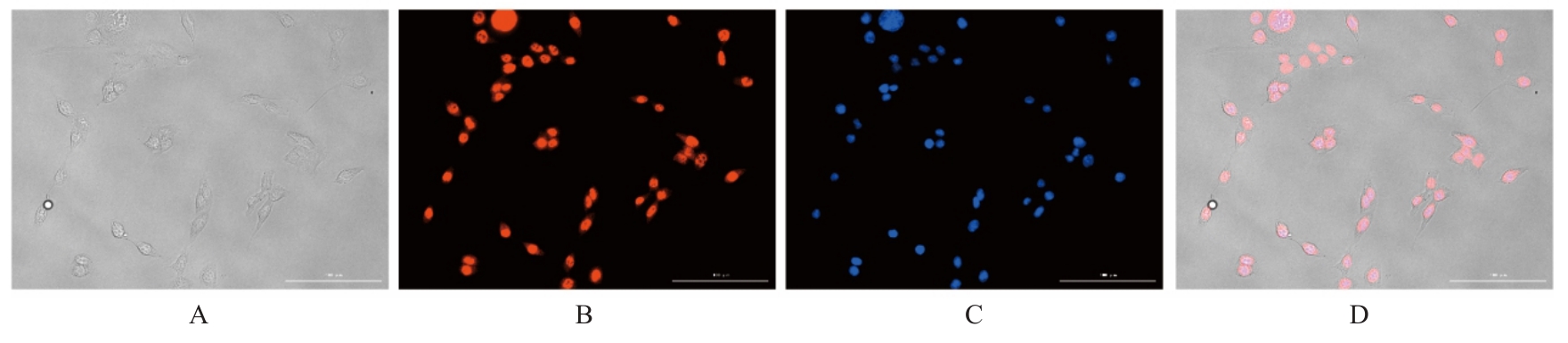
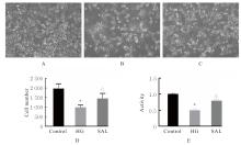
Fig. 2
Bright-field images of RG cells in various groups (A-C) and number (D) and activity (E) of RG cells after treated with salidroside"
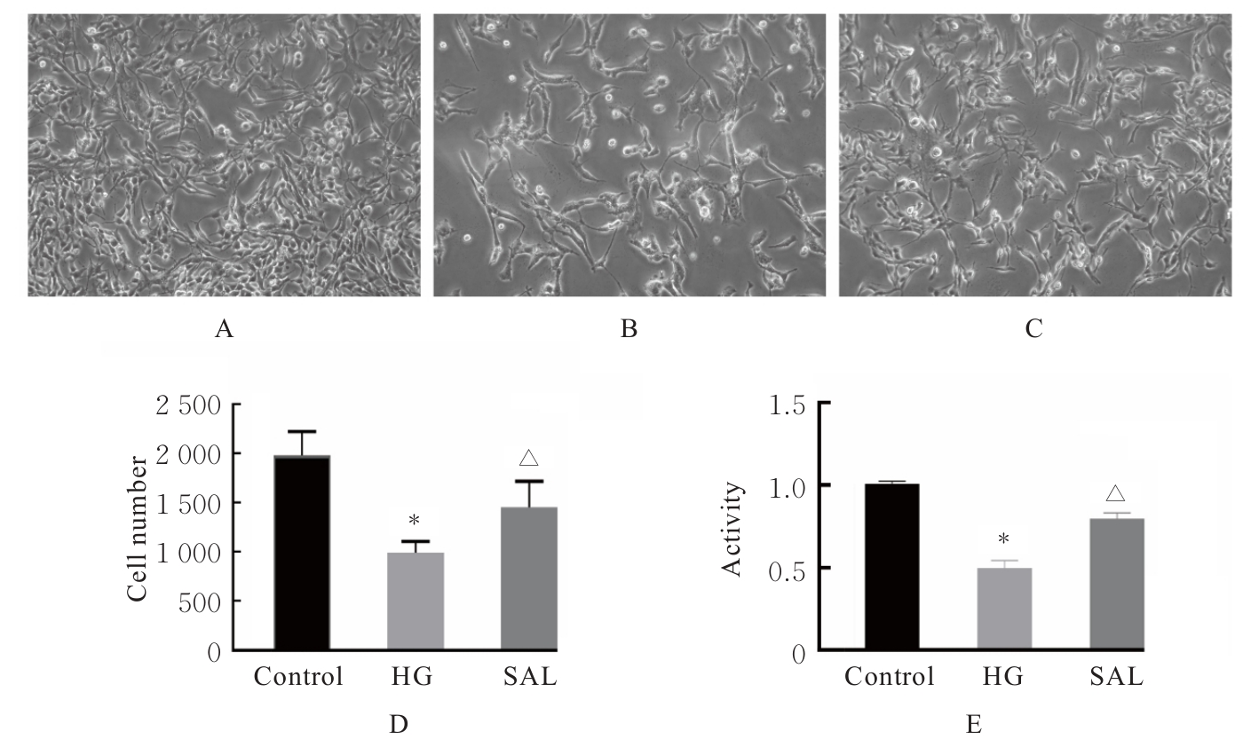
Fig. 3
Retinal images(A) and histogram(B) of rats in various groups"
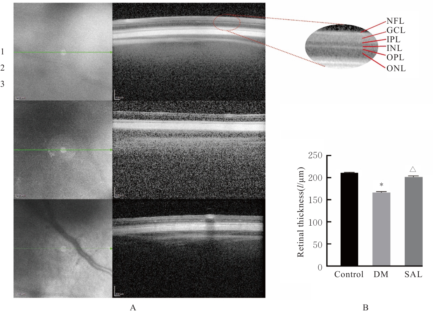
Fig. 4
Morphology of retina of rats in various groups (HE, ×100)"
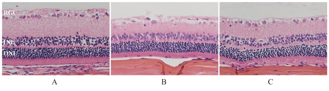
Fig. 5
Venn diagram (A), PPI network diagram (B), MCC core target (C), GO functional enrichment diagram (D), and KEGG signaling pathway enrichment diagram (E) of potential targets for salidroside in treatment of RG cell injury induced by HG"
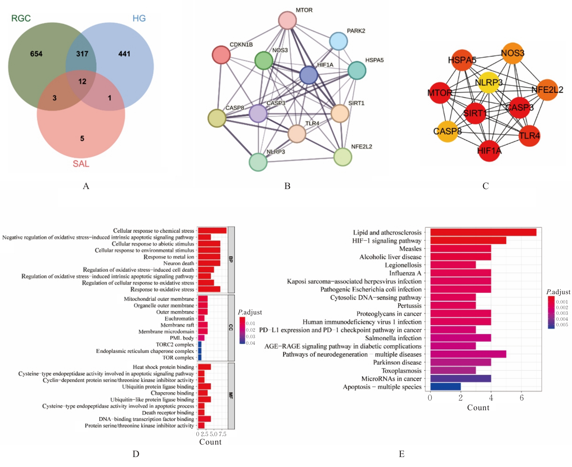
Fig. 6
Levels of ROS in RG cells in various groups"
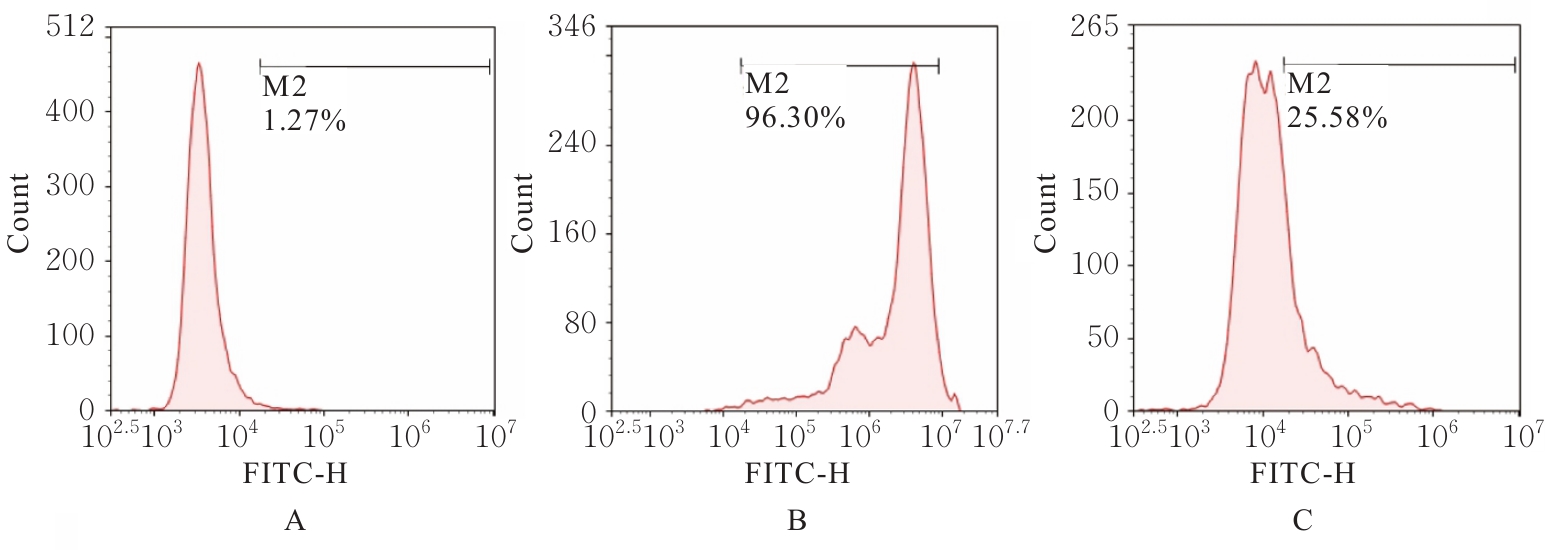
Fig. 7
Core targets of salidroside in treatment of RG cell injury induced by HG verificated by RT-qPCR method"
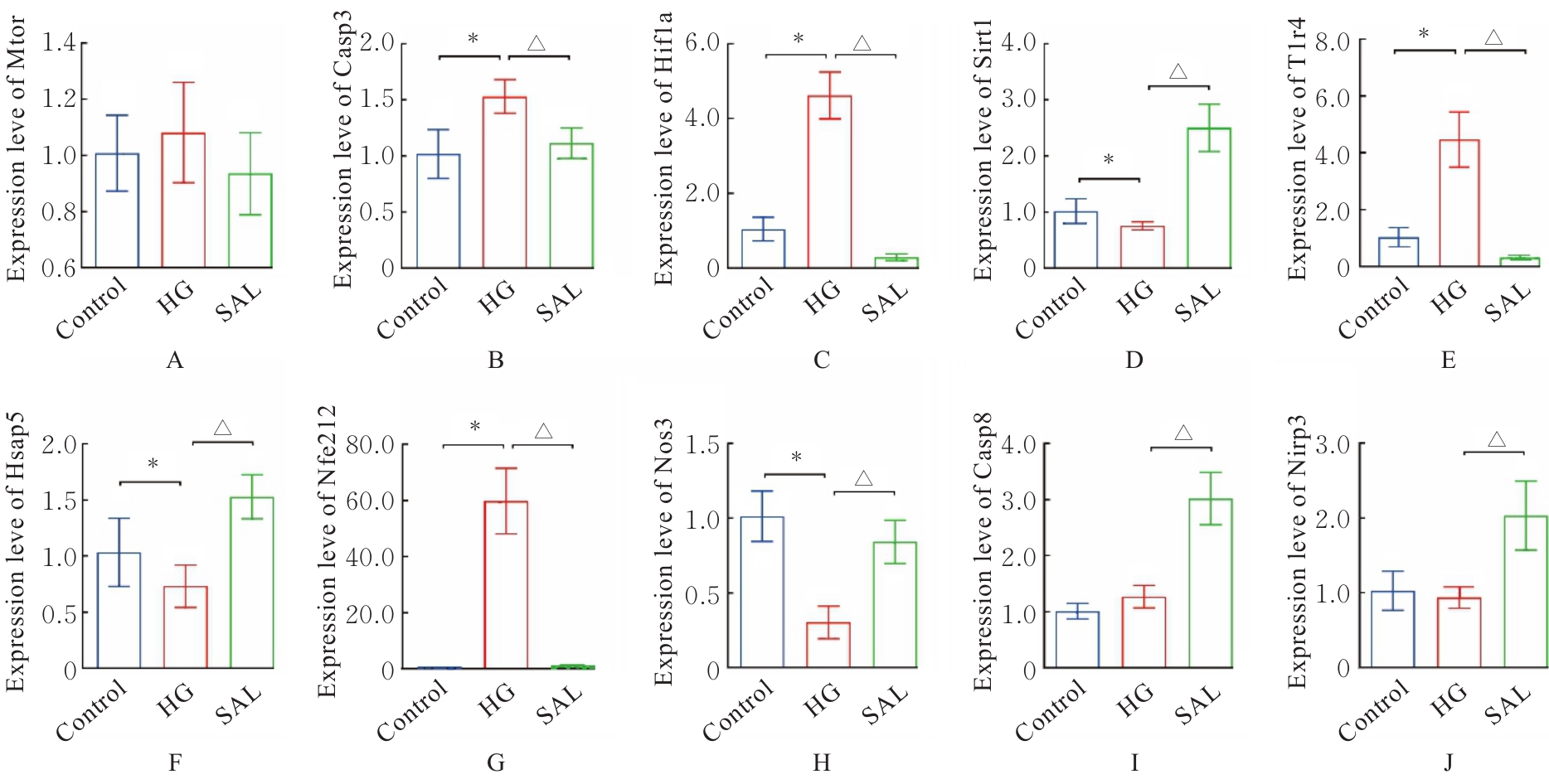
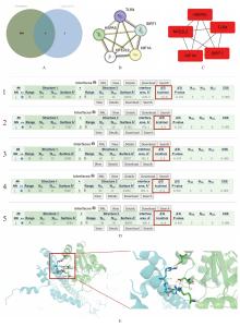
Fig. 8
Venn diagram (A), PPI network diagram (B), core target of MCC (C), protein docking analysis results (D), and NFE2L2 expression (E)"
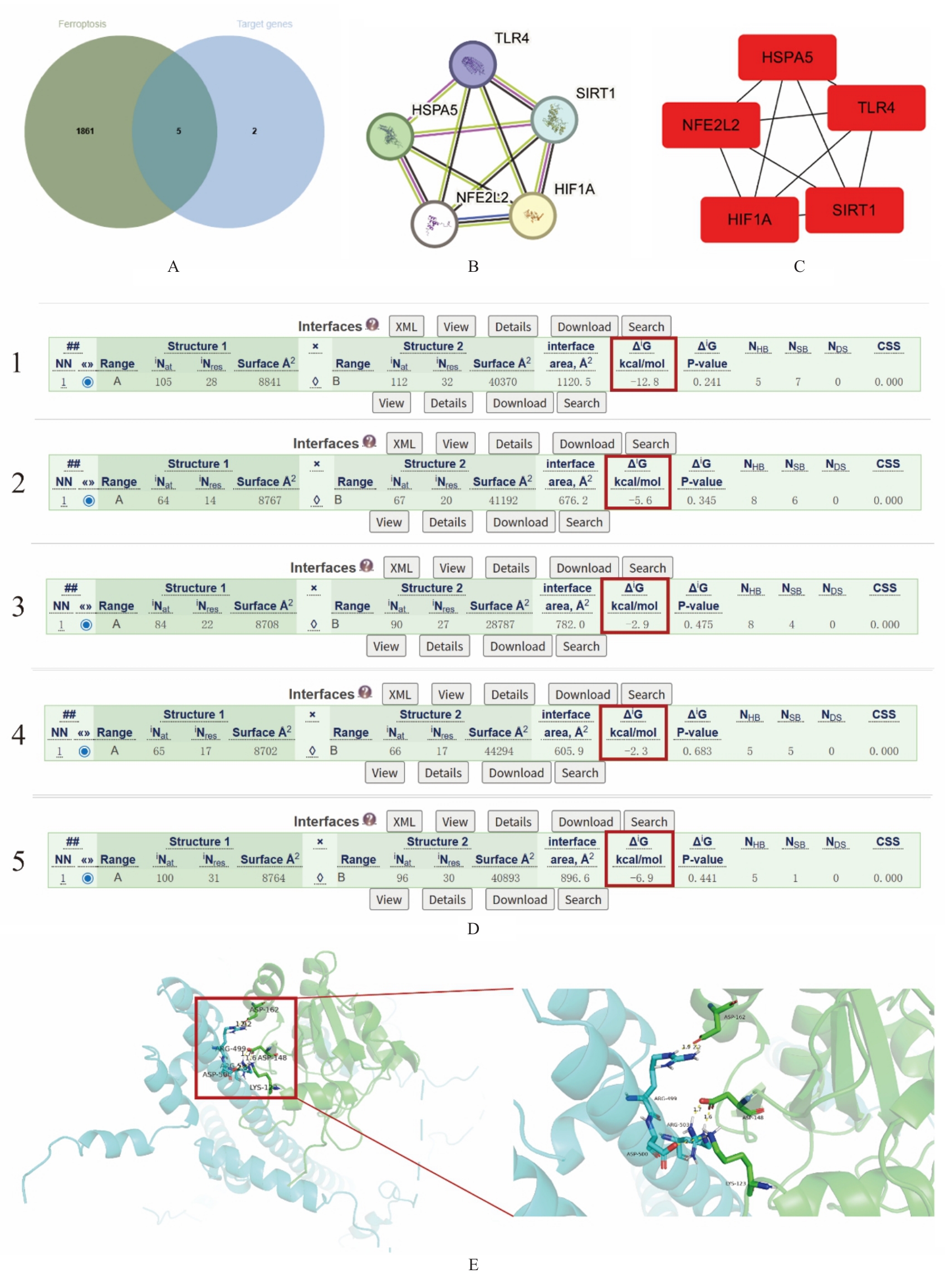
Fig. 9
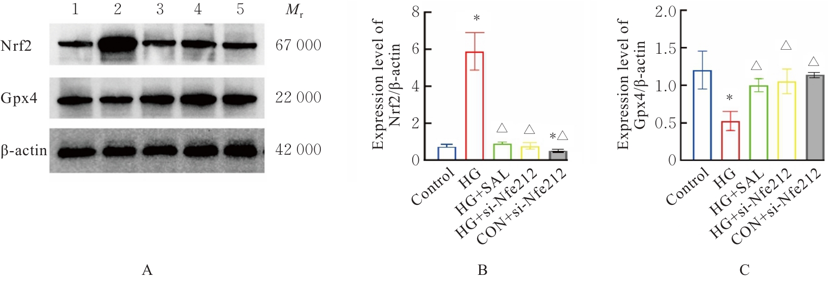
Electrophoregram(A) and histograms (B,C) of expressions of Nfe2l2 and Gpx4 proteins in RG cells in various groups"
| [1] | SUN H, SAEEDI P, KARURANGA S, et al. IDF Diabetes Atlas: Global, regional and country-level diabetes prevalence estimates for 2021 and projections for 2045[J]. Diabetes Res Clin Pract, 2022, 183: 109119. |
| [2] | KOUR V, SWAIN J, SINGH J, et al. A review on diabetic retinopathy[J]. Curr Diabetes Rev, 2024, 20(6): e201023222418. |
| [3] | SOHN E H, VAN DIJK H W, JIAO C H, et al. Retinal neurodegeneration may precede microvascular changes characteristic of diabetic retinopathy in diabetes mellitus[J]. Proc Natl Acad Sci U S A, 2016, 113(19): E2655-E2664. |
| [4] | BIKBOVA G, OSHITARI T, BABA T, et al. Mechanisms of neuronal cell death in AGE-exposed retinas - research and literature review[J]. Curr Diabetes Rev, 2017, 13(3): 280-288. |
| [5] | EL-FAYOUMI D, BADR ELDINE N M, ESMAEL A F, et al. Retinal nerve fiber layer and ganglion cell complex thicknesses are reduced in children with type 1 diabetes with No evidence of vascular retinopathy[J]. Invest Ophthalmol Vis Sci, 2016, 57(13): 5355-5360. |
| [6] | DIXON S J, LEMBERG K M, LAMPRECHT M R, et al. Ferroptosis: an iron-dependent form of nonapoptotic cell death[J]. Cell, 2012, 149(5): 1060-1072. |
| [7] | LEI G, MAO C, YAN Y L, et al. Ferroptosis, radiotherapy, and combination therapeutic strategies[J]. Protein Cell, 2021, 12(11): 836-857. |
| [8] | ZHOU R P, CHEN Y, WEI X, et al. Novel insights into ferroptosis: Implications for age-related diseases[J]. Theranostics, 2020, 10(26): 11976-11997. |
| [9] | ZHANG J F, QIU Q H, WANG H Y, et al. TRIM46 contributes to high glucose-induced ferroptosis and cell growth inhibition in human retinal capillary endothelial cells by facilitating GPX4 ubiquitination[J]. Exp Cell Res, 2021, 407(2): 112800. |
| [10] | YANG D W, KANG O H, LEE Y S, et al. Anti-inflammatory effect of salidroside on phorbol-12-myristate-13-acetate plus A23187-mediated inflammation in HMC-1 cells[J]. Int J Mol Med, 2016, 38(6): 1864-1870. |
| [11] | YUAN Y, WU S J, LIU X, et al. Antioxidant effect of salidroside and its protective effect against furan-induced hepatocyte damage in mice[J]. Food Funct, 2013, 4(5): 763-769. |
| [12] | SHATI A A, ALFAIFI M Y. Salidroside protects against diabetes mellitus-induced kidney injury and renal fibrosis by attenuating TGF-β1 and Wnt1/3a/β-catenin signalling[J]. Clin Exp Pharmacol Physiol, 2020, 47(10): 1692-1704. |
| [13] | ZHANG X M, XIE L, LONG J Y, et al. Salidroside: a review of its recent advances in synthetic pathways and pharmacological properties[J]. Chem Biol Interact, 2021, 339: 109268. |
| [14] | TAN C B, GAO M, XU W R, et al. Protective effects of salidroside on endothelial cell apoptosis induced by cobalt chloride[J]. Biol Pharm Bull, 2009, 32(8): 1359-1363. |
| [15] | YANG W S, STOCKWELL B R. Ferroptosis: death by lipid peroxidation[J]. Trends Cell Biol, 2016, 26(3): 165-176. |
| [16] | URSINI F, MAIORINO M. Lipid peroxidation and ferroptosis: The role of GSH and GPx4[J]. Free Radic Biol Med, 2020, 152: 175-185. |
| [17] | BELI E, YAN Y Q, MOLDOVAN L, et al. Restructuring of the gut microbiome by intermittent fasting prevents retinopathy and prolongs survival in db/db mice[J]. Diabetes, 2018, 67(9): 1867-1879. |
| [18] | WONG T Y, CHEUNG C M G, LARSEN M, et al. Diabetic retinopathy[J]. Nat Rev Dis Primers, 2016, 2: 16012. |
| [19] | SIMÓ R, STITT A W, GARDNER T W. Neurodegeneration in diabetic retinopathy: does it really matter?[J]. Diabetologia, 2018, 61(9): 1902-1912. |
| [20] | VILLARROEL M, CIUDIN A, HERNÁNDEZ C, et al. Neurodegeneration: an early event of diabetic retinopathy[J]. World J Diabetes, 2010, 1(2): 57-64. |
| [21] | CARELLI V, LA MORGIA C, VALENTINO M L, et al. Retinal ganglion cell neurodegeneration in mitochondrial inherited disorders[J]. Biochim Biophys Acta, 2009, 1787(5): 518-528. |
| [22] | KOWLURU R A, CHAN P S. Oxidative stress and diabetic retinopathy[J]. J Diabetes Res, 2007, 2007: 043603. |
| [23] | HAMMES H P. Diabetic retinopathy: hyperglycaemia, oxidative stress and beyond[J]. Diabetologia, 2018, 61(1): 29-38. |
| [24] | KANG Q Z, YANG C X. Oxidative stress and diabetic retinopathy: Molecular mechanisms, pathogenetic role and therapeutic implications[J]. Redox Biol, 2020, 37: 101799. |
| [25] | YANG S X, WANG L S, ZENG Y, et al. Salidroside alleviates cognitive impairment by inhibiting ferroptosis via activation of the Nrf2/GPX4 axis in SAMP8 mice[J]. Phytomedicine, 2023, 114: 154762. |
| [26] | HUANG X Y, ZOU L Z, YU X M, et al. Salidroside attenuates chronic hypoxia-induced pulmonary hypertension via adenosine A2a receptor related mitochondria-dependent apoptosis pathway[J]. J Mol Cell Cardiol, 2015, 82: 153-166. |
| [27] | YAMAMOTO M, KENSLER T W, MOTOHASHI H. The KEAP1-NRF2 system: a thiol-based sensor-effector apparatus for maintaining redox homeostasis[J]. Physiol Rev, 2018, 98(3): 1169-1203. |
| [28] | LEONARD G. Franklin H. Epstein lecture: sirtuins, aging, and medicine[J]. N Engl J Med, 2011, 364(23): 2235-2244. |
| [29] | HAIGIS M C, SINCLAIR D A. Mammalian sirtuins: biological insights and disease relevance[J]. Annu Rev Pathol, 2010, 5: 253-295. |
| [30] | MCILWAIN D R, BERGER T, MAK T W. Caspase functions in cell death and disease[J]. Cold Spring Harb Perspect Biol, 2013, 5(4): a008656. |
| [31] | BRENTNALL M, RODRIGUEZ-MENOCAL L, DE GUEVARA R L, et al. Caspase-9, caspase-3 and caspase-7 have distinct roles during intrinsic apoptosis[J]. BMC Cell Biol, 2013, 14: 32. |
| [32] | GHAREGHOMI S, HABIBI-REZAEI M, ARESE M, et al. Nrf2 modulation in breast cancer[J]. Biomedicines, 2022, 10(10): 2668. |
| [33] | ZHONG Q, MISHRA M, KOWLURU R A. Transcription factor Nrf2-mediated antioxidant defense system in the development of diabetic retinopathy[J]. Invest Ophthalmol Vis Sci, 2013, 54(6): 3941-3948. |
| [34] | INGOLD I, BERNDT C, SCHMITT S, et al. Selenium utilization by GPX4 is required to prevent hydroperoxide-induced ferroptosis[J]. Cell, 2018, 172(3): 409-422.e21. |
| [35] | YATES A D, ACHUTHAN P, AKANNI W, et al. Ensembl 2020[J]. Nucleic Acids Res, 2020, 48(D1): D682-D688. |
| [1] | Xin LIU,Yuqing LIU,Hui LI. Research progress in effect of chronic periodontitis on occurrence and development of metabolic fatty liver disease [J]. Journal of Jilin University(Medicine Edition), 2026, 52(4): 1187-1194. |
| [2] | Xiaowei HUANG,Pengyu ZHENG,Xin LI,Chuanbin HONG,Mengmeng LI,Guangfu LYU,Yan XU,Zhe LIN,Jiaming XU. Promotion effect of velvet antler polypeptide on myocardial injury in diabetic cardiomyopathy mice by regulating Keap1/Nrf2/HO-1 signaling pathway [J]. Journal of Jilin University(Medicine Edition), 2026, 52(4): 942-952. |
| [3] | Lingyu WANG,Ruili LI,Yu ZHANG,Meitong GUO,Yalong LIANG,Xu HAN,Xiaowei HUANG,Yang ZHANG,Xiuhua YU. Improvement effect of Renshen Jianzhi Tablets on learning and memory ability in cognitive impairment model rats and its protective effect on hippocampal neurons and its mechanism [J]. Journal of Jilin University(Medicine Edition), 2026, 52(3): 719-729. |
| [4] | Xiaohui LIANG,Wenna LIU,Jianbo ZHAO. Bioinformatic analysis on PAQR3 expression in hepatocellular carcinoma and its correlations with T lymphocyte infiltration, immune checkpoint genes, and patient survival [J]. Journal of Jilin University(Medicine Edition), 2026, 52(3): 781-790. |
| [5] | Zhipeng PAN,Le WANG,Weiwen HUANG,Zhen YI,Yuxuan GAO. Bioinformatics analysis and experimental validation based on screening and identification of diagnostic candidate genes of bone marrow mononuclear cells of acute myeloid leukemia patients and their impact on prognosis [J]. Journal of Jilin University(Medicine Edition), 2026, 52(3): 791-805. |
| [6] | Jing YU,Yinzhuo HUANG,Zhongwei ZHOU,Yuxin XIE,Qian MAO. Analysis on influencing factors in type 2 diabetes mellitus patients complicated with lower limb atherosclerosis and construction of nomogram model [J]. Journal of Jilin University(Medicine Edition), 2026, 52(3): 806-812. |
| [7] | Zheng TANG,Wei LIU,Hongmin HU,Qi LAI,Yan SHI,Shengquan LIU,Jun YANG,Chun CHU. Improvement effect of Qishen Yiqi dropping pills on myocardium injury in rats induced by sorafenib and its mechanism [J]. Journal of Jilin University(Medicine Edition), 2026, 52(2): 308-317. |
| [8] | Ning MA,Shuang LIU,Chengyao WANG,Jiajun LU,Linyu CHEN,Ziyi WANG,Weiqin ZHAO,Weitong WANG,Pengcheng CHE,Hong SUN. Improvement effect of topical application of exosome loaded hydrogel DCC on peritoneal adhesion in mice and its mechanism [J]. Journal of Jilin University(Medicine Edition), 2026, 52(2): 318-329. |
| [9] | Shu LI,Jiaqi GUO,Wansong LI,Yanfeng ZHEN,Hongjia ZHAI,Jie LI,Hui FANG. Improvement effect of fingolimod on hepatic fibrosis in type 2 diabetes mellitus mice and its mechanism [J]. Journal of Jilin University(Medicine Edition), 2026, 52(2): 330-339. |
| [10] | Ruihan GE,Chen LI,Shengpeng WANG,Yang LU,Caixia TAN,Haotian CUI,Xinmin WANG,Le ZHANG. Bioinformatic analysis on regulatory mechanism of MAPK-Mcl-1 signaling pathway and macrophage polarization during Bacillus Calmette-Guérin infection and its experimental validation [J]. Journal of Jilin University(Medicine Edition), 2026, 52(2): 440-450. |
| [11] | Yuanshuang JIANG,Yang YU,Ruopu LI,Qinyan HUANG,Yunwei SUN,Yan CHEN. Relationship between glucose coefficient of variation and hyperuricemia in early-onset type 2 diabetes mellitus [J]. Journal of Jilin University(Medicine Edition), 2026, 52(2): 513-522. |
| [12] | Ziyi TANG,Shiying YANG,Tianzhen YANG,Wenqiang LIU,Jiangxue ZHONG,Li YIN. Induction effect of pesticide pyraclostrobin on ferroptosis of spermatocytes GC-2 of mice [J]. Journal of Jilin University(Medicine Edition), 2026, 52(1): 18-25. |
| [13] | Sitong CHEN,Dan YANG,Qingjie LI,Xiaolei TANG,Han WANG,Yangyang LIU,Tiejun LIU. Ameliorative effect of puerarin on non-alcoholic fatty liver disease induced by high-fat diet in mice and its mechanism [J]. Journal of Jilin University(Medicine Edition), 2026, 52(1): 44-55. |
| [14] | Xiao LIN,Meng ZHOU,Fan LIN,Xiujuan YAO. Bioinformatics analysis and experimental validation of fatty acid metabolism-related genes in idiopathic pulmonary fibrosis tissue [J]. Journal of Jilin University(Medicine Edition), 2026, 52(1): 93-104. |
| [15] | Yi ZHAO,Bing ZHOU,Huirui QIU,Xuan LI,Xiangli CUI. Improvement effect of lovastatin on hyperlipidemia-induced liver injury in rats and its mechanism [J]. Journal of Jilin University(Medicine Edition), 2025, 51(5): 1155-1164. |
|
||