Journal of Jilin University(Medicine Edition) ›› 2026, Vol. 52 ›› Issue (5): 1377-1389.doi: 10.13481/j.1671-587X.20260519
• Research in basic medicine • Previous Articles
Ameliorative effect of aurone on glucocorticoid-induced inhibition of osteoblast differentiation and osteoporosis in rats and its mechanism
Xiaofei HAN1,Yunfeng MA2( )
- 1.Department of Orthopedics,Henan Provincial Hospital of Traditional Chinese Medicine,Second;Affiliated Hospital,Henan University of Chinese Medicine,Zhengzhou 450002,China
2.Department of Orthopedics,Third Affiliated Hospital,Henan University of Traditional Chinese Medicine,Zhengzhou 450003,China
-
Received:2025-11-20Accepted:2026-01-14Online:2026-09-28Published:2026-10-15 -
Contact:Yunfeng MA E-mail:weiminwanghb@sina.com

CLC Number:
- R681.1
Cite this article
Xiaofei HAN,Yunfeng MA. Ameliorative effect of aurone on glucocorticoid-induced inhibition of osteoblast differentiation and osteoporosis in rats and its mechanism[J].Journal of Jilin University(Medicine Edition), 2026, 52(5): 1377-1389.
share this article
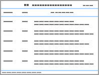
Tab. 1
Cell activities of MC3T3-E1 cells after treated with different concentrations of Dex (n=10, x±s, η/%)"
| Group | Cell activity |
|---|---|
| 0 μmol·L-1 Dex | 100.00±4.52 |
| 1×10⁻³ μmol·L-1 Dex | 99.15±8.11 |
| 1×10⁻² μmol·L-1 Dex | 97.92±4.08 |
| 1×10⁻¹ μmol·L-1 Dex | 75.16±6.33* |
| 1 μmol·L-1 Dex | 48.29±3.81** |
Tab. 2
Cell activities of MC3T3-E1 cells after treated with different concentrations of aurone (n=10, x±s, η/%)"
| Group | Cell activity |
|---|---|
| Control | 100.00±8.65 |
| Dex | 40.26±4.24* |
| Dex+2.5 μmol·L-1 aurone | 65.38±6.08△ |
| Dex+5.0 μmol·L-1 aurone | 98.85±10.05△△ |
| Dex+10.0 μmol·L-1 aurone | 90.71±7.26△△ |
Tab.3
Levels of MDA and ROS, activities of SOD in MC3T3-E1 cells and levels of TNF-α, IL-1β and IL-6 in supernatant in various groups"
| Group | MDA [cB/(μmol·L-1)] | ROS | TNF-α [ρB/(ng·L-1)] | IL-1β [ρB/(ng·L-1)] | IL-6 [ρB/(ng·L-1)] | SOD [λB/(U·mg-1)] |
|---|---|---|---|---|---|---|
| Control | 6.35±0.64 | 1.00±0.38 | 115.38±10.81 | 180.88±18.62 | 250.36±20.49 | 35.22±3.15 |
| Dex | 18.21±2.16* | 15.82±1.54* | 350.48±30.55* | 420.62±45.37* | 518.81±41.29* | 15.38±1.54* |
| Dex+aurone | 9.42±0.95△ | 2.65±0.59△ | 150.29±15.62△ | 210.67±20.38△ | 283.65±20.98△ | 37.71±3.54△ |
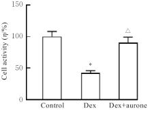
Fig. 1
Cell activities of MC3T3-E1 cells in various groups"
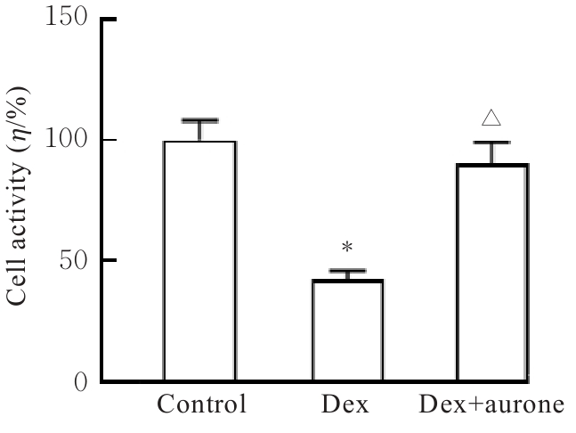
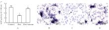
Fig. 2
ALP activities(A) and ALP staining(B-D) of MC3T3-E1 cells in various groups"
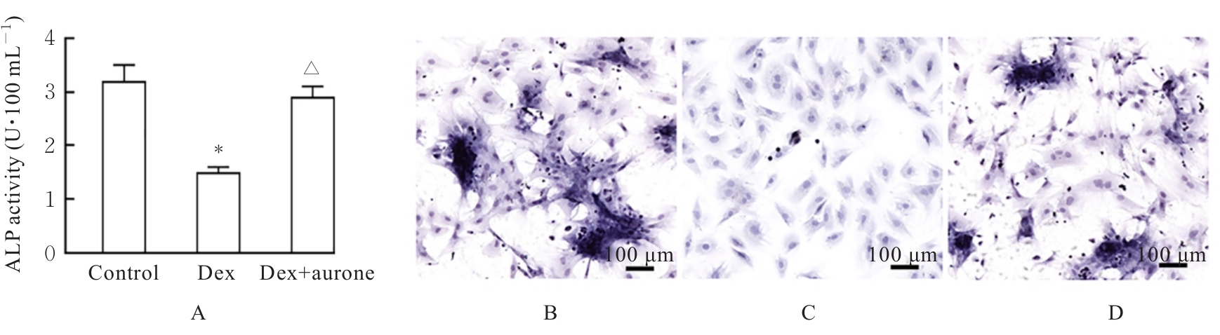
Fig. 3
Electrophoregram(A) and histograms(B-E) of expressions of RUNX-2, OCN, COL1A1 and OPN proteins in MC3T3-E1 cells in various groups"
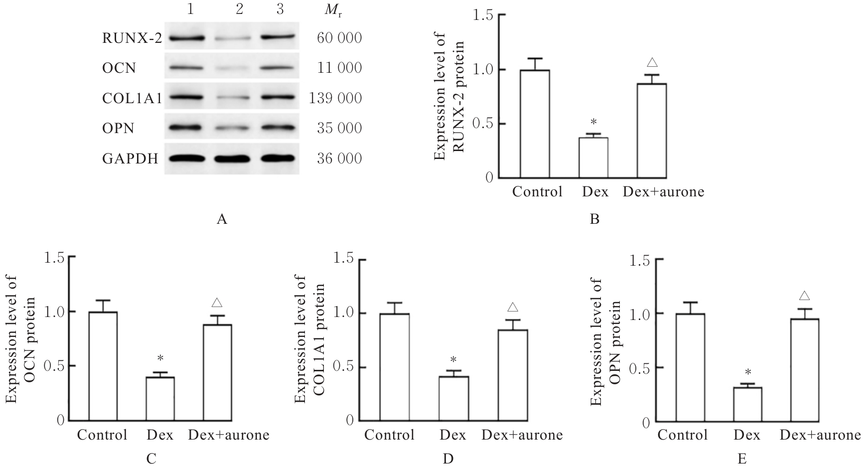
Fig. 4
Electrophoregrams(A-C) and histograms(D-F) of expressions of AHR and NRF2 proteins in nucleus and NQO1 protein in MC3T3-E1 cells in various groups"
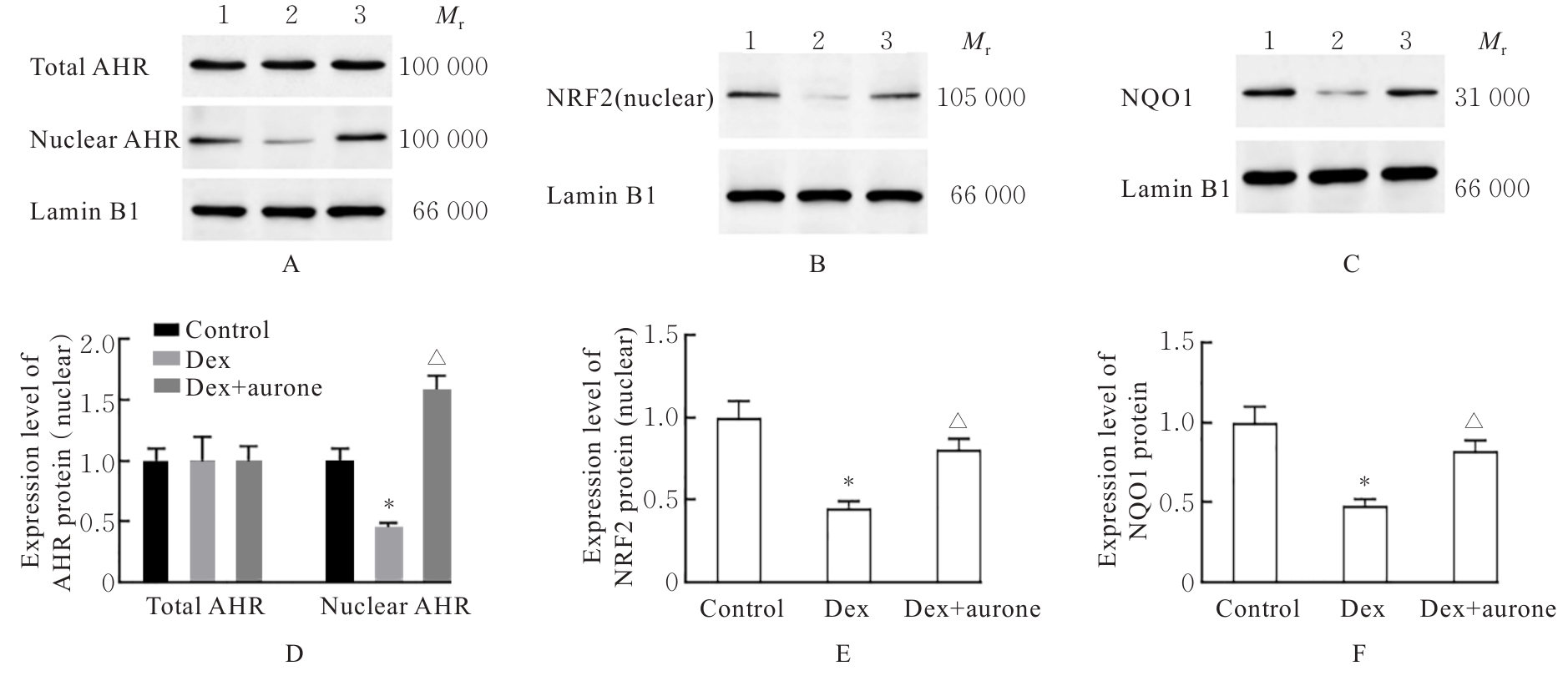
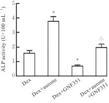
Fig. 5
ALP activities of MC3T3-E1 cells in various groups"
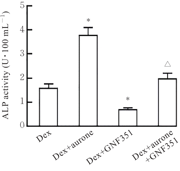
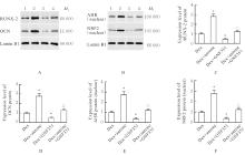
Fig. 6
Electrophoregrams(A,B) and histograms(C-F) of expressions of AHR and NRF2 proteins in nucleus and RUNX-2 and OCN proteins in MC3T3-E1 cells in various groups"
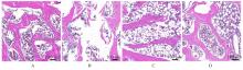
Fig. 7
Pathomorphology of femoral tissue of rats in various groups(HE)"
Fig. 8
BMD(A) of femoral tissue and trabecular BV/TV(B), Tb.N(C), Tb.Sp(D) and Tb.N(E) of rats in various groups"
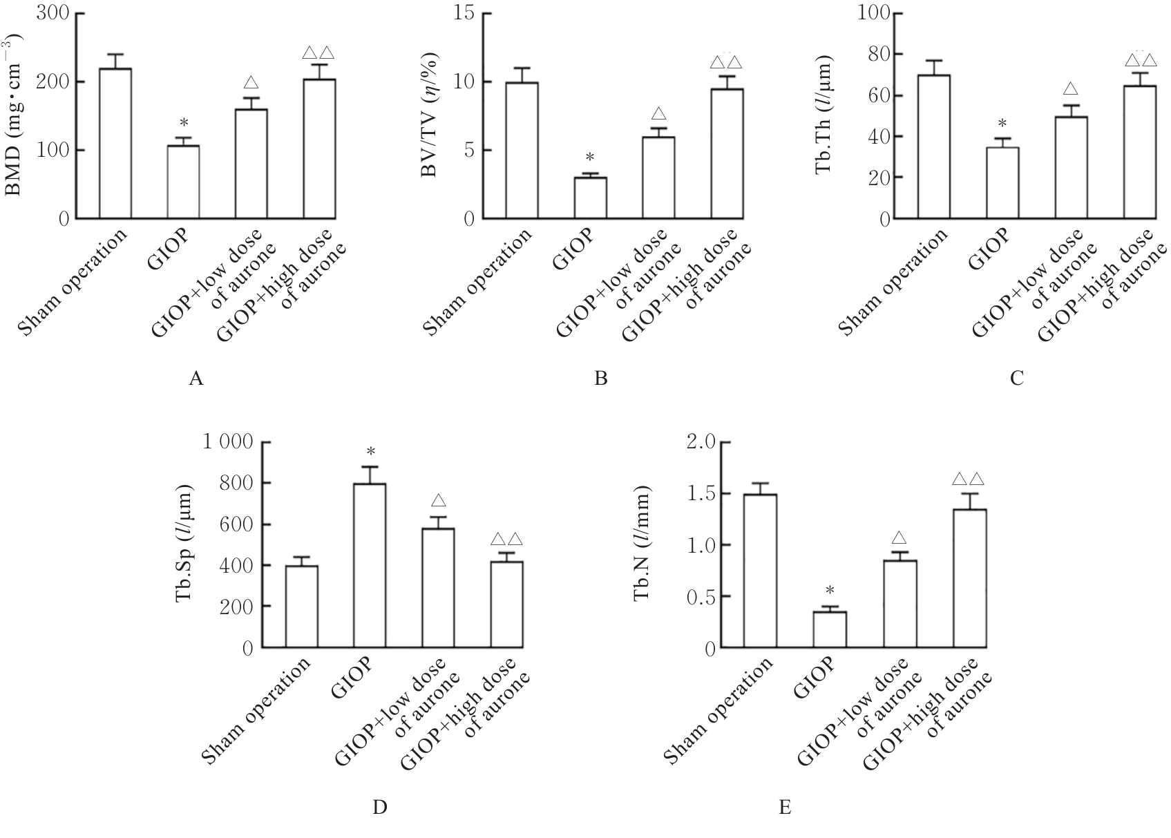
Fig. 9
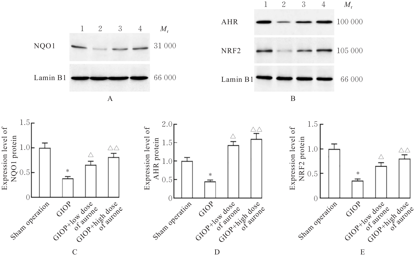
Electrophoregrams(A,B) and histograms(C-E) of expressions of NQO1, AHR and NRF2 proteins in femoral tissue of rats in various groups"
| [1] | LI X M, LIN H Y, ZHANG X R, et al. Notoginsenoside R1 attenuates oxidative stress-induced osteoblast dysfunction through JNK signalling pathway[J]. J Cell Mol Med, 2021, 25(24): 11278-11289. |
| [2] | WANG W, ZHANG X, et al. An immunomodulating regenerating hydrogel that rescues the oxidative microenvironment and reverses cell senescence for osteoporotic bone defects[J]. ACS Nano, 2025, 19(31): 28353-28371. |
| [3] | LIVSHITS G, KALINKOVICH A. Targeting chronic inflammation as a potential adjuvant therapy for osteoporosis[J]. Life Sci, 2022, 306: 120847. |
| [4] | XU W N, ZHENG H L, YANG R Z, et al. HIF-1α regulates glucocorticoid-induced osteoporosis through PDK1/AKT/mTOR signaling pathway[J]. Front Endocrinol, 2020, 10: 922. |
| [5] | FAN S L, GAO X, CHEN P, et al. Myricetin ameliorates glucocorticoid-induced osteoporosis through the ERK signaling pathway[J]. Life Sci, 2018, 207: 205-211. |
| [6] | WANG L F, HECKMANN B L, YANG X R, et al. Osteoblast autophagy in glucocorticoid-induced osteoporosis[J]. J Cell Physiol, 2019, 234(4): 3207-3215. |
| [7] | 杨斌全, 刘 熙, 雷 婷. 抗氧化橙酮类化合物的合成研究[J]. 广东化工, 2023, 50(3): 48-51. |
| [8] | CHIA S Y, KHOR B K, TAY Y J, et al. Discovery of blood-brain barrier permeant amine-functionalized aurones as inhibitors of activated microglia[J]. Bioorg Chem, 2023, 135: 106509. |
| [9] | SUN Y J, GAO X Y, et al. Stilbene dimers, phenylpropanoids, and aurones from the rhizomes of Heterosmilax japonica and their anti-inflammatory activities[J]. J Nat Prod, 2025, 88(6): 1481-1490. |
| [10] | KAUSHAL S K, SINGH D P, PAUL A, et al. Naringenin prevents osteoblast senescence in d-galactose-induced aging conditions via estrogen receptor-mediated pathway[J]. J Nutr Biochem, 2026, 150: 110238. |
| [11] | LIAO Y L, CHEN J Q, ZHAO Y Y, et al. The novel Nrf2 agonist neobavaisoflavone attenuates periodontitis by inhibiting osteoclastogenesis through iron homeostasis regulation and senescence suppression[J]. Free Radic Biol Med, 2026, 245: 168-184. |
| [12] | ALSAYARI A, MUHSINAH ABIN, HASSAN M Z, et al. Aurone: a biologically attractive scaffold as anticancer agent[J]. Eur J Med Chem, 2019, 166: 417-431. |
| [13] | ALURU R, MUKHERJEE A, ZYRYANOV G V, et al. Recent trends in the antidiabetic prominence of natural and synthetic analogues of aurones[J]. Curr Issues Mol Biol, 2023, 45(10): 8461-8475. |
| [14] | LIM P C, YAP B K, TAY Y J, et al. Discovery of aurones bearing two amine functionalities as SHIP2 inhibitors with insulin-sensitizing effect in rat myotubes[J]. RSC Med Chem, 2024, 15(6): 2179-2195. |
| [15] | SHI Y F, TANG Q, SHENG S R, et al. PSMD14 stabilizes SLC7A11 to ameliorate glucocorticoid-induced osteoporosis by suppressing osteocyte ferroptosis[J]. Adv Sci, 2025, 12(31): e14902. |
| [16] | ZHANG D M, DING F, WANG Y Z, et al. TIGAR promotes osteogenic differentiation and ameliorates glucocorticoid-induced osteoporosis via autophagy-Nrf2-ROS axis[J]. Genes Dis, 2026, 13(2): 101735. |
| [17] | XUE F, ZHAO Z L, GU Y P, et al. 7,8-Dihydroxyflavone modulates bone formation and resorption and ameliorates ovariectomy-induced osteoporosis[J]. eLife, 2021, 10: e64872. |
| [18] | XU Y X, WU C L, WU Y, et al. Epimedium-derived flavonoids modulate the balance between osteogenic differentiation and adipogenic differentiation in bone marrow stromal cells of ovariectomized rats via Wnt/β-catenin signal pathway activation[J]. Chin J Integr Med, 2012, 18(12): 909-917. |
| [19] | SONG Q, XIE X G, HU Z, et al. (Z)-7, 4'-dimethoxy-6-hydroxy-aurone-4-O-β-glucopyranoside attenuates lipoteichoic acid-induced damage in rat cardiomyoblast cells[J]. J Int Med Res, 2020, 48(8): 0300060519889716. |
| [20] | DU R, ZHOU X, YANG D, et al. (Z)-7, 4'- Dimethoxy-6-hydroxy-aurone-4-O-β-glucopyranoside alleviates cerebral ischemia-reperfusion injury in rats associating with the regulation of JAK1/STAT1 signaling pathway[J]. Hum Exp Toxicol, 2020, 39(11): 1507-1517. |
| [21] | LARIGOT L, JURICEK L, DAIROU J, et al. AhR signaling pathways and regulatory functions[J]. Biochim Open, 2018, 7: 1-9. |
| [22] | ALHAMAD D W, BENSRETI H, DORN J, et al. Aryl hydrocarbon receptor (AhR)-mediated signaling as a critical regulator of skeletal cell biology[J]. J Mol Endocrinol, 2022, 69(3): R109-R124. |
| [23] | LEE C Y, CHEW E H, GO M L. Functionalized aurones as inducers of NAD(P)H: quinone oxidoreductase 1 that activate AhR/XRE and Nrf2/ARE signaling pathways: Synthesis, evaluation and SAR[J]. Eur J Med Chem, 2010, 45(7): 2957-2971. |
| [24] | WANG K, LV Q, MIAO Y M, et al. Cardamonin, a natural flavone, alleviates inflammatory bowel disease by the inhibition of NLRP3 inflammasome activation via an AhR/Nrf2/NQO1 pathway[J]. Biochem Pharmacol, 2018, 155: 494-509. |
| [25] | 李 杰, 范永升, 丁兴红. 基于“肾主骨”理论探讨解毒祛瘀滋阴方治疗糖皮质激素性骨质疏松的作用机制[J]. 中国中西医结合杂志, 2023, 43(7): 846-850. |
| [26] | 胡 勇, 吴 雪, 张惜燕, 等. 固本活血壮骨方经Nrf2/ARE轴调节糖皮质激素性骨质疏松症[J]. 中国骨质疏松杂志, 2025, 31(11): 1568-1574+1588. |
| [27] | YANG F Y, WU F, LIU J Y, et al. The N-butanol extract of modified Yanghe decoction alleviates ferroptosis in bone marrow mesenchymal stem cells in glucocorticoid-induced osteoporosis by activating the HIF-1α/GPX4 signaling pathway[J]. J Orthop Surg Res, 2025, 20(1): 1049. |
| [1] | Xueting CHI,Fangyuan CHEN,Zifeng PI,Guangfu LYU,Yuchen WANG,Yinqing LI,Xiaowei HUANG,Zhe LIN. Improvement effect of velvet antler polypeptide on postmenopausal osteoporosis in rats and its mechanism [J]. Journal of Jilin University(Medicine Edition), 2024, 50(4): 963-969. |
| [2] | Xueting CHI,Xiaowei HUANG,Fangyuan CHEN,Gaofeng ZHOU,Jinji WANG,Guangfu LYU,Zhe LIN,Qing GONG. Improvement effect of velvet antler polypeptide in osteoporosis model rats and its effect on SIRT1/FOXO1 signaling pathway [J]. Journal of Jilin University(Medicine Edition), 2024, 50(1): 120-127. |
| [3] | Yunfeng MA,Xiaofei HAN. Effects of salvianolate on osteoclast differentiation and bone resorption in osteoporotic rats by regulating SMAD2/FKBP1A/NF-κB axis [J]. Journal of Jilin University(Medicine Edition), 2022, 48(1): 111-121. |
| [4] | Leihua CUI,Yubo HOU,Chang SU,Minghe LI,Xin NIE. Effect of N-acetylcysteine on apoptosis of MC3T3-E1 cells induced by nicotine and its mechanism [J]. Journal of Jilin University(Medicine Edition), 2022, 48(1): 26-32. |
| [5] | Xuanchen LIU,Xiaoying TIE,Yulin LIU, WangNing. Effect of polygonum multiflorum extract on osteoporosis and differentiation of bone marrow mesenchymal stem cells in agravic mice [J]. Journal of Jilin University(Medicine Edition), 2021, 47(6): 1386-1396. |
| [6] | Xining LI,Wei WENG,Zheyuan SHEN,Xiaojie DOU,Jikang MIN. Therapeutic effect of estradiol combined with 1,25-dihydroxyvitamin D3 on postmenopausal osteoporosis in rats [J]. Journal of Jilin University(Medicine Edition), 2021, 47(4): 857-864. |
| [7] | Tianjiao MAO,Duo SUN,Xing GAO,Wei WEI,Xiheng LI,Kexin JIANG,Qiu JIANG,Jiang LI. [J]. Journal of Jilin University(Medicine Edition), 2021, 47(4): 896-903. |
| [8] | Qian MAO, Cuicui CHEN, Huankun LIANG, Penge LIU, Shuhai ZHONG, Laiqing LI. Establishment and evaluation of a double?labeled time? resolved immunofluorescence analysis method for detecting levels of β?CTX and N?MID [J]. Journal of Jilin University(Medicine Edition), 2021, 47(3): 770-776. |
| [9] | SUN Jingchun, JIN Hui, YANG Wenqi, XU Hui. Effect of total flavonids of Rhizomadrynariae on sclerostin expression in bone tissue of osteoporosis rats and its mechanism [J]. Journal of Jilin University(Medicine Edition), 2020, 46(05): 911-916. |
| [10] | XU Dongliang, PENG Zhaohui, XIONG Meicai. Promotion effect of curcumin on implant osseointegration in osteoporosis rats [J]. Journal of Jilin University(Medicine Edition), 2019, 45(04): 877-881. |
| [11] | LI Xining, CHEN Xiaochun, ZHU Yunfei, CAO Jingnan, ZOU Yiting, DU Xiao, WANG Qichun. Effect of PERK signaling pathway on osteoblast differentiation of female rats with experimental postmenopausal osteoporosis [J]. Journal of Jilin University Medicine Edition, 2018, 44(02): 260-264. |
| [12] | FANG Tengjiaozi, LIU Jie, GU Zhongyi, GONG Haihuan, BU Wenhuan, DONG Yue, SUN Hongchen. Osteogenesis differentiation of MC3T3-E1 cells induced by miRNA-2861 mimic transfection mediated by polyethylenimine [J]. Journal of Jilin University Medicine Edition, 2016, 42(05): 848-854. |
| [13] | ZHU Tongtong, HUANG Liandi, LI Junwei, ZHAO Benzheng, JIANG Mengyang, LI Na, ZHAO Fan. Protective effect of icariin on osteoporosis in ovariectomized rats and its mechanism [J]. Journal of Jilin University Medicine Edition, 2016, 42(05): 915-919. |
| [14] | Temuribagen,Tubuxin,QIN Xiong,Hanbagenna,BAI Hai-hua. Preventive effect of Mongolian medicine Tonglaga -5 Pill on osteoporosis induced by retinoic acid in rats and its mechanism [J]. Journal of Jilin University Medicine Edition, 2014, 40(04): 763-767. |
| [15] | ZHANG Lei,ZHANG Shi-tao,ZENG Xian-yang,ZHANG Xiao-ping,HAO Fan-fan,WANG Feng,LI Wan-nan,FU Xue-qi. Cloning and expression of megakaryocyte protein tyrosine phosphatase 2 and its relationship with osteoporosis [J]. Journal of Jilin University Medicine Edition, 2013, 39(2): 227-231. |
|
||