吉林大学学报(医学版) ›› 2026, Vol. 52 ›› Issue (3): 672-679.doi: 10.13481/j.1671-587X.20260310

• 基础研究 • 上一篇    下一篇

急性冷暴露/复温所致大鼠心肌损伤模型的建立

左顺舫1,徐华丽1,王荟沣1,冯淑婷1,孙菡1,张子昊1,李天朗1,李琳玉1,薛岩1,2(),付雯雯1,3()   

  1. 1.吉林大学药学院药理教研室,吉林 长春 130021
    2.吉林大学第一医院烧伤与皮肤创面修复外科,吉林 长春 130021
    3.吉林大学药学院药理实验中心,吉林 长春 130021
  • 收稿日期:2025-08-07 接受日期:2025-09-26 出版日期:2026-05-28 发布日期:2026-06-08
  • 通讯作者: 薛岩,付雯雯 E-mail:xueyan0905@jlu.edu.cn;fww@jlu.edu.cn
  • 作者简介:左顺舫(2001-),女,福建省武夷山市人,在读硕士研究生,主要从事心血管药理学方面的研究。
  • 基金资助:
    吉林省科技厅自然科学基金项目(YDZJ202201ZYTS246);吉林大学实验技术项目(SYXM2025a012)

Establishment of myocardial injury model of rats induced by acute cold exposure/rewarming

Shunfang ZUO1,Huali XU1,Huifeng WANG1,Shuting FENG1,Han SUN1,Zihao ZHANG1,Tianlang LI1,Linyu LI1,Yan XUE1,2(),Wenwen FU1,3()   

  1. 1.Department of Pharmacology,School of Pharmacy,Jilin University,Changchun 130021,China
    2.Department of Burns and Skin Would Repair Surgery,First Hospital,Jilin University,Changchun 130021,China
    3.Pharmacology Laboratory Center,School of Pharmacy,Jilin University,Changchun 130021,China
  • Received:2025-08-07 Accepted:2025-09-26 Online:2026-05-28 Published:2026-06-08
  • Contact: Yan XUE,Wenwen FU E-mail:xueyan0905@jlu.edu.cn;fww@jlu.edu.cn

摘要:

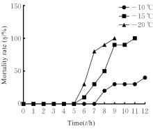
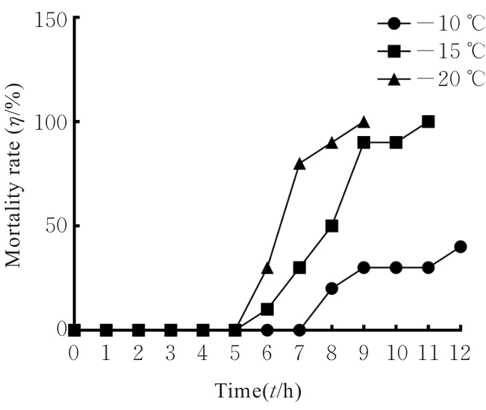
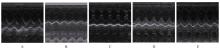
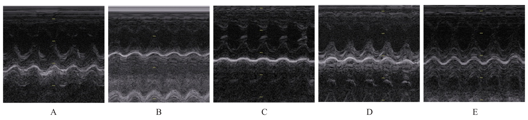
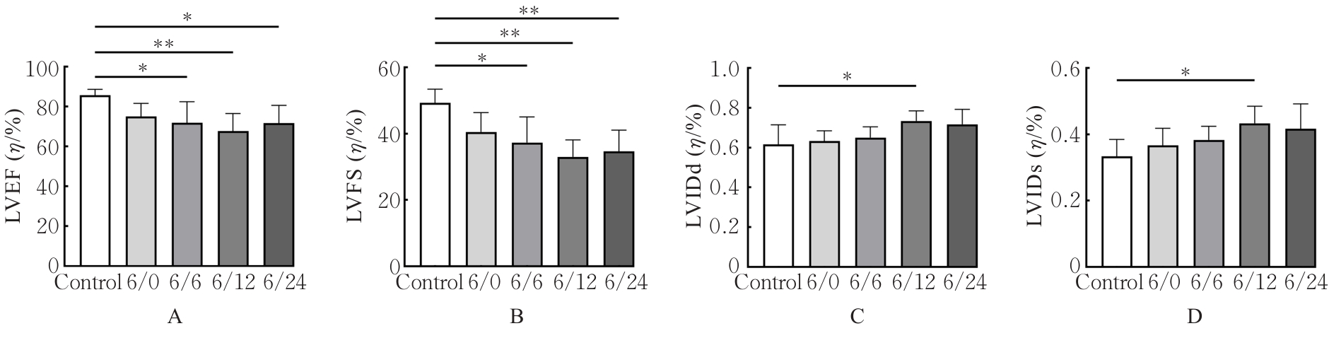
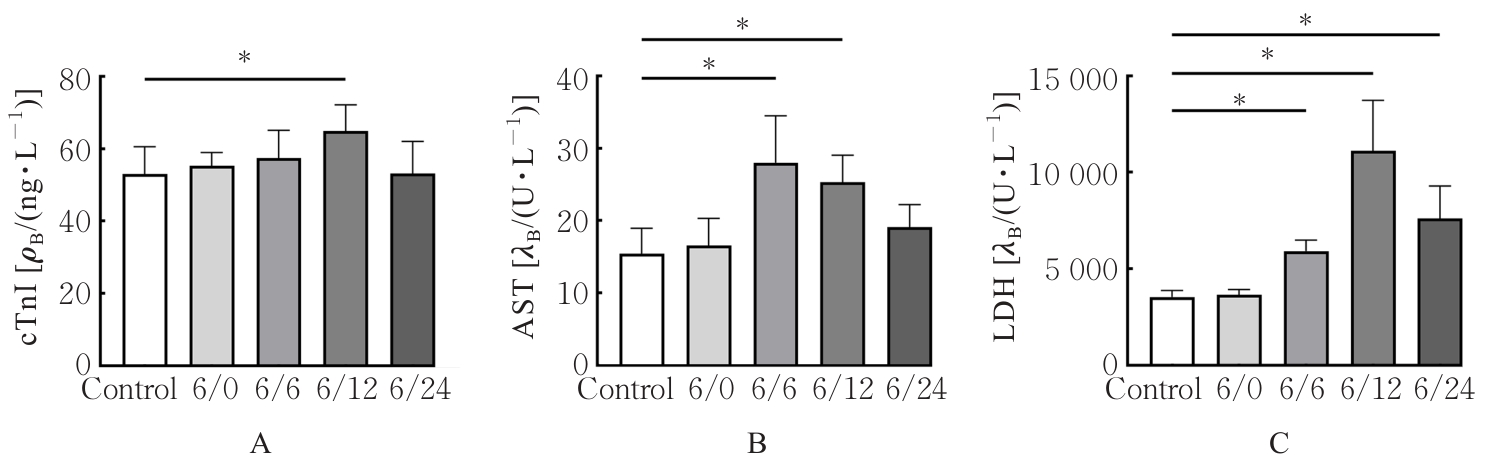
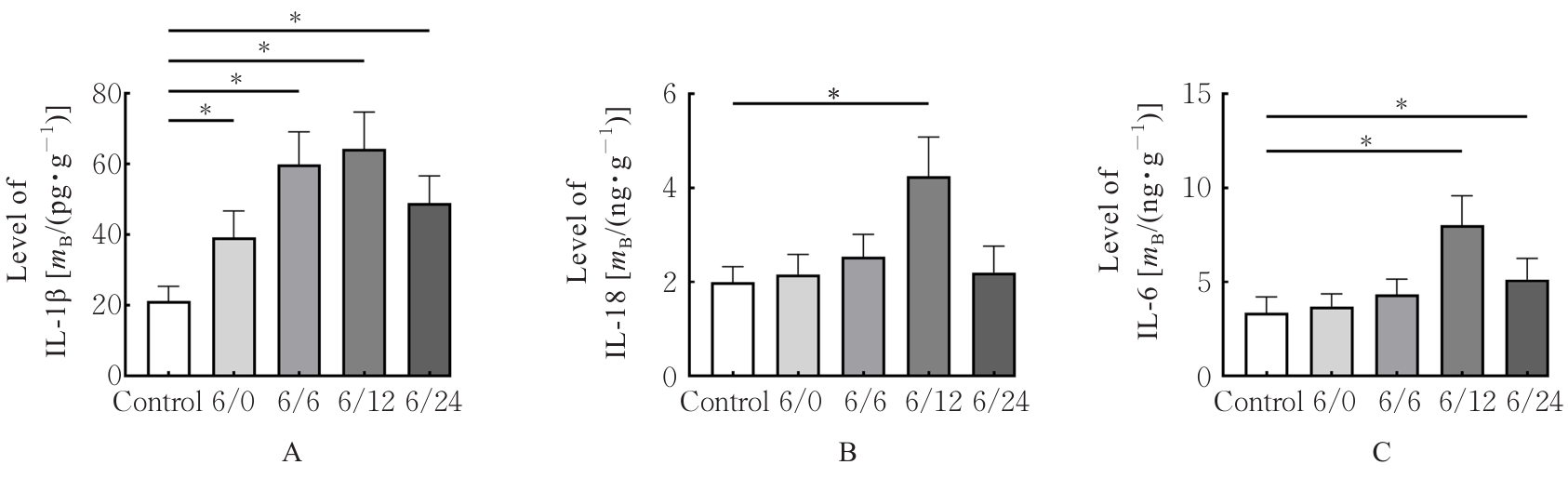
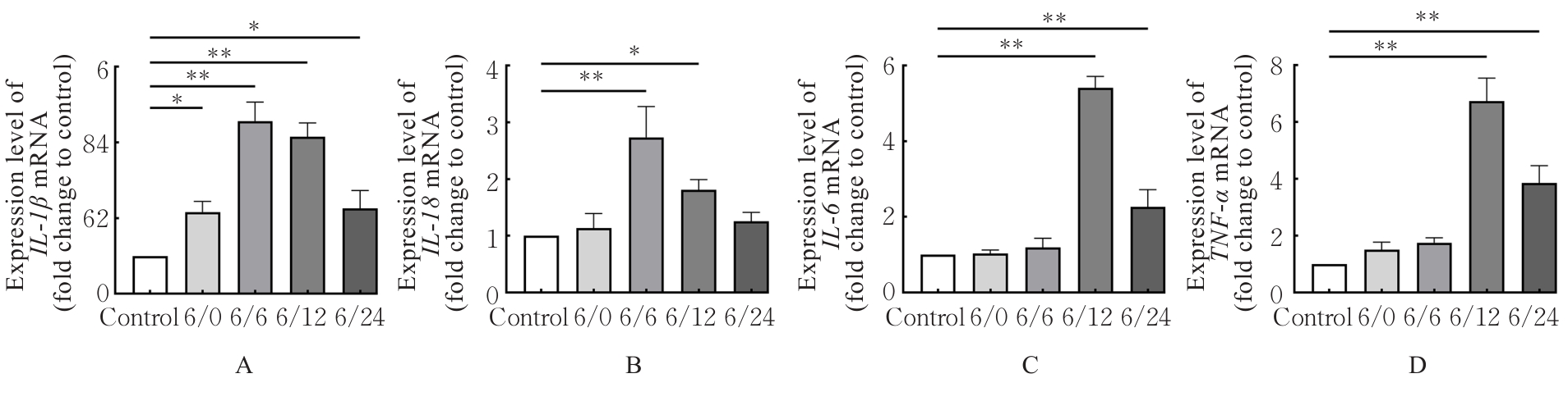
目的 筛选适宜的急性冷暴露(ACE)温度和复温时间,建立其诱导的大鼠心肌损伤模型,并通过血液生化学、病理形态学和分子生物学等指标综合评价模型建立条件的合理性及可行性。 方法 30只雄性Wistar大鼠随机分为-10 ℃、-15 ℃和-20 ℃冷暴露组,每组10只。通过观察各组大鼠死亡率确定最佳造模参数(温度和时间)。另取50只雄性Wistar大鼠随机分为对照组和急性冷暴露/复温(ACE/R)组,根据复温时间分为6/0、6/6、6/12和6/24 h组,每组10只。采用-15 ℃冷暴露6 h后复温相应时间建立ACE/R模型。检测各组大鼠核心体温,并评估各组大鼠心脏功能。HE染色观察各组大鼠心肌组织病理形态表现,测定各组大鼠血清中心肌肌钙蛋白I(cTnI)水平和天冬氨酸氨基转移酶(AST)及乳酸脱氢酶(LDH)活性,酶联免疫吸附试验(ELISA)法检测各组大鼠心肌组织中白细胞介素1β(IL-1β)、白细胞介素18(IL-18)和白细胞介素6(IL-6)水平,实时荧光定量PCR(RT-qPCR)法检测各组大鼠心肌组织中IL-IL-18IL-6和肿瘤坏死因子α(TNF-α) mRNA表达水平。 结果 与对照组比较,不同时间点ACE/R组大鼠核心体温均明显降低(P<0.05或P<0.01)。与对照组比较,6/6、6/12和6/24组大鼠左室射血分数(LVEF)和左室短轴缩短率(LVFS)均明显降低(P<0.05或P<0.01),6/12组大鼠左心室舒张末期内径(LVIDd)和左心室收缩末期内径(LVIDs)明显升高(P<0.05)。与对照组比较,6/12组大鼠血清中cTnI水平和AST及LDH活性均明显升高(P<0.01);HE染色,与对照组比较,6/12组大鼠心肌纤维排列紊乱、间隙增宽,伴大量炎症细胞浸润。ELISA法,与对照组比较,6/12组大鼠心肌组织中IL-1β、IL-18及IL-6水平明显升高(P<0.01)。RT-qPCR法,与对照组比较,6/12组大鼠心肌组织中IL-IL-18IL-6TNF-α mRNA表达水平明显升高(P<0.05或P<0.01)。 结论 采用雄性Wistar大鼠于-15 ℃冷空气暴露6 h后室温复温12 h可建立ACE/R心肌损伤模型。

关键词: 急性冷暴露, 心肌损伤, 复温, Wistar大鼠, 疾病模型, 动物

Abstract:

Objective To screen the appropriate temperature and rewarming time of acute cold exposure (ACE) for establishing the rat model of myocardial injury induced by ACE, and to comprehensively evaluate the rationality and feasibility of the model establishment conditions through blood biochemistry, pathomorphology and molecular biology indicators. Methods Thirty male Wistar rats were randomly divided into -10 ℃, -15 ℃ and -20 ℃ cold exposure groups, with 10 rats in each group. The optimal modeling parameters (temperature and time) were determined by observing the mortality rates of the rats in various groups. Another fifty male Wistar rats were randomly divided into control group and rewarming (ACE/R) groups, which were further divided into 6/0, 6/6, 6/12 and 6/24 h groups according to rewarming time, with 10 rats in each group. The ACE/R model was established by cold exposure at -15 ℃ for 6 h followed by rewarming for corresponding time. The core body temperatures of the rats in various groups were detected, and the cardiac function of the rats in various groups was evaluated. HE staining was used to observe the pathological morphology of myocardium tissue of the rats in various groups; the serum levels of cardiac troponin I (cTnI) and the activities of aspartate aminotransferase (AST) and lactate dehydrogenase (LDH) of the rats in various groups were measured; enzyme-linked immunosorbent assay (ELISA) was used to detect the levels of interleukin-1β (IL-1β), interleukin-18 (IL-18) and interleukin-6 (IL-6) in the myocardium tissue of the rats in various groups; real-time fluorescence quantitative PCR (RT-qPCR) was used to detect the expression levels of IL-IL-18IL-6 and tumor necrosis factor-α (TNF-α) mRNA in the myocardium tissue of the rats in various groups. Results Compared with control group, the core body temperatures of the rats in ACE/R groups at different time points were significantly decreased (P<0.05 or P<0.01). Compared with control group, the left ventricular ejection fraction (LVEF) and left ventricular fractional shortening (LVFS) of the rats in 6/6, 6/12 and 6/24 groups were significantly decreased (P<0.05 or P<0.01), and the left ventricular internal diameter in diastole (LVIDd) and left ventricular internal diameter in systole (LVIDs) of the rats in 6/12 group were significantly increased (P<0.05). Compared with control group, the serum level of cTnI and the activities of AST and LDH of the rats in 6/12 group were significantly increased (P<0.01). HE staining results showed that compared with control group, the myocardium tissue of the rats in 6/12 group showed disordered arrangement of myocardial fibers, widened intercellular spaces, and a large number of inflammatory cell infiltration. The ELISA results showed that compared with control group, the levels of IL-1β, IL-18 and IL-6 in the myocardium tissue of the rats in 6/12 group were significantly increased (P<0.01). The RT-qPCR results showed that compared with control group, the expression levels of IL-IL-18IL-6 and TNF-α mRNA in the myocardium tissue of the rats in 6/12 group were significantly increased (P<0.05 or P<0.01). Conclusion The ACE/R myocardial injury model can be established in the male Wistar rats by exposure of cold air at -15 ℃ for 6 h followed by rewarming at room temperature for 12 h.

Key words: Acute cold exposure, Myocardial injury, Rewarming, Wistar rats, Disease model, animal

中图分类号: 

  • R331