吉林大学学报(医学版) ›› 2026, Vol. 52 ›› Issue (5): 1377-1389.doi: 10.13481/j.1671-587X.20260519

• 基础研究 • 上一篇    

橙酮对糖皮质激素诱导的成骨细胞分化抑制和大鼠骨质疏松症的改善作用及其机制

韩小飞1,马运锋2()   

  1. 1.河南省中医院 河南中医药大学第二附属医院骨病一科, 河南 郑州 450002
    2.河南中医药大学 第三附属医院骨病科,河南 郑州 450003
  • 收稿日期:2025-11-20 接受日期:2026-01-14 出版日期:2026-09-28 发布日期:2026-10-15
  • 通讯作者: 马运锋 E-mail:weiminwanghb@sina.com
  • 作者简介:韩小飞(1982-),女,河南省济源市人,讲师,医学博士,主要从事中医骨伤科学基础与临床等方面的研究。
  • 基金资助:
    河南省卫健委中医药科学研究专项(20-21ZY3013)

Ameliorative effect of aurone on glucocorticoid-induced inhibition of osteoblast differentiation and osteoporosis in rats and its mechanism

Xiaofei HAN1,Yunfeng MA2()   

  1. 1.Department of Orthopedics,Henan Provincial Hospital of Traditional Chinese Medicine,Second;Affiliated Hospital,Henan University of Chinese Medicine,Zhengzhou 450002,China
    2.Department of Orthopedics,Third Affiliated Hospital,Henan University of Traditional Chinese Medicine,Zhengzhou 450003,China
  • Received:2025-11-20 Accepted:2026-01-14 Online:2026-09-28 Published:2026-10-15
  • Contact: Yunfeng MA E-mail:weiminwanghb@sina.com

摘要:

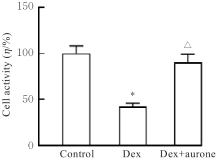
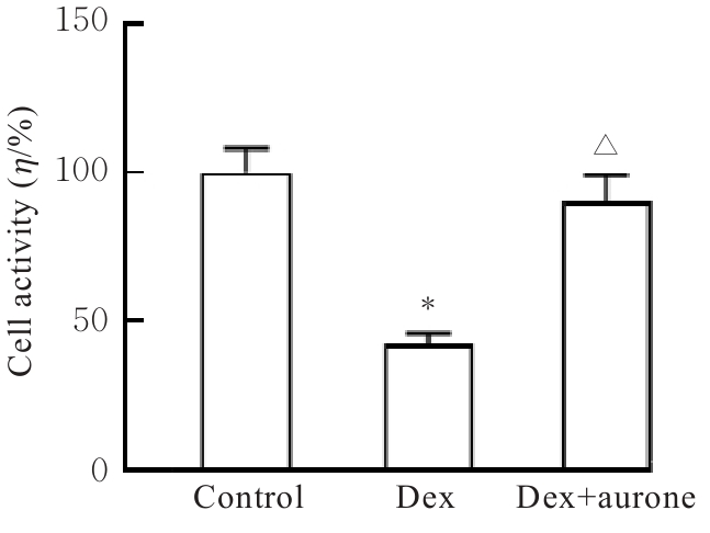
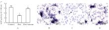
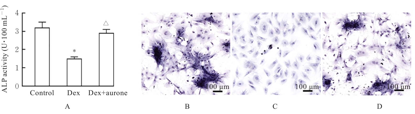
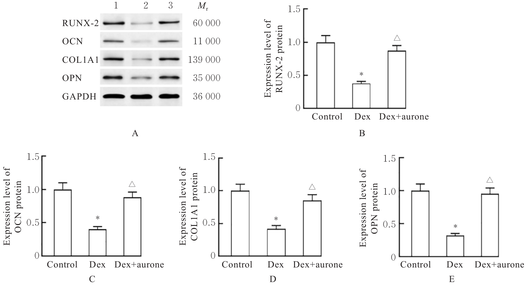
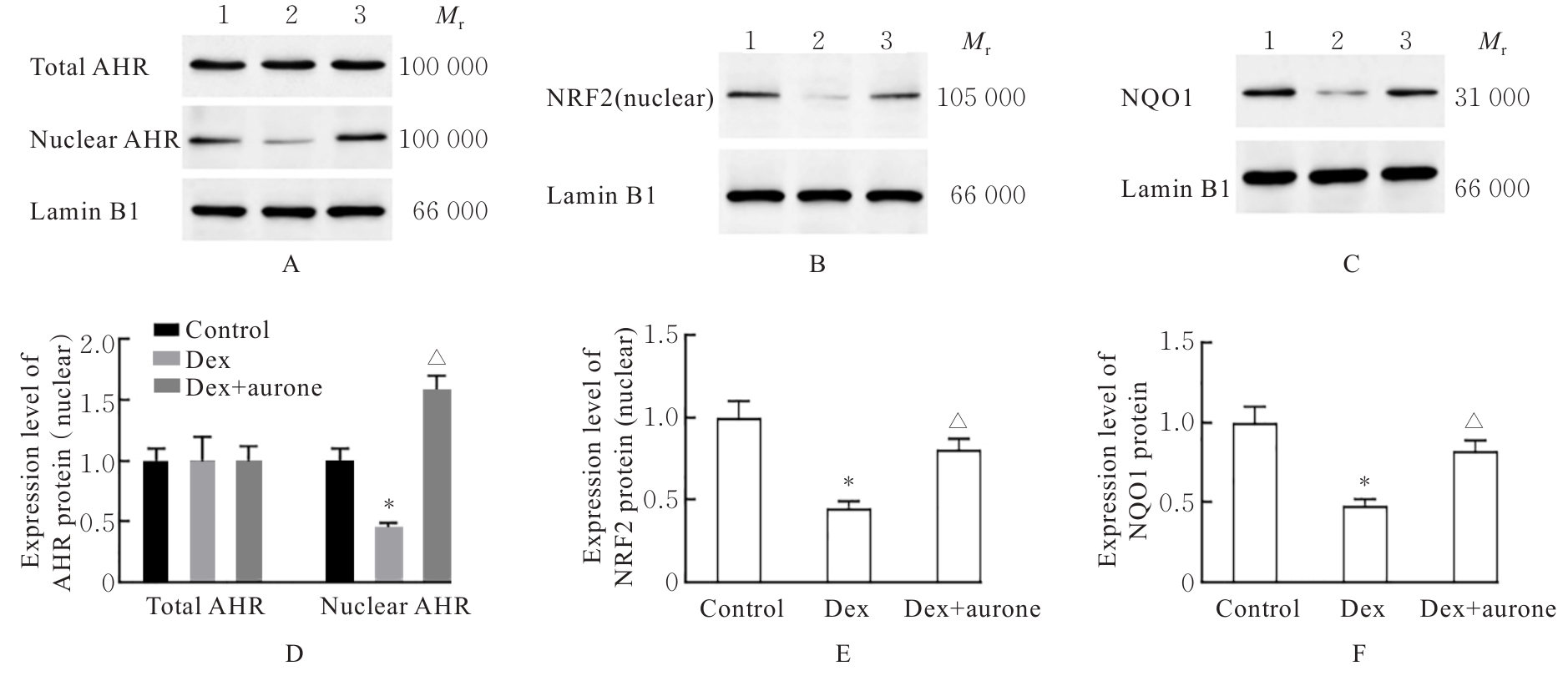
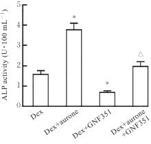
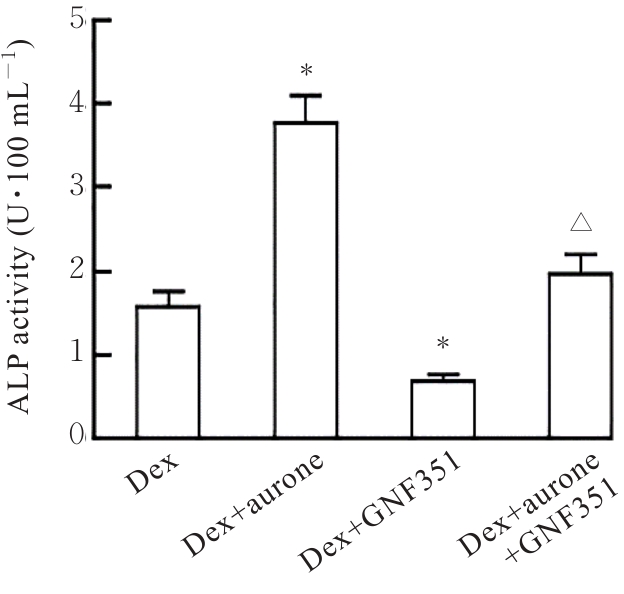
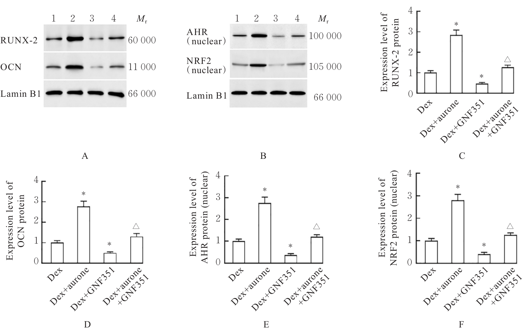
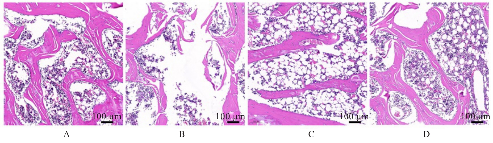
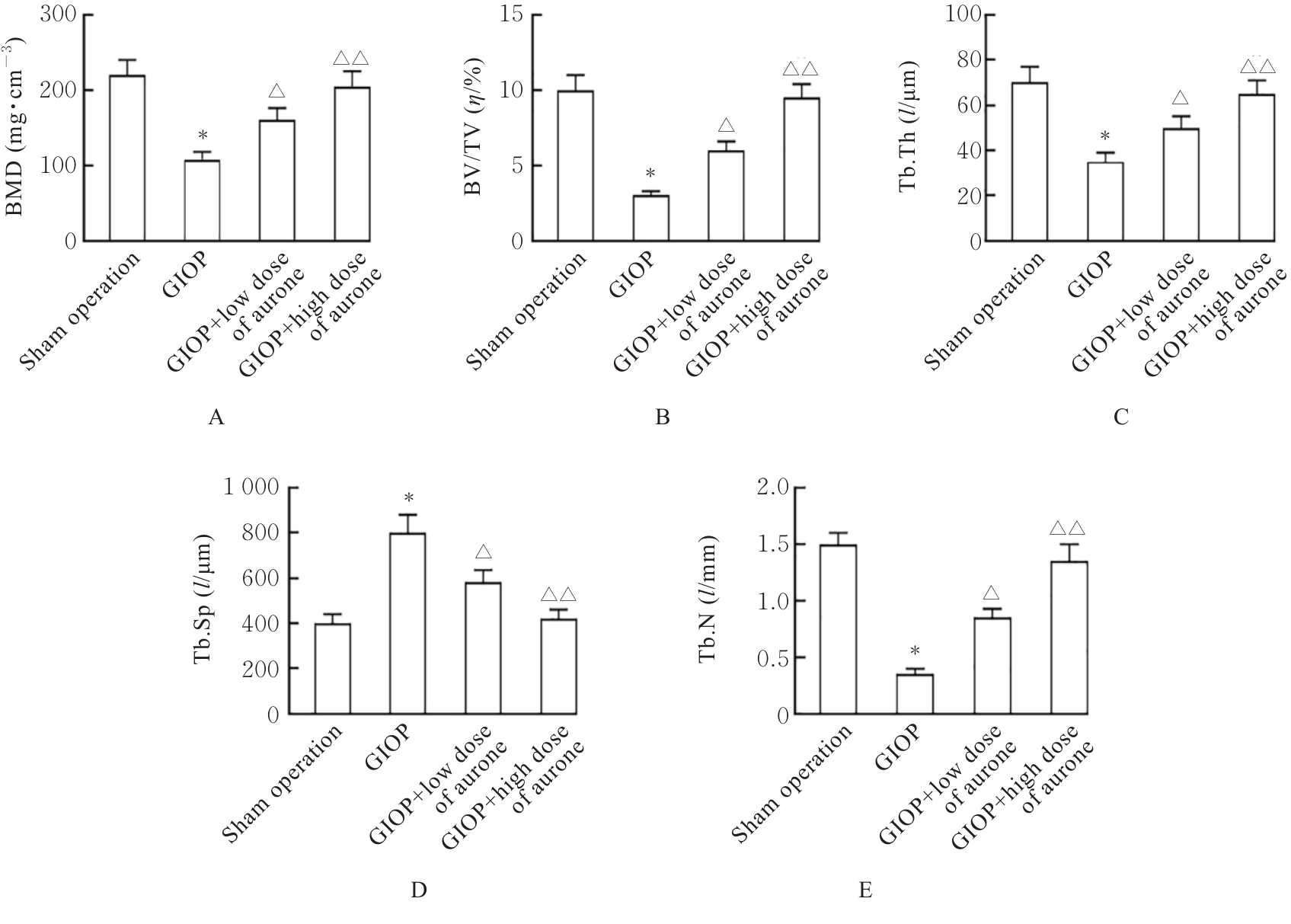
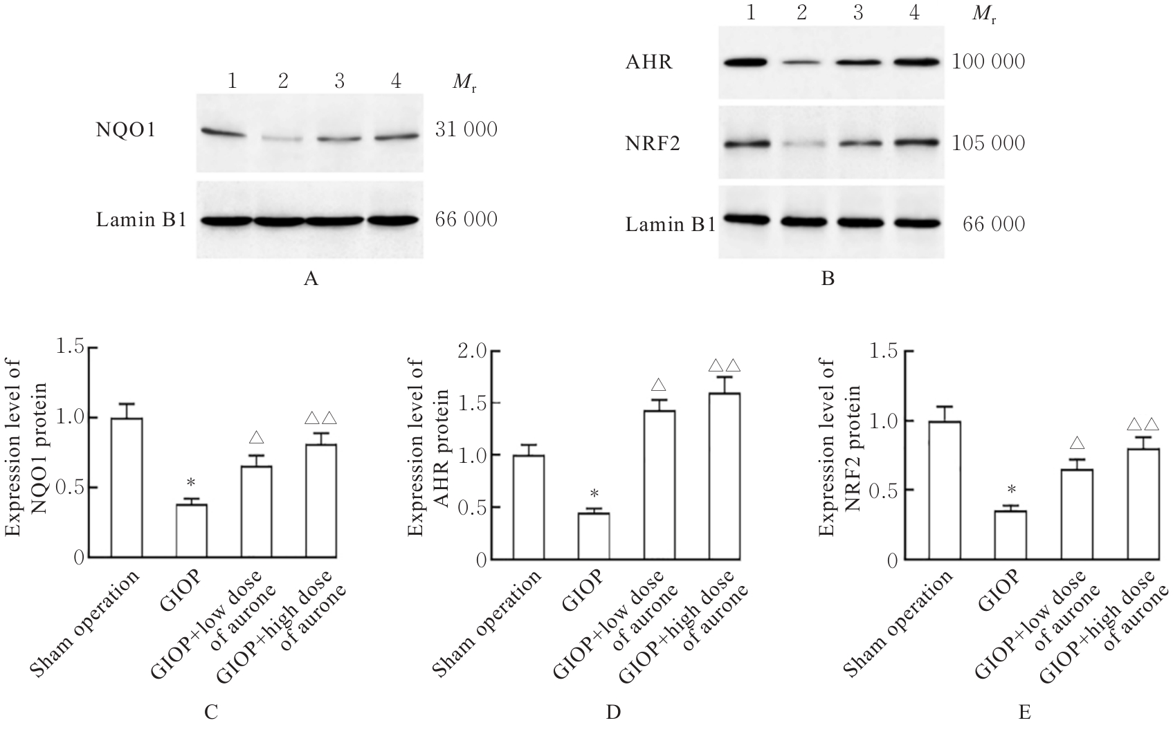
目的 探讨橙酮对成骨细胞分化的影响和对大鼠糖皮质激素性骨质疏松症(GIOP)的改善作用,并阐明其作用机制。 方法 体外实验选取成骨细胞MC3T3-E1,采用细胞计数试剂盒8(CCK-8)法检测不同浓度地塞米松(Dex)和橙酮处理后各组细胞活性,确定Dex和橙酮的最佳处理浓度。采用1 μmol·L-1 Dex诱导成骨细胞MC3T3-E1,建立细胞损伤模型。将细胞分为对照组、Dex组、Dex+橙酮组、Dex+GNF351组和Dex+橙酮+GNF351组。采用试剂盒检测各组细胞中丙二醛(MDA)水平和超氧化物歧化酶(SOD)活性,2',7'-二氯荧光素二乙酸酯(DCFH-DA)荧光法检测各组细胞中活性氧(ROS)水平,酶联免疫吸附试验(ELISA)法检测各组细胞上清液中肿瘤坏死因子α(TNF-α)、白细胞介素(IL)-1β和IL-6水平,碱性磷酸酶(ALP)试剂盒检测各组细胞ALP活性,Western blotting法检测各组细胞中Runt相关转录因子2(RUNX-2)、骨钙素(OCN)、Ⅰ型胶原α1链(COL1A1)、骨桥蛋白(OPN)、芳香烃受体(AHR)、核因子红细胞系2相关因子2(NRF2)和还原型烟酰胺腺嘌呤二核苷酸(磷酸):醌氧化还原酶1(NQO1)蛋白表达水平。体内实验选取20只雌性大鼠随机分为假手术组、GIOP组、GIOP+低剂量橙酮(7 mg·kg-1)组和GIOP+高剂量橙酮(14 mg·kg?1)组,每组5只,除假手术组外各组大鼠采用1 mg·kg-1 Dex灌胃构建GIOP模型。各组大鼠于造模后2 h起给予药物灌胃处理,连续干预7周后采血并处死,收集股骨组织。采用HE染色法观察各组大鼠股骨组织病理形态表现,组织形态计量学检测各组大鼠股骨组织骨密度(BMD)和骨小梁体积分数(BV/TV)、骨小梁厚度(Tb.Th)、骨小梁数目(Tb.N)及骨小梁分离度(Tb.Sp),Western blotting法检测各组大鼠股骨组织中AHR、NRF2和NQO1蛋白表达水平。 结果 与0 μmol·L?1 Dex比较,1 μmol·L?1 Dex组MC3T3-E1细胞活性明显降低(P<0.01),确定后续采用1 μmol·L?1 Dex处理细胞。与Dex组比较,Dex+5 μmol·L?1橙酮组MC3T3-E1细胞活性明显升高(P<0.01),后续采用5 μmol·L?1橙酮处理细胞。与对照组比较,Dex组MC3T3-E1细胞活性明显降低(P<0.01),细胞中MDA和ROS水平及上清液中TNF-α、IL-1β和IL-6水平均明显升高(P<0.01),SOD活性明显降低(P<0.01),细胞ALP活性明显降低(P<0.01),细胞中RUNX-2、OCN、COL1A1和OPN蛋白表达水平均明显降低(P<0.01),细胞核中AHR和NRF2蛋白表达水平及细胞中NQO1蛋白表达水平均明显降低(P<0.01)。与Dex组比较,Dex+橙酮组MC3T3-E1细胞活性明显升高(P<0.01),细胞中MDA和ROS水平及上清液中TNF-α、IL-1β和IL-6水平均明显降低(P<0.01),SOD活性明显升高(P<0.01),细胞ALP活性明显升高(P<0.01),细胞中RUNX-2、OCN、COL1A1和OPN蛋白表达水平均明显升高(P<0.01),细胞核中AHR和NRF2蛋白及细胞中NQO1蛋白表达水平均明显升高(P<0.01)。与Dex+橙酮组比较,Dex+橙酮+GNF351组细胞ALP活性明显降低(P<0.01),细胞核中AHR和NRF2蛋白表达水平以及细胞中NQO1、RUNX-2和OCN蛋白表达水平均明显降低(P<0.01)。体内实验,与假手术组比较,GIOP组大鼠股骨组织骨小梁变薄、数量减少且面积缩小,大鼠股骨组织中BMD、BV/TV、Tb.Th和Tb.N均明显降低(P<0.01),Tb.Sp明显升高(P<0.01),大鼠股骨组织中AHR、NRF2和NQO1蛋白表达水平均明显降低(P<0.01)。与GIOP组比较,GIOP+低剂量橙酮组和GIOP+高剂量橙酮组大鼠股骨组织病理损伤均改善,股骨组织中BMD、BV/TV、Tb.N和Tb.Th均明显升高(P<0.05),Tb.Sp明显降低(P<0.05),股骨组织中AHR、NRF2和NQO1蛋白表达水平均明显升高(P<0.05或P<0.01)。 结论 橙酮可促进大鼠成骨细胞分化,缓解大鼠GIOP,其作用机制可能与促进AHR核易位和激活AHR/NRF2/NQO1信号通路有关。

关键词: 骨质疏松症, 成骨分化, 橙酮, MC3T3-1细胞, 芳香烃受体

Abstract:

Objective To discuss the effects of aurone on osteoblast differentiation and its ameliorative effect on glucocorticoid-induced osteoporosis (GIOP) in rats, and to clarify its mechanism. Methods For in vitro experiments, MC3T3-E1 osteoblasts were selected. CCK-8 method was used to detect the cell activities in various groups after treatment with different concentrations of dexamethasone (Dex) and aurone to determine the optimal treatment concentrations of Dex and aurone. MC3T3-E1 osteoblasts were induced with 1 μmol·L-1 Dex to establish a cell injury model. The cells were divided into control group, Dex group, Dex+aurone group, Dex+GNF351 group, and Dex+aurone+GNF351 group. Kit method was used to detect the level of malondialdehyde (MDA) and the activity of superoxide dismutase (SOD) in the cells in various groups; 2',7'-dichlorofluorescein diacetate (DCFH-DA) fluorescence method was used to detect the level of reactive oxygen species (ROS) in the cells in various groups; enzyme-linked immunosorbent assay (ELISA) method was used to detect the levels of tumor necrosis factor-α (TNF-α), interleukin-1β (IL-1β), and interleukin-6 (IL-6) in the supernatants of the cells in various groups; alkaline phosphatase (ALP) kit method was used to detect the ALP activity in the cells in various groups; Western blotting method was used to detect the expression levels of Runt-related transcription factor 2 (RUNX-2), osteocalcin (OCN), collagen type Ⅰ alpha 1 chain (COL1A1), osteopontin (OPN), aromatic hydrocarbon receptor (AHR), nuclear factor erythroid 2-related factor 2 (NRF2), and NAD(P)H: quinone oxidoreductase 1 (NQO1) proteins in the cells in various groups. For in vivo experiments, 20 female rats were selected and randomly divided into sham operation group, GIOP group, GIOP+low dose of aurone (7 mg·kg-1) group, and GIOP+high dose of aurone (14 mg·kg-1) group, with 5 rats in each group. Except for sham operation group, the rats in various groups were intragastrically administered with 1 mg·kg-1 Dex to establish GIOP model. The rats in various groups were intragastrically administered with drugs from 2 h after modeling; after continuous intervention for 7 weeks, blood was collected and the rats were sacrificed, and femoral tissue was collected. HE staining method was used to observe the pathological morphology of femoral tissue of the rats in various groups. Histomorphometry method was used to detect the bone mineral density (BMD) of the femoral tissue and trabecular bone volume fraction (BV/TV), trabecular thickness (Tb.Th), trabecular number (Tb.N), and trabecular separation (Tb.Sp) of the rats in various groups. Western blotting method was used to detect the expression levels of AHR, NRF2, and NQO1 proteins in the femoral tissue of the rats in various groups. Results Compared with 0 μmol·L-1 Dex group, the cell activity of MC3T3-E1 cells in 1 μmol·L-1 Dex group was significantly decreased (P<0.01), and 1 μmol·L-1 Dex was determined for subsequent cell treatment. Compared with Dex group, the cell activity of MC3T3-E1 cells in Dex+5 μmol·L-1 aurone group was significantly increased (P<0.01), and 5 μmol·L-1 aurone was used for subsequent cell treatment. Compared with control group, the cell activity of MC3T3-E1 cells in Dex group was significantly decreased (P<0.01); the levels of MDA and ROS in the cells and the levels of TNF-α, IL-1β, and IL-6 in the supernatants were significantly increased (P<0.01); the SOD activity was significantly decreased (P<0.01); the ALP activity in the cells was significantly decreased (P<0.01); the expression levels of RUNX-2, OCN, COL1A1, and OPN proteins in the cells were significantly decreased (P<0.01); and the expression levels of AHR and NRF2 proteins in the nucleus and the expression level of NQO1 protein in the cells were significantly decreased (P<0.01). Compared with Dex group, the cell activity of MC3T3-E1 cells in Dex+aurone group was significantly increased (P<0.01); the levels of MDA and ROS in the cells and the levels of TNF-α, IL-1β, and IL-6 in the supernatants were significantly decreased (P<0.01); the SOD activity was significantly increased (P<0.01); the ALP activity in the cells was significantly increased (P<0.01); and the expression levels of RUNX-2, OCN, COL1A1, and OPN proteins in the cells were significantly increased (P<0.01). Compared with Dex+aurone group, the ALP activity in the cells in Dex+aurone+GNF351 group was significantly decreased (P<0.01), and the expression levels of AHR and NRF2 proteins in the nucleus as well as the expression levels of NQO1, RUNX-2, and OCN proteins in the cells were significantly decreased (P<0.01). The in vivo experiment results showed that compared with sham operation group, the trabecular bones in the femoral tissue of the rats in GIOP group were thinned, reduced in number, and diminished in area, the BMD of the femoral tissue, trabecular BV/TV, Tb.Th, and Tb.N of the rats were significantly decreased (P<0.01), the Tb.Sp was significantly increased (P<0.01), and the expression levels of AHR, NRF2, and NQO1 proteins in the femoral tissue of the rats were significantly decreased (P<0.01). Compared with GIOP group, the pathological damage in femoral tissue of the rats in GIOP+low dose of aurone group and GIOP+high dose of aurone group was alleviated, the BMD of the femoral tissue, trabecular BV/TV, Tb.N, and Tb.Th were significantly increased (P<0.05), the Tb.Sp was significantly decreased (P<0.05), and the expression levels of AHR, NRF2, and NQO1 proteins in the femoral tissue were significantly increased (P<0.05 or P<0.01). Conclusion Aurone promotes osteoblast differentiation and alleviates GIOP in rats, and its mechanism is potentially associated with promoting the nuclear translocation of AHR and activating the AHR/NRF2/NQO1 signaling pathway.

Key words: Osteoporosis, Osteoblast differentiation, Aurone, MC3T3-E1 cell, Aromatic hydrocarbon receptor

中图分类号: 

  • R681.1