吉林大学学报(医学版) ›› 2026, Vol. 52 ›› Issue (5): 1453-1459.doi: 10.13481/j.1671-587X.20260526

• 临床医学 • 上一篇    

非综合征型上颌恒中切牙融合患者1例报告及文献复习

李欣鹏,康芙嘉,常路广达,贺赞然,王柏力,朱宪春()   

  1. 吉林大学口腔医院正畸科,吉林 长春 130021
  • 收稿日期:2025-10-25 接受日期:2026-01-05 出版日期:2026-09-28 发布日期:2026-10-15
  • 通讯作者: 朱宪春 E-mail:zhuxc@jlu.edu.cn
  • 作者简介:李欣鹏(2000-),男,内蒙古自治区呼伦贝尔市人,在读硕士研究生,主要从事口腔正畸学临床方面的研究。
  • 基金资助:
    吉林省科技厅自然科学基金项目(YDZJ202201ZYTS057)

Patient with non-syndromic fusion of permanent maxillary central incisor:A case report and literature review

Xinpeng LI,Fujia KANG,Luguangda CHANG,Zanran HE,Boli WANG,Xianchun ZHU()   

  1. Department of Orthodontics,Stomatology Hospital,Jilin University,Changchun 130021,China
  • Received:2025-10-25 Accepted:2026-01-05 Online:2026-09-28 Published:2026-10-15
  • Contact: Xianchun ZHU E-mail:zhuxc@jlu.edu.cn

摘要:

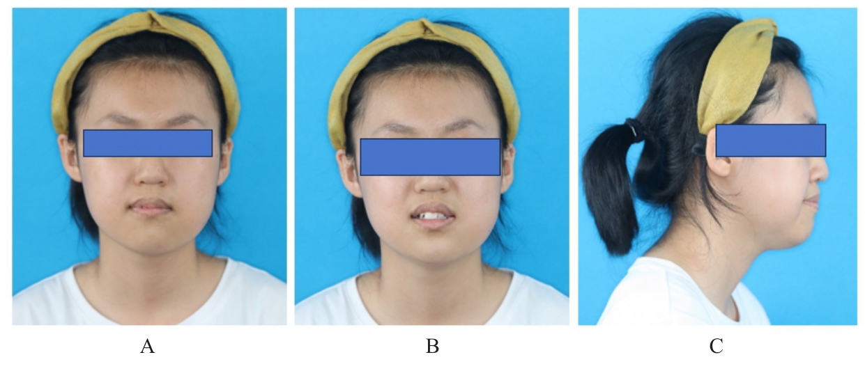
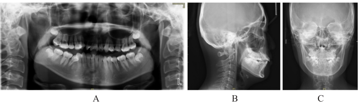
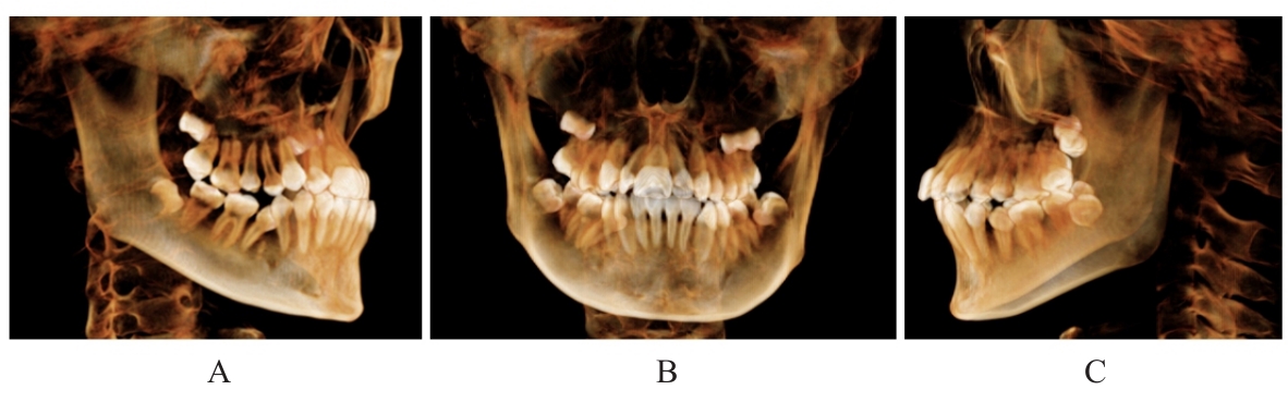
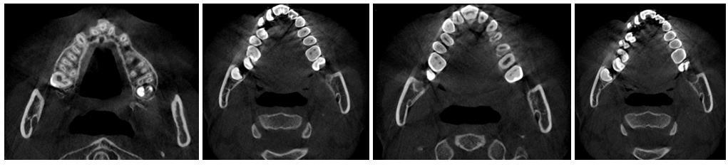
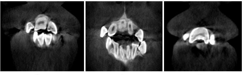
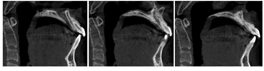
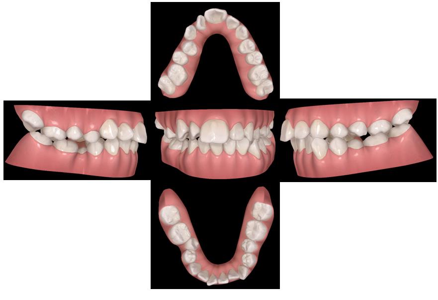
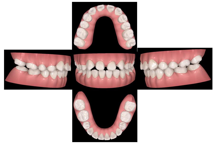
非综合征型恒牙融合牙(NSFPT)是一种由非系统性疾病因素导致的牙齿发育异常。恒牙融合牙好发于上颌中切牙与上颌侧切牙,由上颌恒中切牙融合而成的NSFPT极为罕见。本文作者报道1例NSFPT患者,制定正畸治疗方案,并结合相关文献进行复习。患者,女性,15岁,主诉牙不齐,嘴突。患者既往体健,否认系统性疾病史。影像学检查示患者蝶鞍-鼻根-上牙槽座角(SNA)、蝶鞍-鼻根-下牙槽座角(SNB)和上中切牙-前颅底平面角(U1-SN)偏大,上颌前牙区双侧恒中切牙冠根完全融合。诊断为11与21融合牙,安氏Ⅱ类1分类,凸面型。治疗方案为拔除融合牙和双侧下颌第二前磨牙,采用正畸-修复联合治疗以恢复正常面型和牙列形态。患者因个人原因未接受治疗。融合牙患者临床上需通过口内检查结合影像学检查明确诊断。依据病因、牙齿萌出状态及发生来源可将融合牙分为不同类型,本病例为NSFPT且非多生牙来源。正畸治疗需综合考量牙列拥挤度和侧貌等多种因素,以决定是否进行拔牙矫治。临床上应早发现、早干预,以减少对患者颜面部和牙列的影响,降低后续矫治难度。

关键词: 恒牙融合牙, 牙融合, 正畸治疗, 影像学诊断, 病例报告

Abstract:

Non?syndromic fused permanent teeth (NSFPT) is a dental developmental anomaly caused by non?systemic factors. Fused permanent teeth commonly occur between the maxillary central incisor and lateral incisor, while NSFPT resulting from fusion of bilateral maxillary permanent central incisors is extremely rare. This study reported a case of NSFPT, formulated an orthodontic treatment plan, and reviewed the relevant literature. The patient was a 15-year-old female whose chief complaint was irregular teeth and protruded lips. She had no history of systemic diseases. Imaging examination revealed increased sella-nasion-subspinal angle(SNA), sella-nasion-supramental angle(SNB), and upper incisor to sella-nasion plane angle(U1-SN) in the patient, as well as complete crown?root fusion of the bilateral maxillary permanent central incisors. The diagnoses were fused teeth of 11 and 21, Angle Class Ⅱ division 1 malocclusion, and convex facial profile. The proposed treatment plan involved extraction of the fused teeth and bilateral mandibular second premolars, followed by combined orthodontic?prosthodontic treatment to restore normal facial profile and dental arch morphology. However, the patient declined treatment due to personal reasons. For patients with fused teeth, diagnosis should be established through intraoral examination combined with imaging studies. Orthodontic treatment planning for such cases requires comprehensive consideration of multiple factors, including the degree of dental crowding and the facial profile, to determine whether extraction-based treatment is indicated. Early detection and intervention are essential to minimize the impact on the patient’s facial aesthetics and dentition, and to reduce the complexity of subsequent orthodontic treatment.

Key words: Fused permanent teeth, Tooth fusion, Orthodontic treatment, Imaging diagnosis, Case report

中图分类号: 

  • R781.2