吉林大学学报(医学版) ›› 2026, Vol. 52 ›› Issue (4): 1022-1033.doi: 10.13481/j.1671-587X.20260414

• 基础研究 • 上一篇    

基质细胞衍生因子1对雪旺细胞铁死亡促进大鼠周围神经损伤修复的抑制作用及其机制

袁野1,姜宇1,杜赛赛1,杨振军2,王培3()   

  1. 1.承德医学院研究生院,河北 承德 067000
    2.承德医学院基础医学院人体解剖学教研室,河北 承德 067000
    3.承德医学院附属医院手足外科,河北 承德 067000
  • 收稿日期:2025-10-24 接受日期:2025-12-03 出版日期:2026-07-28 发布日期:2026-07-27
  • 通讯作者: 王培 E-mail:cdgkwp@sina.com
  • 作者简介:袁 野(1999-),男,河北省承德市人,在读硕士研究生,主要从事周围神经损伤和修复方面的研究。
  • 基金资助:
    河北省科技厅指令性课题项目(142777105D);河北省神经损伤与修复重点实验室开放课题项目(NJKF202404)

Inhibitory effect of stromal cell-derived factor 1 on Schwann cells ferroptosis in promoting peripheral nerve injury repairment and its mechanism

Ye YUAN1,Yu JIANG1,Saisai DU1,Zhenjun YANG2,Pei WANG3()   

  1. 1.Graduate School,Chengde Medical University,Chengde 067000,China
    2.Department of Human Anatomy,School of Basic Medical Sciences,Chengde Medical University,Chengde 067000,China
    3.Department of Hand and Foot Surgery,Affiliated Hospital,Chengde Medical University,Chengde 067000,China
  • Received:2025-10-24 Accepted:2025-12-03 Online:2026-07-28 Published:2026-07-27
  • Contact: Pei WANG E-mail:cdgkwp@sina.com

摘要:

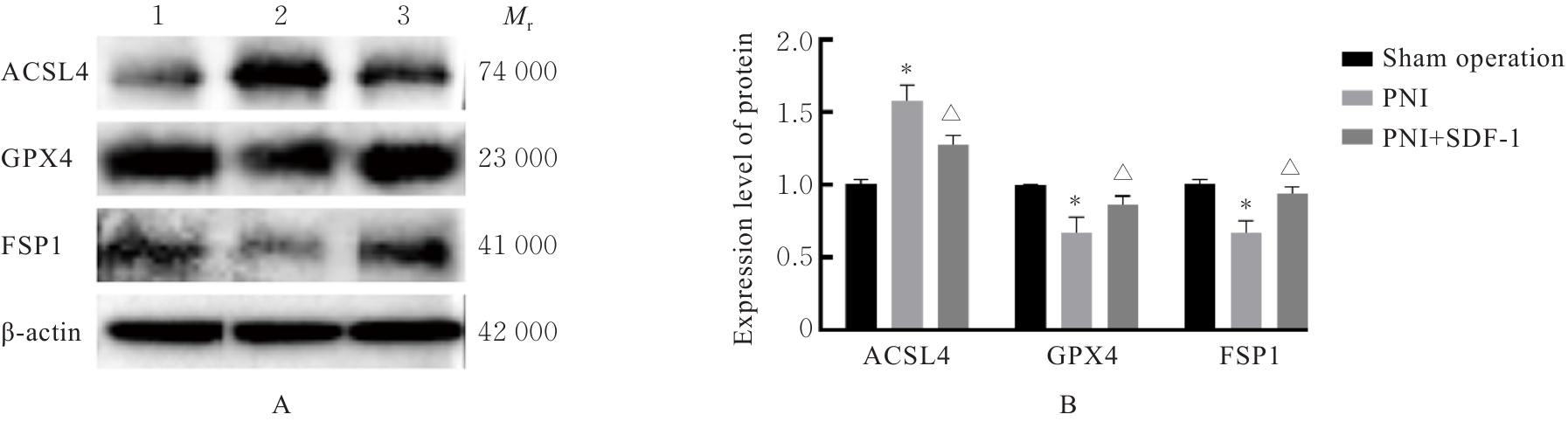
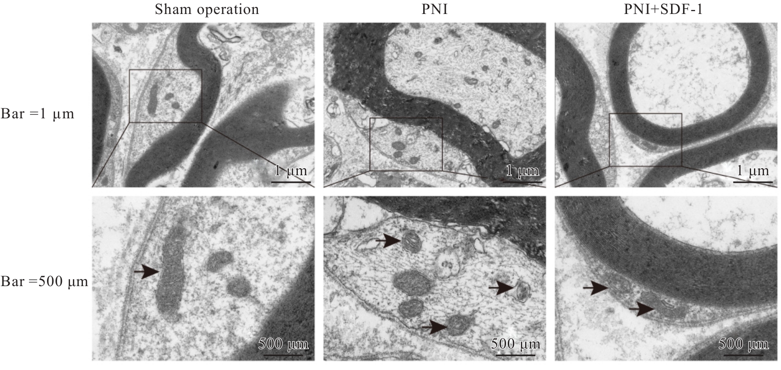
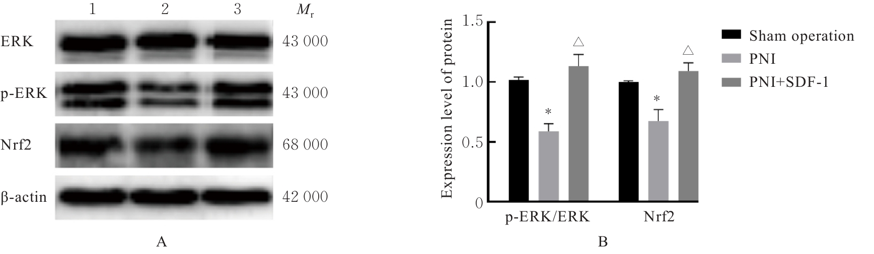
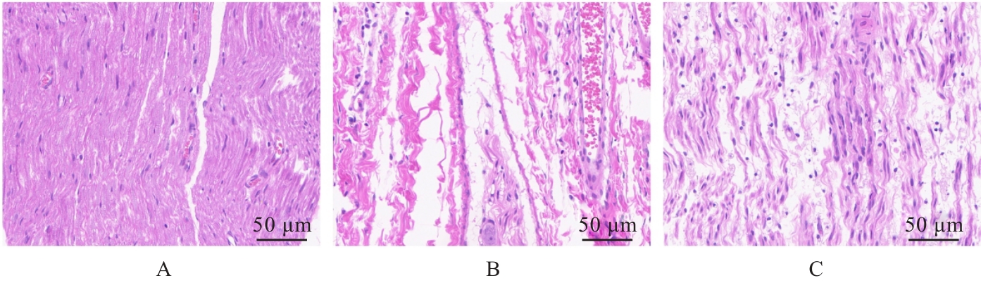
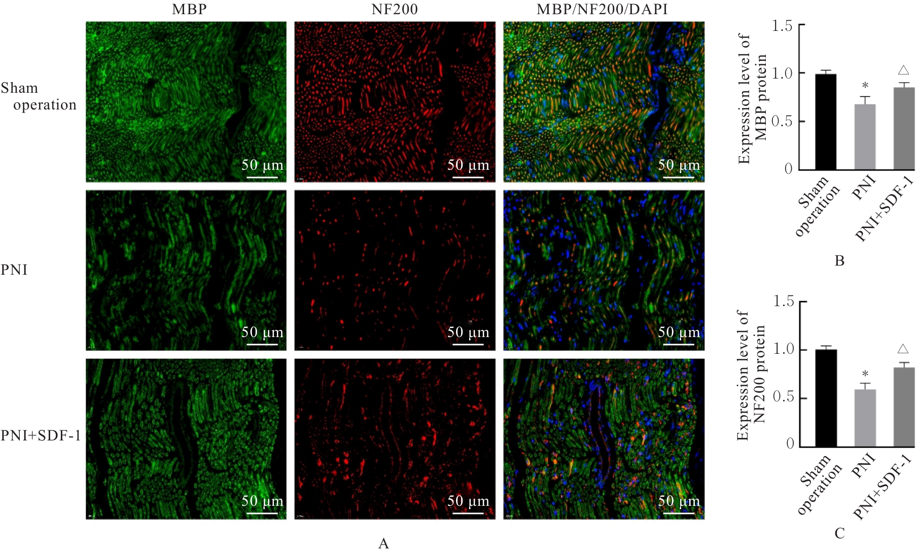
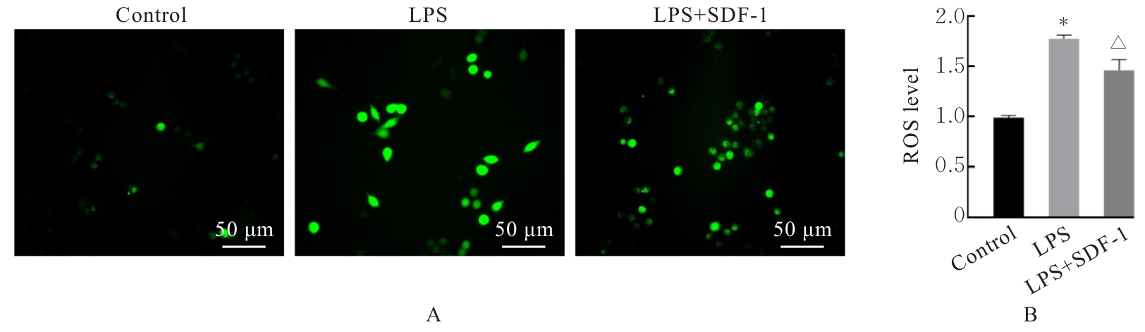
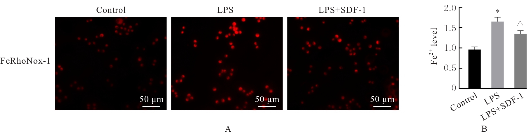
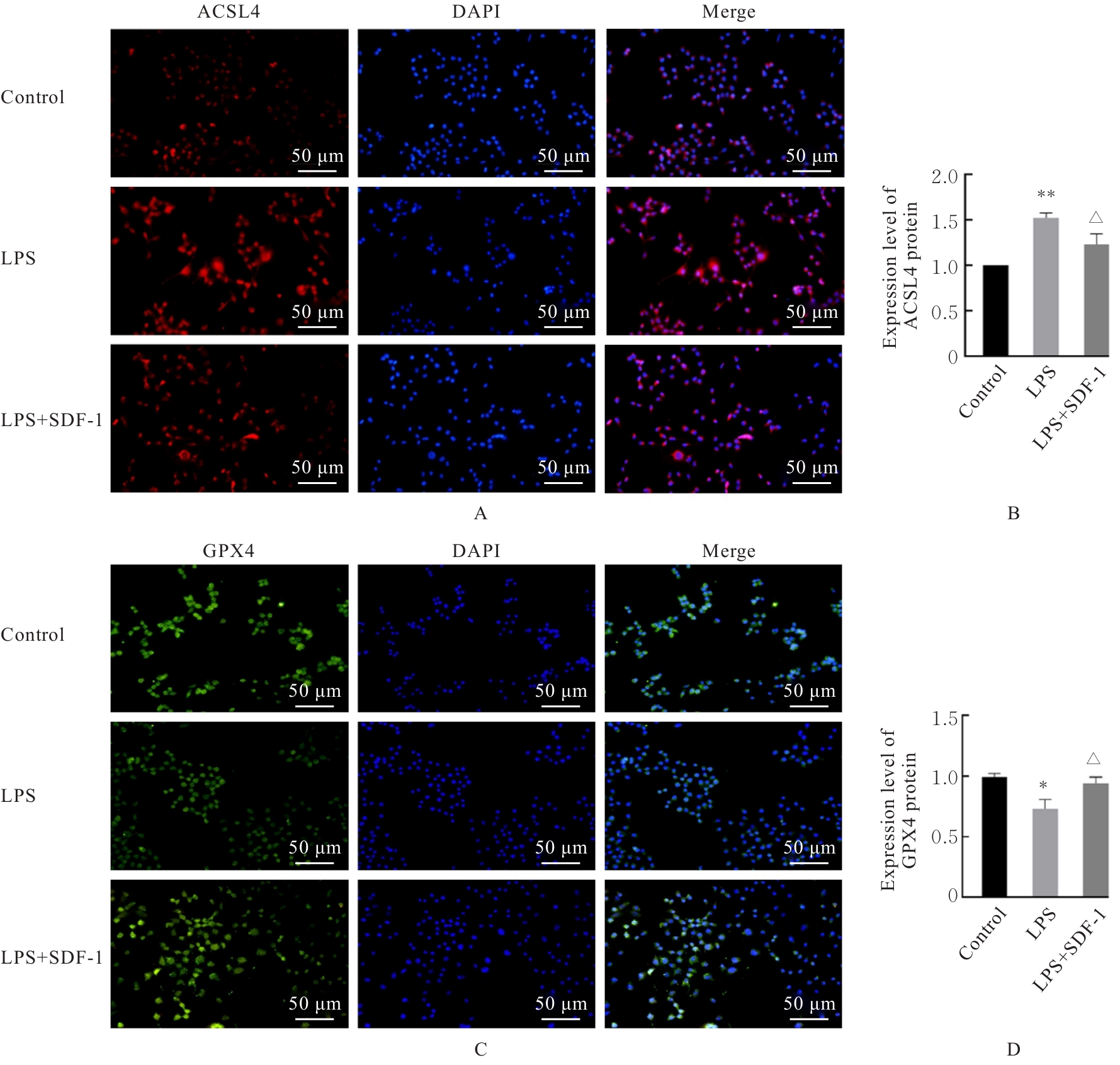
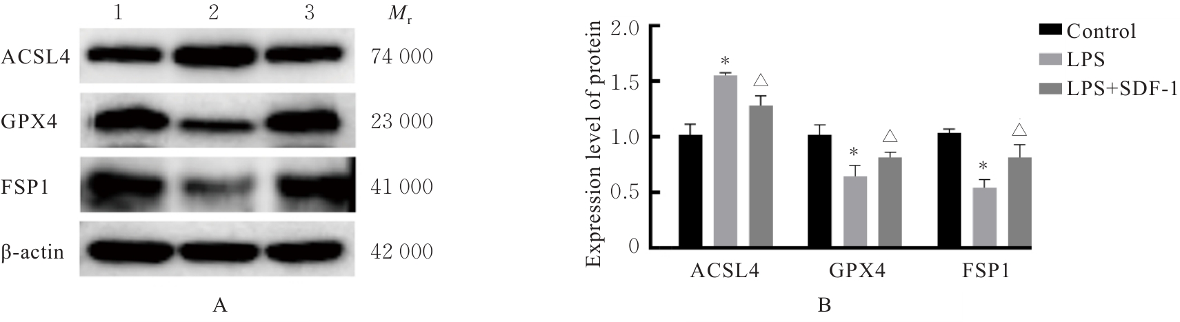
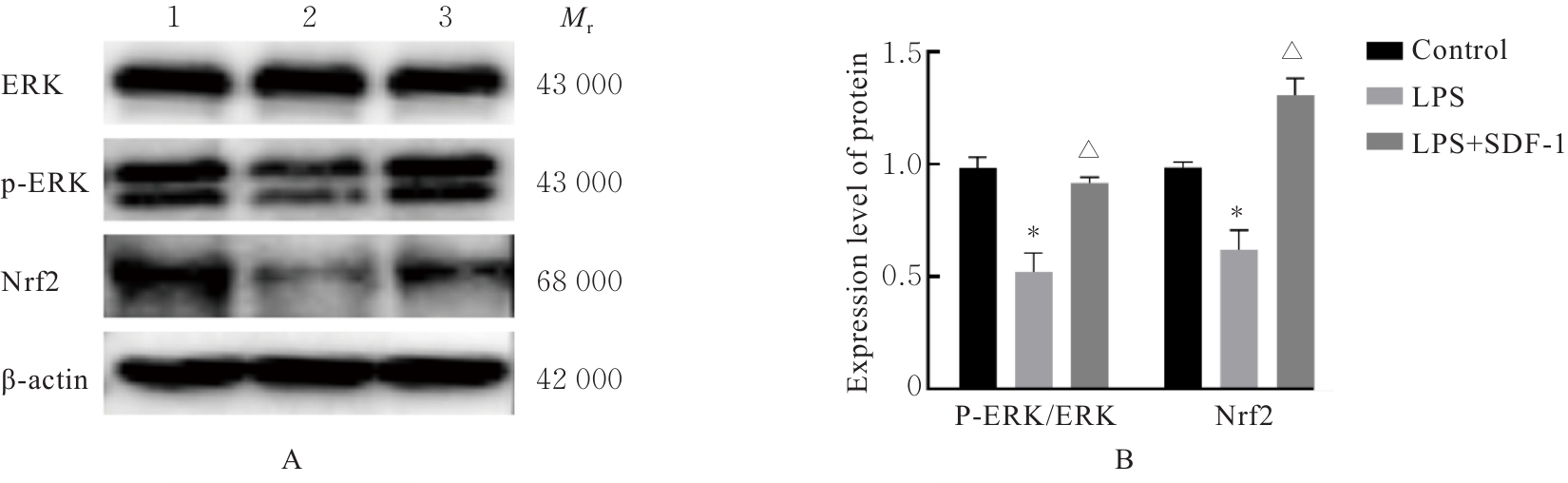
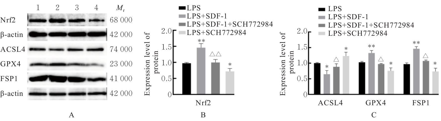
目的 探讨基质细胞衍生因子1(SDF-1)对周围神经损伤(PNI)后雪旺细胞(SCs)铁死亡的影响,并阐明其可能作用机制。 方法 动物实验,45只雄性8周龄SD大鼠随机分为假手术组、PNI组和SDF-1治疗组(PNI+SDF-1组),每组15只。假手术组大鼠暴露坐骨神经后不施加损伤处理;PNI组和SDF-1治疗组大鼠采用坐骨神经钳夹法造模。造模后PNI组大鼠肌注0.2 mL·d-1生理盐水,SDF-1治疗组大鼠肌注4 μg·kg-1·d-1 SDF-1,连续5 d。术后第5天,每组随机取10只大鼠处死,取坐骨神经,采用Western blotting法检测各组大鼠坐骨神经中长链脂酰辅酶A合成酶4(ACSL4)、谷胱甘肽过氧化物酶4(GPX4)和铁死亡抑制蛋白1(FSP1)蛋白表达水平,透射电镜观察坐骨神经SCs中线粒体形态表现,Western blotting法检测坐骨神经中细胞外信号调节激酶(ERK)、磷酸化细胞外信号调节激酶(p-ERK)和核因子红细胞2相关因子2(Nrf2)蛋白表达水平。术后第14天,每组取剩余5只大鼠处死取坐骨神经,采用HE染色法观察坐骨神经纤维形态表现,免疫荧光法检测各组大鼠坐骨神经中神经丝蛋白重链(NF200)与髓鞘碱性蛋白(MBP)表达水平。细胞实验,将SCs分为对照组、脂多糖(LPS)诱导损伤组(LPS组)和SDF-1治疗组(LPS+SDF-1组)。采用2',7'-二氯荧光素二乙酸酯(DCFH-DA)荧光探针法检测各组SCs中活性氧(ROS)水平,FeRhoNox-1荧光探针法检测各组SCs中亚铁离子(Fe2+)水平,免疫荧光法检测各组SCs中ACSL4和GPX4蛋白表达情况,Western blotting法检测各组SCs中ACSL4、GPX4、FSP1、ERK、p-ERK和Nrf2蛋白表达水平。回复实验,将SCs分为LPS组、LPS+SDF-1组、LPS+SDF-1+ SCH772984组和LPS+SCH772984组,采用Western blotting法检测各组SCs中Nrf2、ACSL4、GPX4和FSP1蛋白表达水平。 结果 动物实验,术后第5天,与假手术组比较,PNI组大鼠坐骨神经中ACSL4蛋白表达水平升高(P<0.01),GPX4和FSP1蛋白表达水平均降低(P<0.01);SCs中线粒体体积缩小、嵴结构模糊;p-ERK/ERK比值和Nrf2蛋白表达水平均降低(P<0.01)。与PNI组比较,PNI+SDF-1组大鼠坐骨神经中ACSL4蛋白表达水平降低(P<0.05),GPX4和FSP1蛋白表达水平均升高(P<0.05);SCs中线粒体体积增加、嵴结构趋向正常;p-ERK/ERK比值和Nrf2蛋白表达水平均升高(P<0.01)。术后第14天,与假手术组比较,PNI组大鼠坐骨神经纤维排列混乱,坐骨神经中NF200和MBP蛋白表达水平均降低(P<0.01);与PNI组比较,PNI+SDF-1组大鼠坐骨神经纤维排列趋于正常,坐骨神经中NF200和MBP蛋白表达水平均升高(P<0.05)。细胞实验,与对照组比较,LPS组SCs中ROS与Fe2+水平均升高(P<0.01),ACSL4蛋白表达水平均升高(P<0.01),GPX4蛋白表达水平均降低(P<0.05或P<0.01),FSP1蛋白表达水平降低(P<0.01),p-ERK/ERK比值和Nrf2蛋白表达水平均降低(P<0.01)。与LPS组比较,LPS+SDF-1组SCs中ROS与Fe2+水平均降低(P<0.05),ACSL4蛋白表达水平均降低(P<0.05),GPX4蛋白表达水平均升高(P<0.05),FSP1蛋白表达水平升高(P<0.05),p-ERK/ERK比值和Nrf2蛋白表达水平均升高(P<0.01)。回复实验,与LPS组比较,LPS+SDF-1组SCs中Nrf2蛋白表达水平升高(P<0.01),ACSL4蛋白表达水平降低(P<0.05),GPX4和FSP1蛋白表达水平升高(P<0.01);LPS+SCH772984组SCs中Nrf2蛋白表达水平降低(P<0.05),ACSL4蛋白表达水平升高(P<0.05),GPX4和FSP1蛋白表达水平均降低(P<0.05)。与LPS+SDF-1组比较,LPS+SDF-1+SCH772984组SCs中Nrf2蛋白表达水平降低(P<0.01),ACSL4蛋白表达水平升高(P<0.05),GPX4和FSP1蛋白表达水平均降低(P<0.05)。 结论 SDF-1可抑制PNI后SCs铁死亡,促进受损坐骨神经修复,其机制与激活ERK/Nrf2信号通路有关。

关键词: 神经再生, 基质细胞衍生因子1, 雪旺细胞, 铁死亡, 细胞外信号调节激酶

Abstract:

Objective To investigate the effect of stromal cell-derived factor 1 (SDF-1) on ferroptosis of Schwann cells (SCs) after peripheral nerve injury (PNI), and to elucidate its possible mechanism. Methods Animal experiment, 45 male 8-week-old SD rats were randomly divided into sham operation group, PNI group and SDF-1 treatment group (PNI+SDF-1 group), with 15 rats in each group. The rats in sham operation group were subjected to sciatic nerve exposure without injury; the rats in PNI group and SDF-1 treatment group were subjected to sciatic nerve clamping method to establish the models. After modeling, the rats in PNI group were intramuscularly injected with 0.2 mL·d-1 normal saline, and the rats in SDF-1 treatment group were intramuscularly injected with 4 μg·kg-1·d-1 SDF-1 for 5 consecutive days. On the 5th day after operation, 10 rats from each group were randomly selected and sacrificed to harvest the sciatic nerves. Western blotting method was used to detect the expression levels of long-chain acyl-CoA synthetase 4 (ACSL4), glutathione peroxidase 4 (GPX4), and ferroptosis suppressor protein 1 (FSP1) proteins in sciatic nerve of the rats in various groups; transmission electron microscope was used to observe the morphology of mitochondria in SCs in the sciatic nerve; Western blotting method was used to detect the expression levels of extracellular signal-regulated kinase (ERK), phosphorylated extracellular signal-regulated kinase (p-ERK), and nuclear factor erythroid 2-related factor 2 (Nrf2) proteins in the sciatic nerve. On the 14th day after operation, the remaining 5 rats from each group were sacrificed to harvest the sciatic nerves. HE staining was used to observe the morphology of sciatic nerve fibers; immunofluorescence staining was used to detect the expression levels of neurofilament 200 (NF200) and myelin basic protein (MBP) in sciatic nerve of the rats in various groups. Cell experiment, the SCs were divided into control group, lipopolysaccharide (LPS)- induced injury group (LPS group) and SDF-1 treatment group (LPS+SDF-1 group). 2',7'- Dichlorodihydrofluorescein diacetate (DCFH-DA) fluorescence probe method was used to detect the reactive oxygen species (ROS) levels in the SCs in various groups; FeRhoNox-1 fluorescence probe method was used to detect the ferrous ion (Fe2+)) levels in the SCs in various groups; immunofluorescence staining was used to detect the expression of ACSL4 and GPX4 proteins in the SCs in various groups; Western blotting method was used to detect the protein expression levels of ACSL4, GPX4, FSP1, ERK, p-ERK, and Nrf2 in the SCs in various groups. Rescue experiment, the SCs were divided into LPS group, LPS+SDF-1 group, LPS+SDF-1+SCH772984 group and LPS+SCH772984 group; Western blotting method was used to detect the expression levels of Nrf2, ACSL4, GPX4, and FSP1 proteins in the SCs in various groups. Results In animal experiment, on the 5th day after operation, compared with sham operation group, the expression level of ACSL4 protein in sciatic nerve of the rats in PNI group was increased (P<0.01), while the protein expression levels of GPX4 and FSP1 proteins were decreased (P<0.05); the mitochondria in SCs were shrunken and the cristae structure was blurred; the p-ERK/ERK ratio and the protein expression level of Nrf2 were decreased (P<0.01). Compared with PNI group, the expression level of ACSL4 protein in sciatic nerve of the rats in PNI+SDF-1 group was decreased (P<0.05), while the expression levels of GPX4 and FSP1 proteins were increased (P<0.05); the mitochondria in SCs showed increased volume and cristae structure tended to be normal; the p-ERK/ERK ratio and the protein expression level of Nrf2 were increased (P<0.01). The HE staining results showed that on the 14th day after operation, compared with sham operation group, the sciatic nerve fibers in PNI group were arranged in disorder, and the fluorescence intensities of NF200 and MBP proteins in the sciatic nerve were decreased (P<0.01); compared with PNI group, the sciatic nerve fiber arrangement in PNI+SDF-1 group tended to be normal, and the expression levels of NF200 and MBP proteins in the sciatic nerve were increased (P<0.05). In cell experiment, compared with control group, the levels of ROS and Fe2+ in the SCs in LPS group were increased (P<0.01), the expression level of ACSL4 protein was increased (P<0.01), the expression level of GPX4 protein was decreased (P<0.05 or P<0.01), the expression level of FSP1 protein was decreased (P<0.01), and the p-ERK/ERK ratio and Nrf2 protein expression level were decreased (P<0.01). Compared with LPS group, the levels of ROS and Fe2+ in the SCs in LPS+SDF-1 group were decreased (P<0.05), the expression level of ACSL4 protein was decreased (P<0.05), the expression level of GPX4 protein was increased (P<0.05), the expression level of FSP1 protein was increased (P<0.05), and the p-ERK/ERK ratio and Nrf2 protein expression level were increased (P<0.01). In rescue experiment, compared with LPS group, the expression level of Nrf2 protein in the SCs in LPS+SDF-1 group was increased (P<0.01), the expression level of ACSL4 protein was decreased (P<0.05), and the expression levels of GPX4 and FSP1 proteins were increased (P<0.01); the expression level of Nrf2 protein in the SCs in LPS+SCH772984 group was decreased (P<0.05), the expression level of ACSL4 protein was increased (P<0.05), and the expression levels of GPX4 and FSP1 proteins were decreased (P<0.05). Compared with LPS+SDF-1 group, the expression level of Nrf2 protein in the SCs in LPS+SDF-1+SCH772984 group was decreased (P<0.01), the expression level of ACSL4 protein was increased(P<0.05), and the expression levels of GPX4 and FSP1 proteins were decreased(P<0.05). Conclusion SDF-1 can inhibit ferroptosis of the SCs after PNI and promote the repair of the injured sciatic nerve, and its mechanism is related to the activation of ERK/Nrf2 signaling pathway.

Key words: Nerve regeneration, Stromal cell-derived factor 1, Schwann cells, Ferroptosis, Extracellular signal-regulated kinase

中图分类号: 

  • R338.3