| [1] |
Hui KE,Lizheng YAN,Yu ZHANG,Jilong MAO,Yufei YAO.
Regulation effect of resveratrol on ovarian granulosa cell function in females and mechanism of SIRT1 in regulation process
[J]. Journal of Jilin University(Medicine Edition), 2026, 52(4): 1043-1050.
|
| [2] |
Xin LIU,Yuqing LIU,Hui LI.
Research progress in effect of chronic periodontitis on occurrence and development of metabolic fatty liver disease
[J]. Journal of Jilin University(Medicine Edition), 2026, 52(4): 1187-1194.
|
| [3] |
Shunfang ZUO,Huali XU,Huifeng WANG,Shuting FENG,Han SUN,Zihao ZHANG,Tianlang LI,Linyu LI,Yan XUE,Wenwen FU.
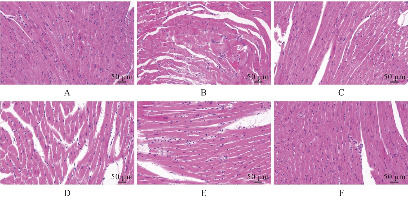
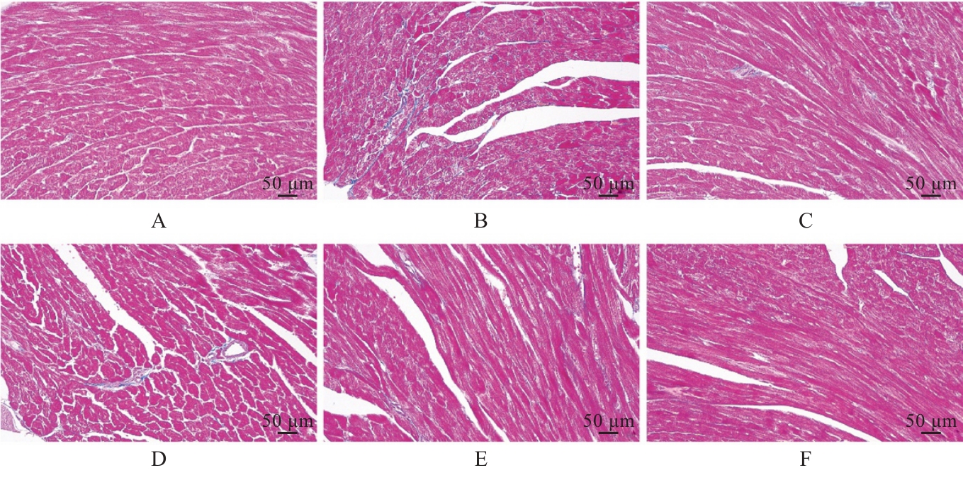
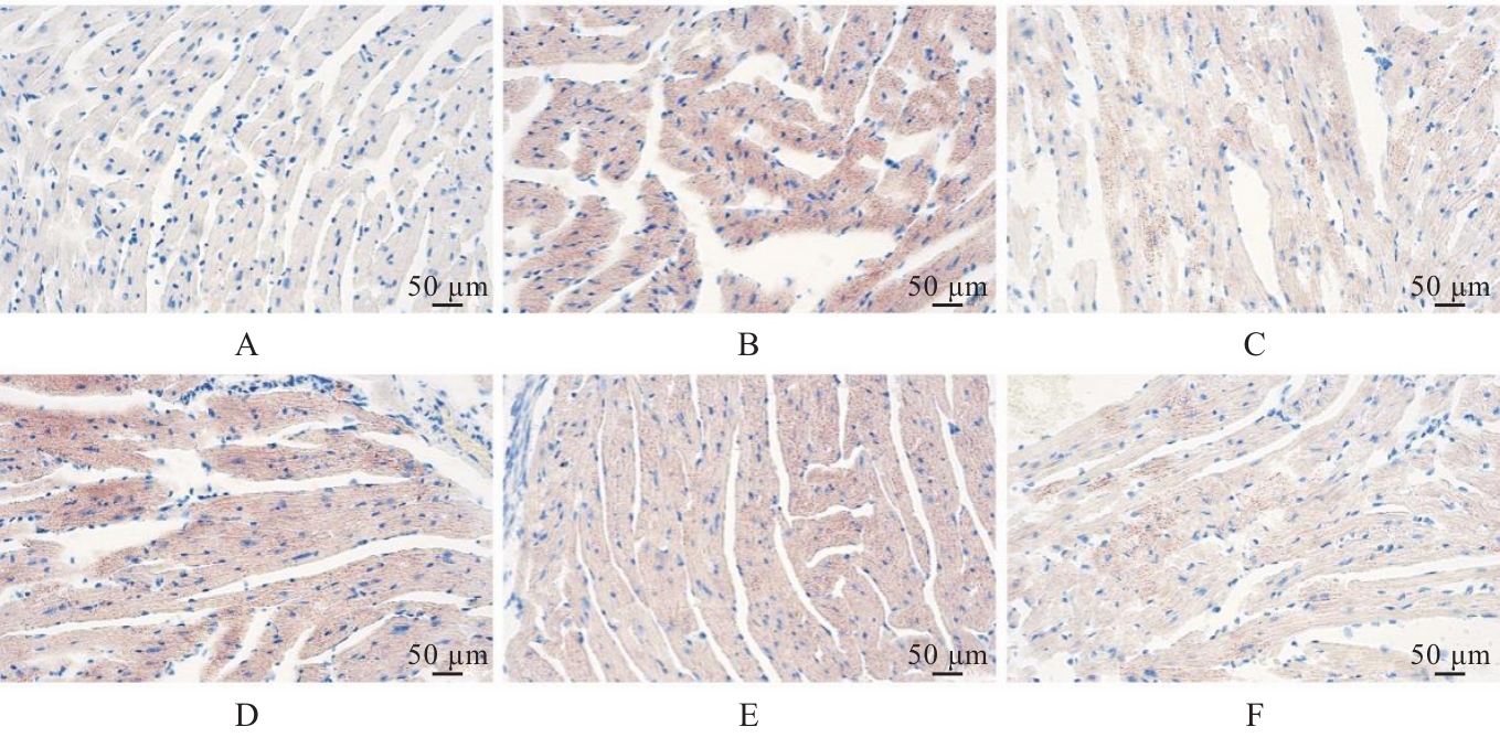
Establishment of myocardial injury model of rats induced by acute cold exposure/rewarming
[J]. Journal of Jilin University(Medicine Edition), 2026, 52(3): 672-679.
|
| [4] |
Yang LI,Shiyang LI,Hongmei ZHANG,Qianqian LIU,Yubing CHEN.
Effect of NGFR gene expression differences on promotion of apoptosis of squamous cell carcinoma and adenocarcinoma cells by PD-1 inhibitor-activated T lymphocytes
[J]. Journal of Jilin University(Medicine Edition), 2026, 52(3): 680-693.
|
| [5] |
Lingyu WANG,Ruili LI,Yu ZHANG,Meitong GUO,Yalong LIANG,Xu HAN,Xiaowei HUANG,Yang ZHANG,Xiuhua YU.
Improvement effect of Renshen Jianzhi Tablets on learning and memory ability in cognitive impairment model rats and its protective effect on hippocampal neurons and its mechanism
[J]. Journal of Jilin University(Medicine Edition), 2026, 52(3): 719-729.
|
| [6] |
Qiaotong REN,Weihua WU,Xingxiang WANG,Shichao WANG,Yipeng CHENG,Chun LI.
Effect of interleukin-17 recepter B gene silencing on proliferation, invasion and apoptosis of lung adenocarcinoma A549 cells and its mechanism
[J]. Journal of Jilin University(Medicine Edition), 2026, 52(3): 738-745.
|
| [7] |
Qiuping SU,Lei XING,Wei ZHANG,Shan JIANG,Yangyang ZHAO,Fangfang CHEN.
Research progress in anti-tumor mechanism of berberine and application of its novel nano-delivery system in tumor therapy
[J]. Journal of Jilin University(Medicine Edition), 2026, 52(3): 863-871.
|
| [8] |
Zheng TANG,Wei LIU,Hongmin HU,Qi LAI,Yan SHI,Shengquan LIU,Jun YANG,Chun CHU.
Improvement effect of Qishen Yiqi dropping pills on myocardium injury in rats induced by sorafenib and its mechanism
[J]. Journal of Jilin University(Medicine Edition), 2026, 52(2): 308-317.
|
| [9] |
Ning MA,Shuang LIU,Chengyao WANG,Jiajun LU,Linyu CHEN,Ziyi WANG,Weiqin ZHAO,Weitong WANG,Pengcheng CHE,Hong SUN.
Improvement effect of topical application of exosome loaded hydrogel DCC on peritoneal adhesion in mice and its mechanism
[J]. Journal of Jilin University(Medicine Edition), 2026, 52(2): 318-329.
|
| [10] |
Jie YU,Zongkang WANG,Xun LIU,Yanan YANG,Jin TAN.
Effect of miR-153-3p/PGC-1α axis on apoptosis of oral squamous cell carcinoma cells by modulation of mitochondrial apoptosis pathway
[J]. Journal of Jilin University(Medicine Edition), 2026, 52(2): 348-358.
|
| [11] |
Jinyu LI,Zhonghui LI,Aibin CHENG,Xuan BU,Jing BAI,Jianjun WANG.
Improvement effect of TREM-1 inhibitory peptide LR12 on myocardial injury in septic mice and its mechanism
[J]. Journal of Jilin University(Medicine Edition), 2026, 52(2): 366-374.
|
| [12] |
Yaping LI,Chunyan TAN,Lujie ZHAO,Jiayi ZHAO,Ting LI,Xiao YANG,Xiaoyun YANG.
Inductive effect of lysophosphatidic acid combined with 6-hydroxydopamine on apoptosis of SH-SY5Y cells and its mechanism
[J]. Journal of Jilin University(Medicine Edition), 2026, 52(1): 152-161.
|
| [13] |
Ziyi TANG,Shiying YANG,Tianzhen YANG,Wenqiang LIU,Jiangxue ZHONG,Li YIN.
Induction effect of pesticide pyraclostrobin on ferroptosis of spermatocytes GC-2 of mice
[J]. Journal of Jilin University(Medicine Edition), 2026, 52(1): 18-25.
|
| [14] |
Jiaxin WANG,Junwen MAO,Xuan ZHANG.
Expression of natural autoantibodies against apoptosis inhibitory genes in plasma of patients with hepatocellular carcinoma and its clinical significance
[J]. Journal of Jilin University(Medicine Edition), 2026, 52(1): 228-235.
|
| [15] |
Sitong CHEN,Dan YANG,Qingjie LI,Xiaolei TANG,Han WANG,Yangyang LIU,Tiejun LIU.
Ameliorative effect of puerarin on non-alcoholic fatty liver disease induced by high-fat diet in mice and its mechanism
[J]. Journal of Jilin University(Medicine Edition), 2026, 52(1): 44-55.
|
 ),Jiaming XU3(