吉林大学学报(医学版) ›› 2026, Vol. 52 ›› Issue (4): 942-952.doi: 10.13481/j.1671-587X.20260407

• 基础研究 • 上一篇    下一篇

鹿茸多肽通过调控Keap1/Nrf2/HO-1信号通路对糖尿病心肌病小鼠心肌损伤的改善作用

黄晓巍1,郑彭予2,李馨3,洪传彬3,李萌萌4,律广富5,徐岩3,林喆3(),许佳明3()   

  1. 1.长春中医药大学东北亚中医药研究院基础研究所,吉林 长春 130117
    2.江苏康缘药业股份有限公司,江苏 连云港 222047
    3.长春中医药大学药学院临床药学与中药药理教研室,吉林 长春 130117
    4.长春中医药大学东北亚中医药研究院中药研究所,吉林 长春 130117
    5.长春中医药大学吉林省 人参科学研究院中药药理组,吉林 长春 130117
  • 收稿日期:2025-09-28 接受日期:2025-12-02 出版日期:2026-07-28 发布日期:2026-07-27
  • 通讯作者: 林喆,许佳明 E-mail:linzhe1228@163.com;5717619@qq.com
  • 作者简介:黄晓巍(1972-),女,吉林省长春市人,主任药师,医学博士,主要从事中药学基础和临床方面的研究。
  • 基金资助:
    吉林省科技厅科技发展计划项目(YDZJ202501ZYTS711);吉林省教育厅大学生创新创业训练计划项目(S202410199038)

Promotion effect of velvet antler polypeptide on myocardial injury in diabetic cardiomyopathy mice by regulating Keap1/Nrf2/HO-1 signaling pathway

Xiaowei HUANG1,Pengyu ZHENG2,Xin LI3,Chuanbin HONG3,Mengmeng LI4,Guangfu LYU5,Yan XU3,Zhe LIN3(),Jiaming XU3()   

  1. 1.Basic Research Institute of Northeast Asia Institute of Traditional Chinese Medicine,Changchun University of Chinese Medicine,Changchun 130117,China
    2.Jiangsu Kangyuan Pharmaceutical Co. ,Ltd,Lianyungang 222047,China
    3.Clinical Pharmacy and Chinese Medicine Pharmacology Teaching and Research Office,School of Pharmacy,Changchun University of Chinese Medicine,Changchun 130117,China
    4.Institute of Chinese Materia Medica,Northeast Asia Institute of Traditional Chinese Medicine,Changchun University of Chinese Medicine,Changchun 130117,China
    5.Chinese Medicine Pharmacology Group,Jilin Ginseng Academy,Changchun University of Chinese Medicine,Changchun 130117,China
  • Received:2025-09-28 Accepted:2025-12-02 Online:2026-07-28 Published:2026-07-27
  • Contact: Zhe LIN,Jiaming XU E-mail:linzhe1228@163.com;5717619@qq.com

摘要:

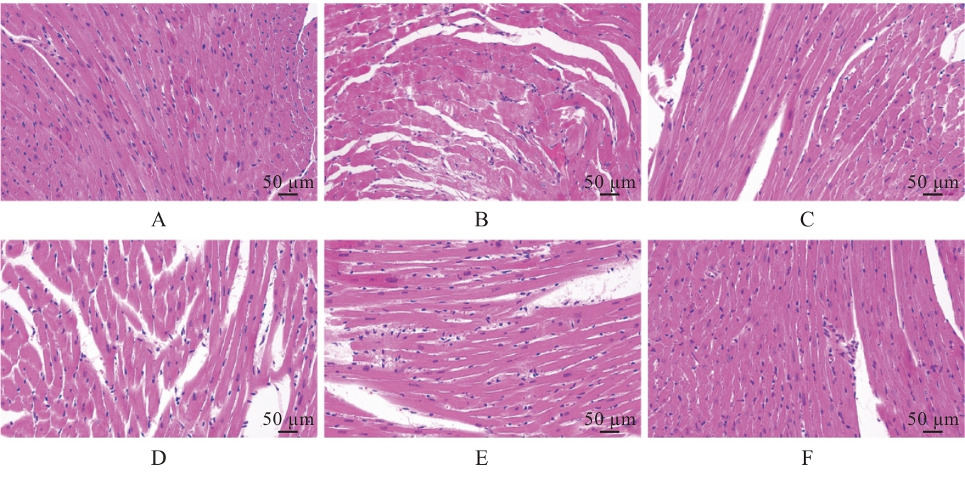
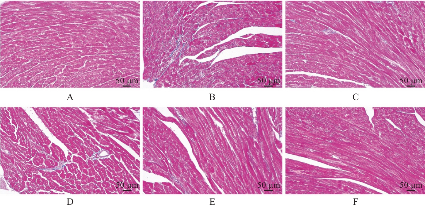
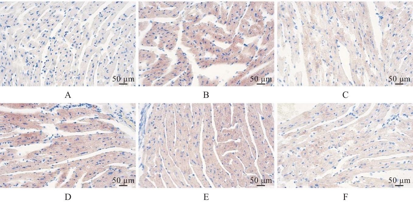
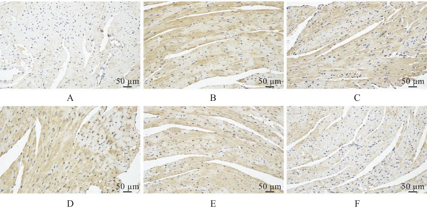
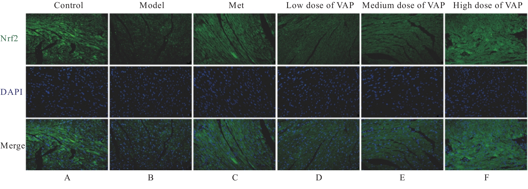
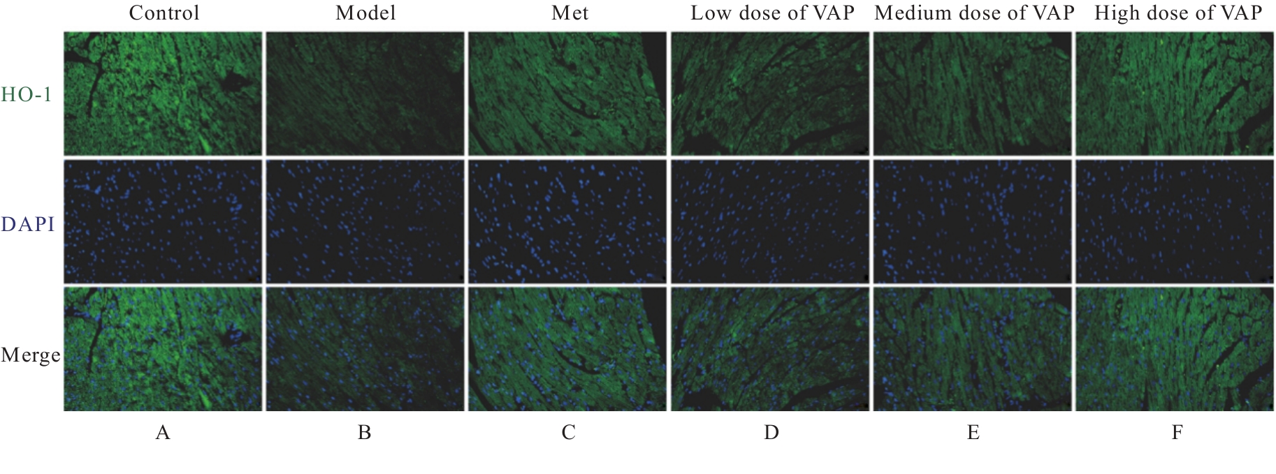
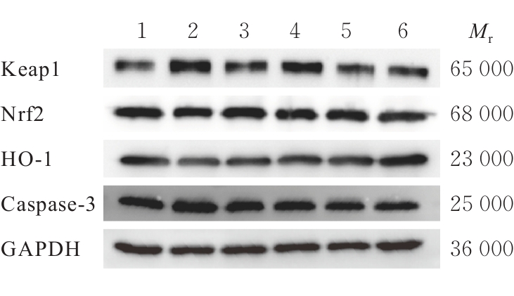
目的 探讨鹿茸多肽(VAP)对糖尿病心肌病(DCM)小鼠心肌损伤的改善作用,并阐明其作用机制。 方法 高糖高脂饲料饲养联合腹腔注射链脲佐菌素(STZ)法建立DCM模型。将50只成功造模C57BL/6J小鼠随机均分为模型组、二甲双胍(Met)组(给予 250 mg·kg-1 Met)、低剂量组VAP(给予100 mg·kg-1 VAP)、中剂量组VAP(给予200 mg·kg-1 VAP)和高剂量组VAP(给予300 mg·kg-1 VAP),每天给药1次,连续给药4周;另取10只C57BL/6J小鼠作为对照组(给予标准饲料饲养)。监测各组小鼠体质量和空腹血糖(FBG)水平,心脏超声检测小鼠心功能,HE染色观察小鼠心肌组织病理形态表现,Masson染色观察小鼠心肌组织纤维化程度,油红O染色观察小鼠心肌组织脂质分布,试剂盒检测小鼠心肌组织中超氧化物歧化酶(SOD)、过氧化氢酶(CAT)和谷胱甘肽过氧化物酶(GSH-Px)活性及丙二醛(MDA)水平,免疫组织化学染色观察小鼠心肌组织中Kelch样ECH关联蛋白1(Keap1)表达情况,免疫荧光染色观察小鼠心肌组织中核因子红细胞2相关因子2(Nrf2)和血红素加氧酶1(HO-1)表达情况,Western blotting法检测小鼠心肌组织中Keap1、Nrf2、HO-1和含半胱氨酸的天冬氨酸蛋白酶3(Caspase-3)蛋白表达水平。 结果 与对照组比较,模型组小鼠心肌损伤严重,心肌细胞间隙增大、胶原纤维增多及脂质分布增加,体质量、收缩末期左心室后壁厚度(LVPWs)和射血分数(EF)明显降低(P<0.05或P<0.01),FBG、左心室舒张末期内径(LVIDd)与收缩末期内径(LVIDs)均明显升高(P<0.05或P<0.01),心肌组织中SOD、CAT和GSH-Px活性明显降低(P<0.01),MDA水平明显升高(P<0.01),心肌组织中Nrf2和HO-1蛋白表达水平明显降低(P<0.01),Keap1和Caspase-3蛋白表达水平明显升高(P<0.01)。与模型组比较,Met组、中剂量VAP组和高剂量VAP组小鼠心肌组织病理形态表现、心肌纤维化及脂质分布均得到不同程度改善,体质量、LVPWs和EF明显升高(P<0.05或P<0.01),FBG、LVIDd和LVIDs明显降低(P<0.05或P<0.01),心肌组织中SOD、CAT和GSH-Px活性明显升高(P<0.05或P<0.01),MDA水平明显降低(P<0.05或P<0.01),心肌组织中Nrf2和HO-1蛋白表达水平明显升高(P<0.01),Keap1和Caspase-3蛋白表达水平明显降低(P<0.01);低剂量VAP组小鼠体质量明显增加(P<0.05)、FBG明显降低(P<0.05),但LVPWs、EF、LVIDd和LVIDs,SOD、CAT和GSH-Px活性,MDA水平,Keap1、Nrf2、HO-1和Caspase-3蛋白表达水平差异均无统计学意义(P>0.05)。 结论 VAP可改善DCM小鼠心肌损伤和心功能,其作用具有剂量依赖性,其机制可能与调控Keap1/Nrf2/HO-1信号通路、降低氧化应激与细胞凋亡有关。

关键词: 鹿茸多肽, 糖尿病心肌病, 心肌损伤, 氧化应激, 细胞凋亡

Abstract:

Objective To investigate the promotion effect of velvet antler polypeptide (VAP) on myocardial injury in the diabetic cardiomyopathy (DCM) mice, and to clarify its mechanism. Methods The DCM model was established by feeding a high-sugar and high-fat diet combined with intraperitoneal injection of streptozotocin (STZ). Fifty successfully modeled C57BL/6J mice were randomly divided into model group, metformin (Met) group (administered 250 mg·kg-1 Met), low dose of VAP group (administered 100 mg·kg-1 VAP), medium dose of VAP group (administered 200 mg·kg-1 VAP), and high dose of VAP group (administered 300 mg·kg-1 VAP), with once daily administration for 4 weeks; another 10 C57BL/6J mice were used as control group (fed a standard diet). The body weights and fasting blood glucose (FBG) levels of the mice in various groups were monitored; cardiac ultrasound was used to detect the cardiac function of the mice; HE staining was used to observe the pathomorphology of myocardium tissue of the mice; Masson staining was used to observe the degree of myocardial fibrosis of the mice; Oil Red O staining was used to observe the lipid distribution in myocardium tissue of the mice; kits were used to detect the activities of superoxide dismutase (SOD), catalase (CAT), and glutathione peroxidase (GSH-Px) and the level of malondialdehyde (MDA) in myocardium tissue of the mice; immunohistochemistry staining was used to observe the expression of Kelch-like ECH-associated protein 1 (Keap1) in myocardium tissue of the mice; immunofluorescence staining was used to observe the expression of nuclear factor erythroid 2-related factor 2 (Nrf2) and heme oxygenase 1 (HO-1) in myocardium tissue of the mice; Western blotting method was used to detect the expression levels of Keap1, Nrf2, HO-1, and cysteinyl aspartate-specific proteinase 3 (Caspase-3) proteins in myocardium tissue of the mice. Results Compared with control group, the myocardial injury of the mice in model group was severe, with enlarged myocardial cell gaps, increased collagen fibers, and increased lipid distribution; the body weight, left ventricular posterior wall thickness at end-systole (LVPWs), and ejection fraction (EF) of the mice were significantly decreased (P<0.05 or P<0.01); FBG, left ventricular end-diastolic diameter (LVIDd), and end-systolic diameter (LVIDs) were significantly increased (P<0.05 or P<0.01); the activities of SOD, CAT, and GSH-Px in myocardium tissue were significantly decreased (P<0.01), and the MDA level was significantly increased (P<0.01); the expression levels of Nrf2 and HO-1 proteins in myocardium tissue were significantly decreased (P<0.01), while the expression levels of Keap1 and Caspase-3 proteins were significantly increased (P<0.01). Compared with model group, the pathomorphology, myocardial fibrosis, and lipid distribution of myocardium tissue of the mice in Met group, medium dose of VAP group, and high dose of VAP group were improved to varying degrees; the body weight, LVPWs, and EF were significantly increased (P<0.05 or P<0.01); FBG, LVIDd, and LVIDs were significantly decreased (P<0.05 or P<0.01); the activities of SOD, CAT, and GSH-Px in myocardium tissue were significantly increased (P<0.05 or P<0.01), and the MDA level was significantly decreased (P<0.05 or P<0.01); the expression levels of Nrf2 and HO-1 proteins in myocardium tissue were significantly increased (P<0.01), and the expression levels of Keap1 and caspase-3 proteins were significantly decreased (P<0.01). Compared with model group, the body weight of the mice in low dose of VAP group was significantly increased (P<0.05) and FBG was significantly decreased (P<0.05), but there were no significant differences in LVPWs, EF, LVIDd, LVIDs, activities of SOD, CAT, and GSH-Px, level of MDA, expression level of Keap1, Nrf2, HO-1, and Caspase-3 proteins (P>0.05). Conclusion VAP can improve myocardial injury and cardiac function in the DCM mice in a dose-dependent manner, and its mechanism may be related to regulating the Keap1/Nrf2/HO-1 signaling pathway and reducing oxidative stress and apoptosis.

Key words: Velvet antler polypeptide, Diabetic cardiomyopathy, Myocardial injury, Oxidative stress, Apoptosis

中图分类号: 

  • R587.2