Journal of Jilin University(Medicine Edition) ›› 2026, Vol. 52 ›› Issue (5): 1309-1321.doi: 10.13481/j.1671-587X.20260513
• Research in basic medicine • Previous Articles
Therapeutic effect of Mishitong capsule on kidney stones induced by lithogenic solution in mice and its mechanism
Lijia SUN1,Chang SHEN1,Libin WANG2,Fanyu MENG2,Tao LU2,Xiaofei SUN2,Anning LI2,Guofeng XIA2( ),Bo HONG3,Chuanbo DING1()
- 1.Department of Chinese Medicinal Resources Development,College of Traditional Chinese Medicine,Jilin Agricultural Science and Technology University,Jilin 132101,China
2.Jilin Aodong Yanbian Pharmaceutical Co. ,Ltd. ,Dunhua 133700,China
3.Department of Chemistry,College of Resources and Environment,Jilin Agricultural University,Changchun 130118,China
-
Received:2025-11-29Accepted:2026-01-24Online:2026-09-28Published:2026-10-15 -
Contact:Guofeng XIA,Chuanbo DING E-mail:xiaodiliqian@163.com;dingchuanbo@jlnku.edu.cn

CLC Number:
- R692
Cite this article
Lijia SUN,Chang SHEN,Libin WANG,Fanyu MENG,Tao LU,Xiaofei SUN,Anning LI,Guofeng XIA,Bo HONG,Chuanbo DING. Therapeutic effect of Mishitong capsule on kidney stones induced by lithogenic solution in mice and its mechanism[J].Journal of Jilin University(Medicine Edition), 2026, 52(5): 1309-1321.
share this article
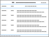
Tab.1
Body weights of mice in various groups (x±s, n=10, m/g)"
| Group | 1 d | 6 d | 11 d | 16 d | 21 d | 28 d |
|---|---|---|---|---|---|---|
| Control | 21.53±0.91 | 25.43±0.92 | 26.40±1.04 | 27.86±1.43 | 30.26±1.56 | 31.86±1.61 |
| Model | 20.85±0.87 | 22.29±1.20 | 24.09±1.42 | 25.17±1.41 | 27.48±1.76 | 29.14±1.89 |
| Low dose of Mishitong capsule | 21.18±0.97 | 23.90±2.07 | 26.09±1.72 | 27.36±2.37 | 29.22±2.36 | 30.12±2.33 |
| High dose of Mishitong capsule | 19.04±0.96 | 21.59±1.70 | 23.74±1.61 | 25.83±2.01 | 28.17±2.28 | 30.02±2.41 |
| Positive drug | 20.68±0.74 | 22.97±1.22 | 24.38±1.10 | 26.67±0.94 | 28.86±1.08 | 30.23±1.26 |
Tab.2
Levels of GSH and MDA, and activities of SOD in kidney tissue of mice in various groups"
| Group | GSH [mB/(μmol·g-1)] | MDA [cB/(nmol·L-1)] | SOD [λB/(U·mg-1)] |
|---|---|---|---|
| Control | 0.91±0.11 | 2.69±0.20 | 18.73±0.40 |
| Model | 0.56±0.02* | 7.97±0.32* | 10.94±0.60* |
| Low dose of Mishitong capsule | 0.60±0.02△ | 5.39±0.11△ | 17.25±0.37△ |
| High dose of Mishitong capsule | 0.79±0.06△ | 2.78±0.10△ | 19.06±0.41△ |
| Positive drug | 0.61±0.07△ | 4.87±0.10△ | 18.23±0.13△ |
Tab.3
Levels of BUN、Cr、Ca、P and Mgin serum of mice in various groups [x±s, n=10, cB/(mmol·L-1)]"
| Group | BUN | Cr | Ca | P | Mg |
|---|---|---|---|---|---|
| Control | 2.51±0.52 | 0.27±0.04 | 1.05±0.14 | 0.75±0.05 | 0.82±0.08 |
| Model | 8.13±0.39** | 0.45±0.01** | 3.63±0.23** | 1.08±0.05* | 0.31±0.05** |
| Low dose of Mishitong capsule | 6.95±0.07△ | 0.42±0.01 | 2.68±0.10△ | 0.98±0.04 | 0.48±0.04△ |
| High dose of Mishitong capsule | 4.21±0.39△△ | 0.31±0.07△ | 2.28±0.07△ | 0.85±0.03△ | 0.60±0.02△ |
| Positive drug | 3.69±0.19△△ | 0.34±0.03△ | 2.27±0.09△△ | 0.92±0.02△ | 0.57±0.06△ |
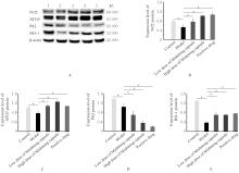
Fig. 1
Electrophoregram(A) and histograms(B-E) of expressions of Nrf2, ATG5, P62 and HO-1 proteins in kidney tissue of mice in various in various groups detected by Western blotting method"
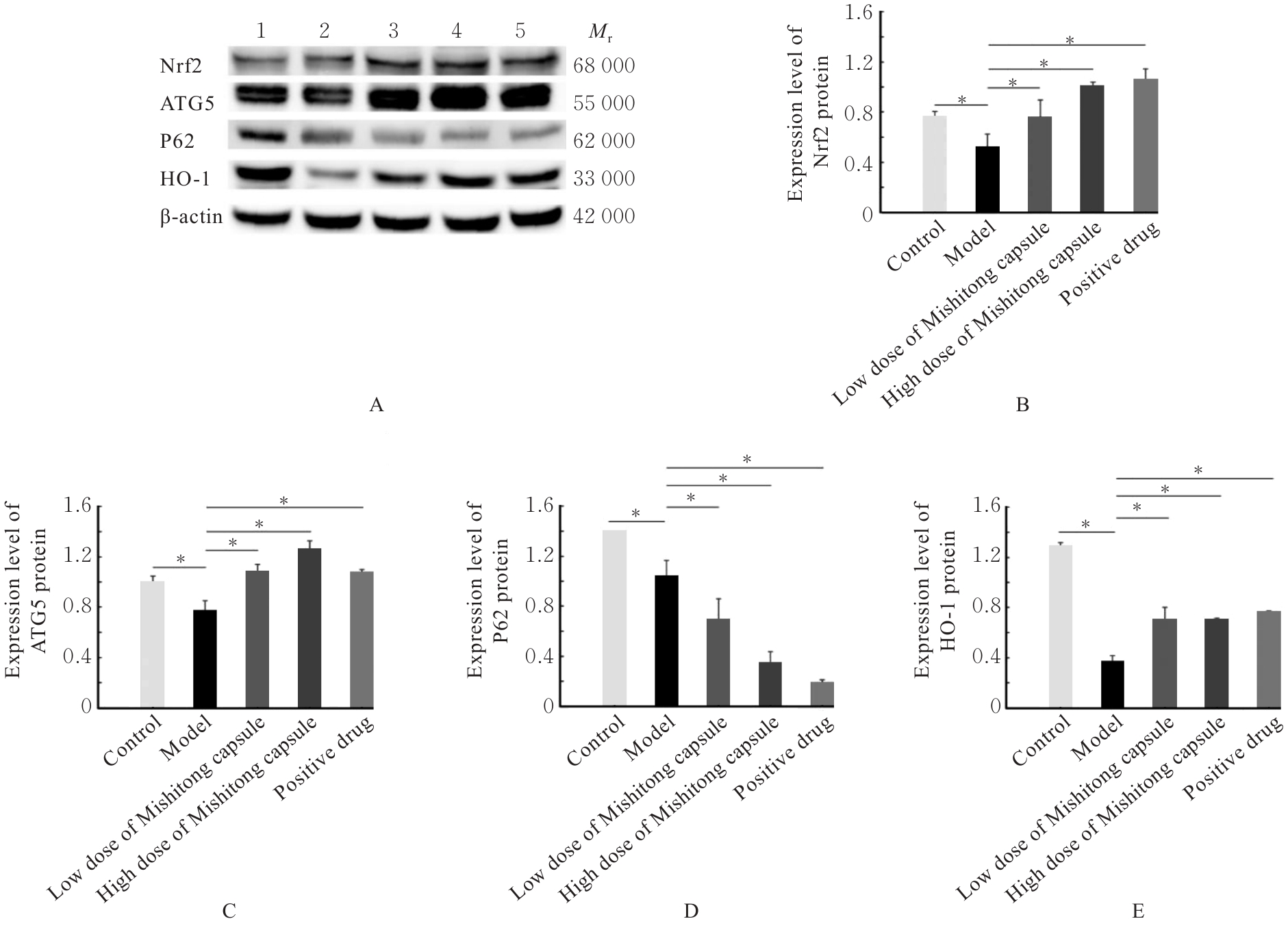
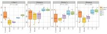
Fig. 2
Alpha diversity of gut microbiota of mice in various groups"
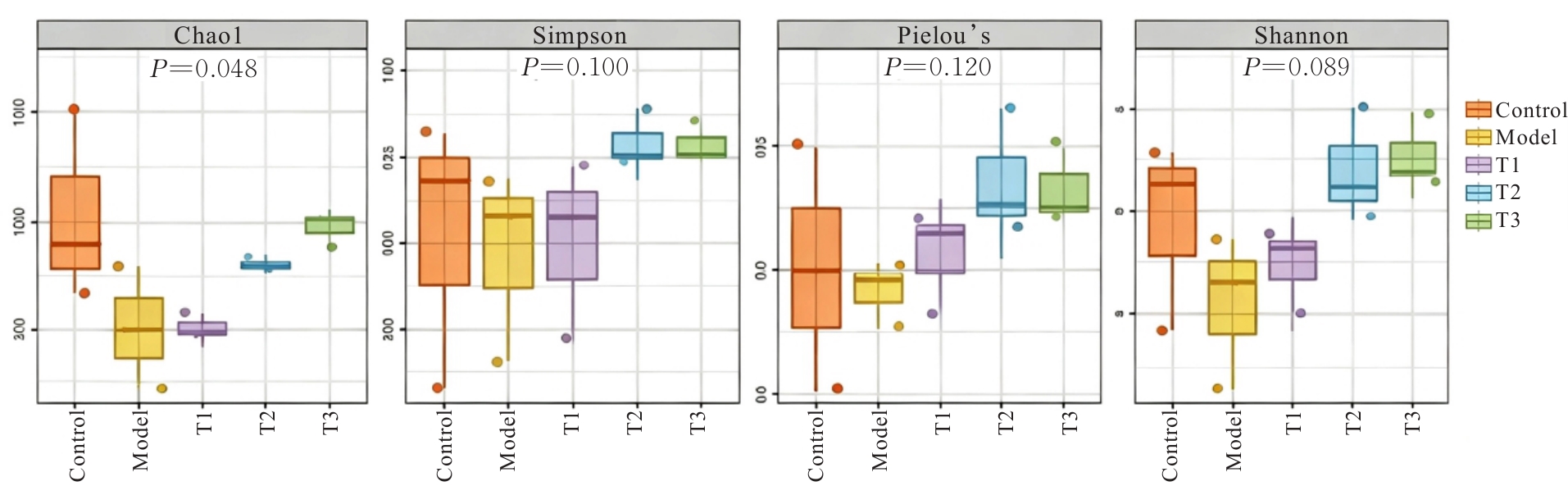
Fig. 3
Hierarchical cluster analysis of gut microbiota of mice in various groups"
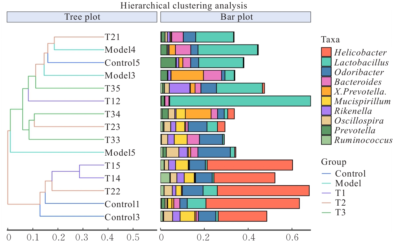
Fig. 4
PCoA analysis of Beta diversity of gut microbiota of mice in various groups"
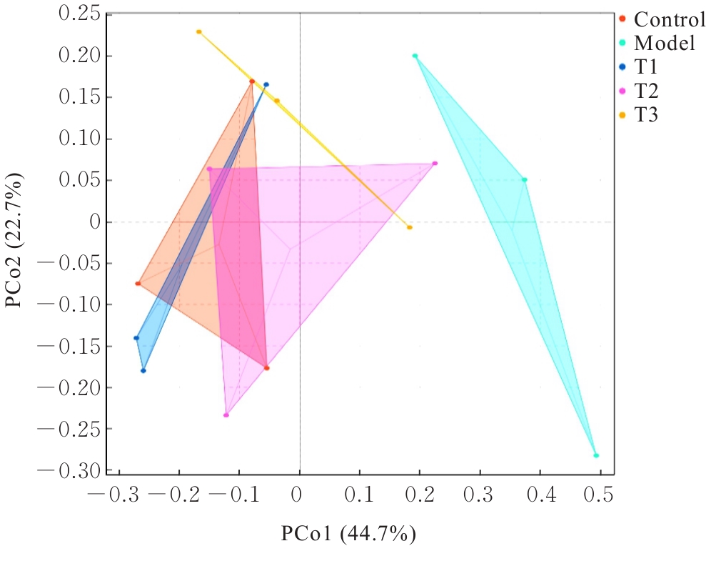
Fig. 5
Structure of gut microbiota of mice in various groups at phylum level"
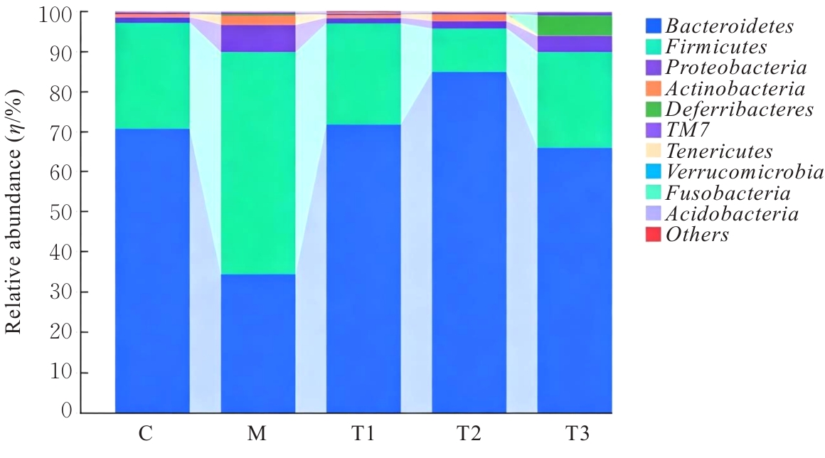
Fig. 6
Structure of gut microbiota of mice in various groups at genus level"
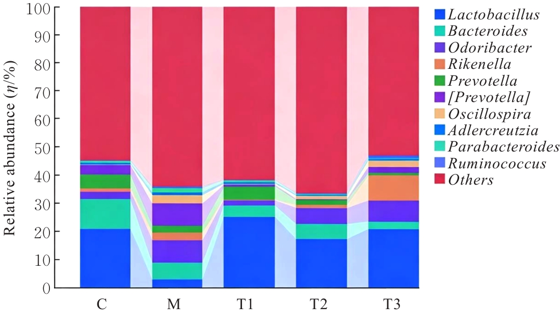
Fig. 7
Venn diagram of OTUs of gut microbiota of mice in various groups"
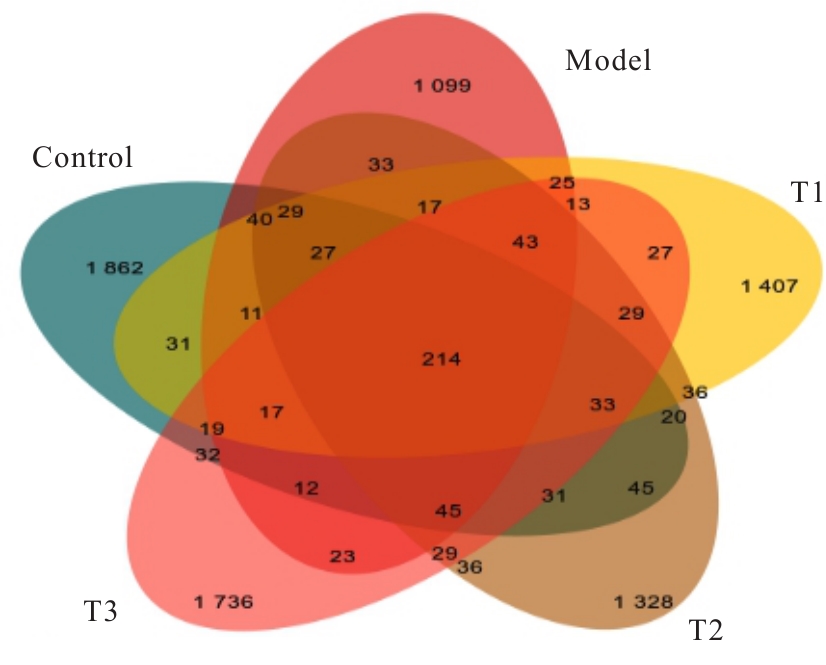
Fig. 8
Heatmap of relative abundance of dominant bacteria in gut microbiota of mice in various groups"
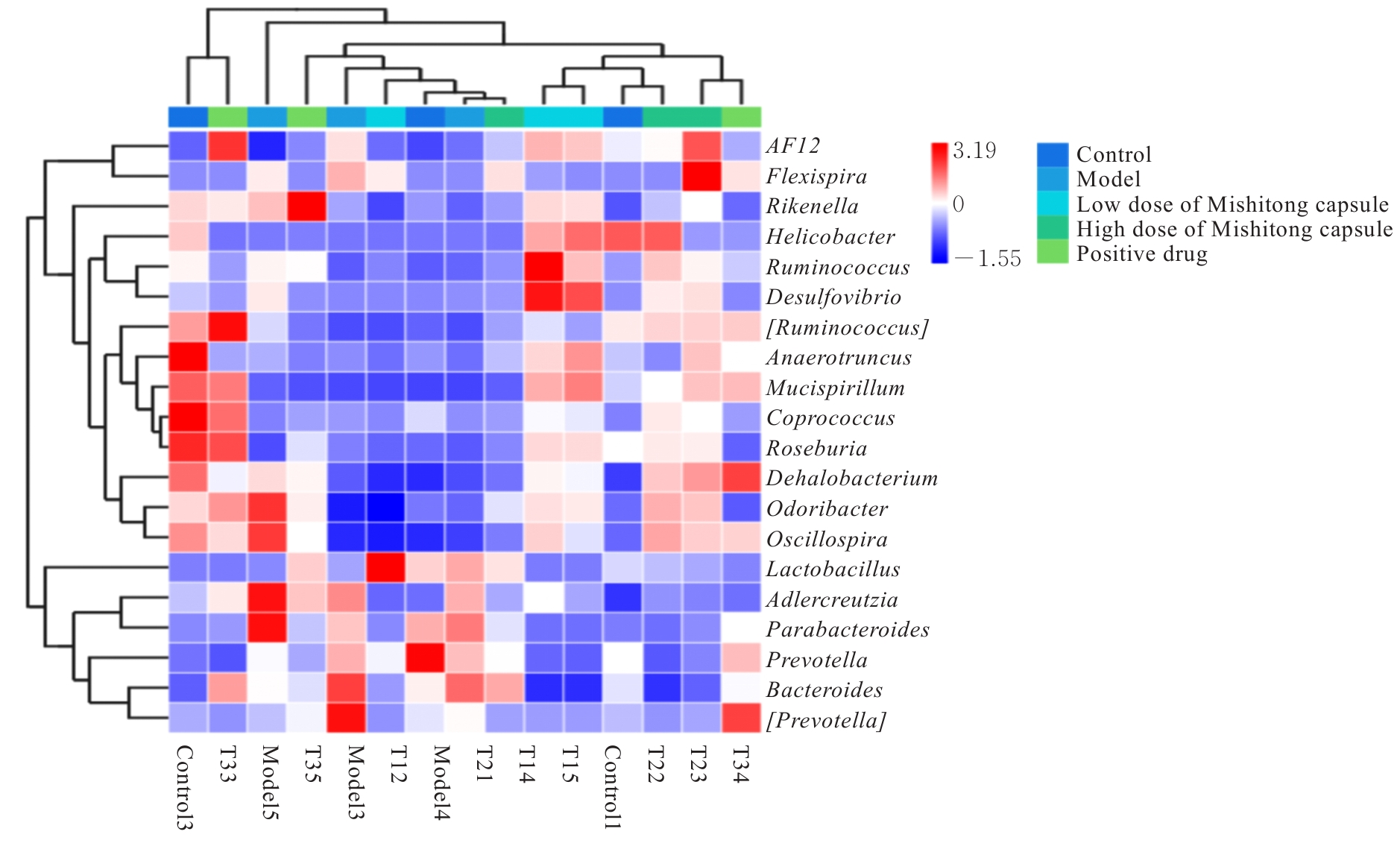
Fig. 9
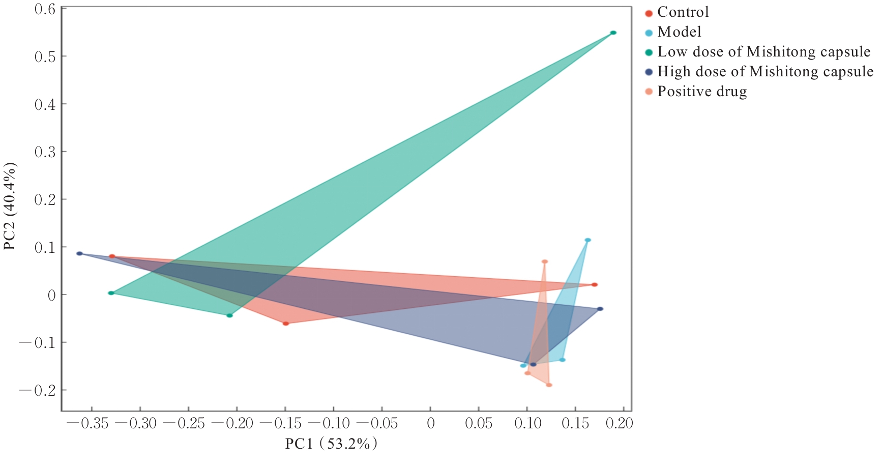
PCA score plot of gut microbiota of mice in various groups"
| [1] | WANG X Y, XIE H, XIE X G, et al. The protective role of cardiovascular health against kidney stones: a study based on LE8 score[J]. Heliyon, 2024, 10(13): e32497. |
| [2] | GAN X Q, LIU M Y, HE P P, et al. Habitual fish oil supplementation, genetic susceptibility of kidney stones and the risk of new-onset kidney stones[J]. J Clin Lipidol, 2024, 18(1): e116-e124. |
| [3] | KHAN S R, GLENTON P A. Experimental induction of calcium oxalate nephrolithiasis in mice[J]. J Urol, 2010, 184(3): 1189-1196. |
| [4] | HU S G, XU C M, ZHANG Y H, et al. Preparation of enteric capsules with pulsatile drug delivery potential using pullulan and polyacrylic acid resin Ⅲ[J]. Arab J Chem, 2024, 17(4): 105691. |
| [5] | AKTAŞ İ, YAHYAZADEH A. Protective potential of misoprostol against kidney alteration via alleviating oxidative stress in rat following exposure to paclitaxel[J]. Tissue Cell, 2022, 79: 101966. |
| [6] | ZHANG Y H, XIAN J Y, LI S J, et al. Hydroxycitric acid inhibits kidney stone through crystallization regulation and reduction of hyperoxalate-induced lipotoxicity[J]. J Funct Foods, 2023, 105: 105561. |
| [7] | LIU L H, PENG L, LI Q Y, et al. Dual-frequency ultrasound-assisted extraction of Pongamia pinnata (L.) Pierre polysaccharides: Process optimization, structural characterization, and anti-nephrolithiatic effects[J]. Ultrason Sonochem, 2026, 129: 107866. |
| [8] | HAJAJI S, JABRI M A, ARRARI F, et al. Myrtle berry juice improves hematological parameters and alleviates oxidative stress and inflammation in the myocardium of rats fed a high-fat diet: a combined in vivo and in silico investigation[J]. J Sci Food Agric, 2026, 106(12): 7023-7033. |
| [9] | ZHAO J W, WU Y X, ZHOU K, et al. Ferroptosis in calcium oxalate kidney stone formation and the possible regulatory mechanism of ANKRD1[J]. Biochim Biophys Acta BBA Mol Cell Res, 2023, 1870(5): 119452. |
| [10] | SU Y, LI S, LI X, et al. Tartronic acid as a potential inhibitor of pathological calcium oxalate crystallization[J]. Adv Sci, 2024, 11(21): 2400642. |
| [11] | YING X L, CHEN Y, HAO Z Y, et al. The significance of reactive oxygen species in the formation of calcium oxalate stones and the protective effects of antioxidants on the kidneys[J]. Front Immunol, 2025, 16: 1540075. |
| [12] | MING S X, TIAN J, MA K, et al. Oxalate-induced apoptosis through ERS-ROS-NF-κB signalling pathway in renal tubular epithelial cell[J]. Mol Med, 2022, 28(1): 88. |
| [13] | SAHA I, SINHA A, KUMAR SADHU A, et al. Renal function dynamics in COVID-19: exploring biomarker interactions with D-dimer and C-reactive proteins[J]. Biosci Rep, 2026, 46(4): BSR20254002. |
| [14] | NING X H, ZHONG Y C, CAI Q, et al. Gold nanoparticle transport in the injured kidneys with elevated renal function biomarkers[J]. Adv Mater, 2024, 36(36): 2402479. |
| [15] | SONG Q L, SONG C, CHEN X, et al. Oxalate regulates crystal-cell adhesion and macrophage metabolism via JPT2/PI3K/AKT signaling to promote the progression of kidney stones[J]. J Pharm Anal, 2024, 14(6): 100956. |
| [16] | ZHOU D H, WU Y, YAN H, et al. Gallic acid ameliorates calcium oxalate crystal-induced renal injury via upregulation of Nrf2/HO-1 in the mouse model of stone formation[J]. Phytomedicine, 2022, 106: 154429. |
| [17] | KULSOOM, AHMAD S, URWA, et al. Associations between clinical, biochemical, and nutritional factors in kidney stone formation and recurrence[J]. Urolithiasis, 2025, 53(1): 112. |
| [18] | YANG X Y, LIU Y C, CAO J H, et al. Targeting epigenetic and post-translational modifications of NRF2: key regulatory factors in disease treatment[J]. Cell Death Discov, 2025, 11: 189. |
| [19] | KUMAR P, YANG Z Q, LEVER J M, et al. Hydroxyproline stimulates inflammation and reprograms macrophage signaling in a rat kidney stone model[J]. Biochim Biophys Acta BBA Mol Basis Dis, 2022, 1868(9): 166442. |
| [20] | GONG Y H, ZHU W, LI Y Q, et al. Dynamic regulation of proximal tubular autophagy from injury to repair after ischemic kidney damage[J]. Cell Mol Biol Lett, 2024, 29(1): 151. |
| [21] | ZENG L Y, DENG Y J, ZHOU X H, et al. Simiao pills alleviates renal injury associated with hyperuricemia: a multi-omics analysis[J]. J Ethnopharmacol, 2024, 333: 118492. |
| [22] | WANG L, TAO Y F, WANG X S, et al. Aqueous extract of Phellinus igniarius ameliorates hyperuricemia and renal injury in adenine/potassium oxonate-treated mice[J]. Biomed Pharmacother, 2024, 177: 116859. |
| [23] | FU Q, ZHANG J J, ZHU Q F, et al. Uric acid-lowering effect of harpagoside and its protective effect against hyperuricemia-induced renal injury in mice[J]. Biochem Biophys Res Commun, 2024, 716: 150038. |
| [24] | RAO Z C, AO G, ZHANG Y M, et al. HNRNP Ⅰ promotes IRAK1 degradation to reduce podocyte apoptosis and inflammatory response alleviating renal injury in diabetic nephropathy[J]. Immunobiology, 2024, 229(5): 152835. |
| [25] | AGUDELO J, CHEN X, MUKHERJEE S D, et al. Cefazolin shifts the kidney microbiota to promote a lithogenic environment[J]. Nat Commun, 2024, 15: 10509. |
| [26] | HU H J, SUN W J, ZHANG L F, et al. Carboxymethylated Abrus cantoniensis polysaccharide prevents CTX-induced immunosuppression and intestinal damage by regulating intestinal flora and butyric acid content[J]. Int J Biol Macromol, 2024, 261: 129590. |
| [27] | PENG Y H, HUANG Y R, LI H, et al. Associations between rheumatoid arthritis and intestinal flora, with special emphasis on RA pathologic mechanisms to treatment strategies[J]. Microb Pathog, 2024, 188: 106563. |
| [28] | JIANG Y N, ZHANG Z H, CHEN H, et al. Mechanism of Guangdong Shenqu in regulating intestinal flora in mice with food stagnation and internal heat based on 16S rDNA sequencing[J]. J Tradit Chin Med Sci, 2024, 11(2): 232-244. |
| [1] | Yu ZHANG,Shuguang LI,Mingfu CAO,Di HUANG. Promotion effect of TXNDC9 gene knockdown on apoptosis and autophagy of colorectal cancer cells and its influence in xenograft tumor in nude mice [J]. Journal of Jilin University(Medicine Edition), 2026, 52(5): 1263-1272. |
| [2] | Dongyu XIANG,Guiyu CHEN,Yue XIANG. Effects of Panax notoginseng saponins on biological behaviors of gastric cancer cells and Keap1-Nrf2 signaling pathway [J]. Journal of Jilin University(Medicine Edition), 2026, 52(5): 1273-1281. |
| [3] | Xin LIU,Yuqing LIU,Hui LI. Research progress in effect of chronic periodontitis on occurrence and development of metabolic fatty liver disease [J]. Journal of Jilin University(Medicine Edition), 2026, 52(4): 1187-1194. |
| [4] | Xiaowei HUANG,Pengyu ZHENG,Xin LI,Chuanbin HONG,Mengmeng LI,Guangfu LYU,Yan XU,Zhe LIN,Jiaming XU. Promotion effect of velvet antler polypeptide on myocardial injury in diabetic cardiomyopathy mice by regulating Keap1/Nrf2/HO-1 signaling pathway [J]. Journal of Jilin University(Medicine Edition), 2026, 52(4): 942-952. |
| [5] | Junqi LI,Jiayun JIANG,Zhongfu ZUO,Hongdan YU. Bioinformatics analysis and in vitro and in vivo experimental verification on protective effect of salidroside on retinal ganglion cells in diabetic rats [J]. Journal of Jilin University(Medicine Edition), 2026, 52(4): 963-976. |
| [6] | Yunxia MA,Dan LIU,Ying ZHANG,Shifeng MA. Protective effect of triptolide on hippocampal neuron injury in neonatal rats with hypoxic-ischemic encephalopathy by regulating PTEN/PI3K/AKT signaling pathway [J]. Journal of Jilin University(Medicine Edition), 2026, 52(4): 977-986. |
| [7] | Jingxin WANG,Yuejuan GAO,Jingnan ZHANG,Jiawen NIU,Chunhua JIN. Effect of sesamin on learning and memory abilities of rats with Alzheimer’s disease model through regulating AMPK/SIRT1 autophagy pathway [J]. Journal of Jilin University(Medicine Edition), 2026, 52(3): 663-671. |
| [8] | Lingyu WANG,Ruili LI,Yu ZHANG,Meitong GUO,Yalong LIANG,Xu HAN,Xiaowei HUANG,Yang ZHANG,Xiuhua YU. Improvement effect of Renshen Jianzhi Tablets on learning and memory ability in cognitive impairment model rats and its protective effect on hippocampal neurons and its mechanism [J]. Journal of Jilin University(Medicine Edition), 2026, 52(3): 719-729. |
| [9] | Zheng TANG,Wei LIU,Hongmin HU,Qi LAI,Yan SHI,Shengquan LIU,Jun YANG,Chun CHU. Improvement effect of Qishen Yiqi dropping pills on myocardium injury in rats induced by sorafenib and its mechanism [J]. Journal of Jilin University(Medicine Edition), 2026, 52(2): 308-317. |
| [10] | Ning MA,Shuang LIU,Chengyao WANG,Jiajun LU,Linyu CHEN,Ziyi WANG,Weiqin ZHAO,Weitong WANG,Pengcheng CHE,Hong SUN. Improvement effect of topical application of exosome loaded hydrogel DCC on peritoneal adhesion in mice and its mechanism [J]. Journal of Jilin University(Medicine Edition), 2026, 52(2): 318-329. |
| [11] | Ling DENG,Shouqing LI,Chunxue NIU,Xiuying LIN,Xuguang MI,Jianhua FU. Effect of bisphenol AF on senescence of human endometrial stromal cells and inhibitory role of rapamycin [J]. Journal of Jilin University(Medicine Edition), 2026, 52(2): 340-347. |
| [12] | Yao ZHENG,Mingxia FU,Weichen WANG,Weiwei CHEN,Yuchen HAN,Yu BAI,Jiajia AN. Effect of sodium selenite on biological behaviors of breast cancer doxorubicin-resistant MCF-7/ADR cells [J]. Journal of Jilin University(Medicine Edition), 2026, 52(2): 410-417. |
| [13] | Yu ZHAO,Xiaoshuang HE,Xiaoyin DONG,Fengyi GAO,Jiageng HE. Regulatory effect of lncRNA urothelial carcinoembryonic antigen 1 on anoikis in human gastrointestinal stromal tumor cells and its mechanism [J]. Journal of Jilin University(Medicine Edition), 2026, 52(2): 429-439. |
| [14] | Huiyan ZHU,Min CHEN,Jinxian LI,Chunli LI. Effect of enriched environment on neurofunctional damage in rats with ischemic stroke via transcription factor EB-mediated autophagy [J]. Journal of Jilin University(Medicine Edition), 2026, 52(1): 116-124. |
| [15] | Ziyi TANG,Shiying YANG,Tianzhen YANG,Wenqiang LIU,Jiangxue ZHONG,Li YIN. Induction effect of pesticide pyraclostrobin on ferroptosis of spermatocytes GC-2 of mice [J]. Journal of Jilin University(Medicine Edition), 2026, 52(1): 18-25. |
|
||