Journal of Jilin University(Medicine Edition) ›› 2026, Vol. 52 ›› Issue (5): 1345-1356.doi: 10.13481/j.1671-587X.20260516
• Research in basic medicine • Previous Articles
Protective effect of human bone marrow mesenchymal stem cell-derived exosomes on neurological injury after intracerebral hemorrhage in rats and its mechanism
Lixin CAO1,Chunxue NIU1,Meng JING2,Xiumei DU2,Xuguang MI1( ),Fankai MENG2,3()
- 1.Central Laboratory,People’s Hospital,Jilin Province,Changchun,130021,China
2.Department of Neurosurgery,People’s Hospital,Jilin Province,Changchun,130021,China
3.Clinical Medical School,Changchun University of Chinese Medicine,Changchun,130117,China
-
Received:2025-12-01Accepted:2026-01-12Online:2026-09-28Published:2026-10-15 -
Contact:Xuguang MI,Fankai MENG E-mail:mixg699@163.com;mfk2003721019@126.com

CLC Number:
- R743.34
Cite this article
Lixin CAO,Chunxue NIU,Meng JING,Xiumei DU,Xuguang MI,Fankai MENG. Protective effect of human bone marrow mesenchymal stem cell-derived exosomes on neurological injury after intracerebral hemorrhage in rats and its mechanism[J].Journal of Jilin University(Medicine Edition), 2026, 52(5): 1345-1356.
share this article
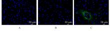
Fig. 1
Distribution of PKH67-labeled Exo in brain tissue of rats in various groups"
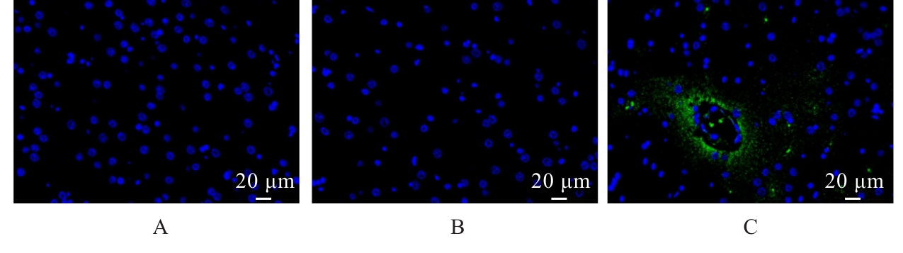
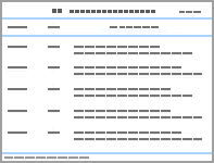
Tab.1
mNSS scores of rats at different time points in various groups"
| mNSS score | |||||
|---|---|---|---|---|---|
| Group | (t/d) 1 | 3 | 7 | 14 | 21 |
Sham operation ICH ICH+Exo | 0.17±0.21 | 0.23±0.41 | 0.12±0.29 | 0.13±0.24 | 0.18±0.30 |
12.50±0.84* 8.83±0.75△ | 11.67±0.82* 8.58±0.66△ | 7.67±1.21* 5.65±0.63△ | 5.67±0.82* 3.80±0.32△ | 3.83±0.41* 2.65±0.54△ | |
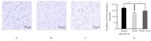
Fig. 2
Morphology of caudate nucleus(A-C) and numbers of positive neurons(D) of rats in various groups(Nissl staining)"
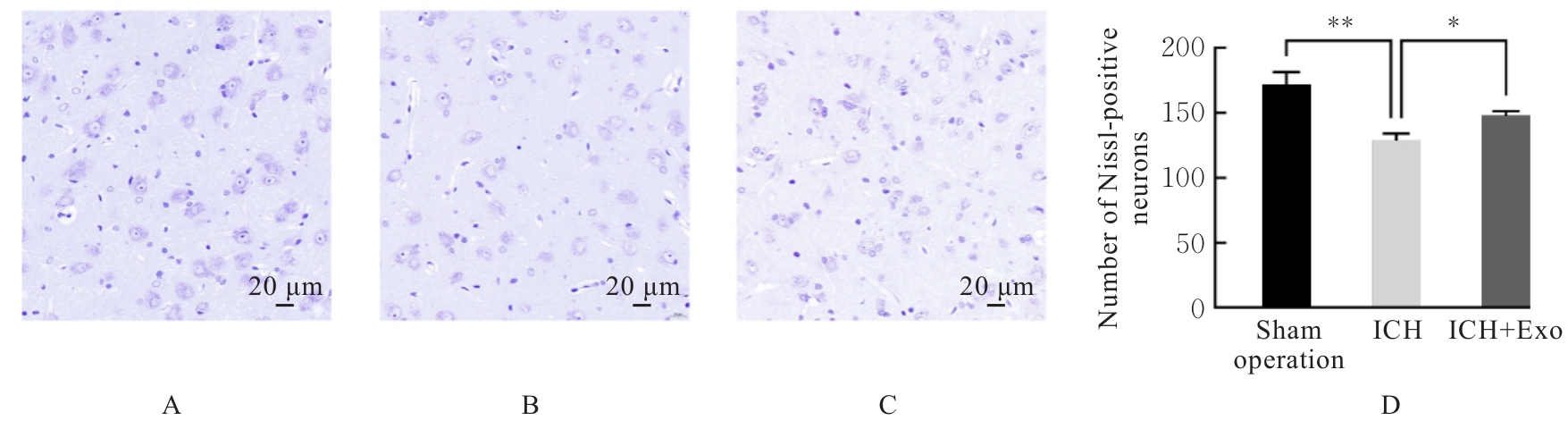
Fig. 3
Morphology of cells(A) and percentages of TUNEL-positive cells(B) in brain tissue of rats in various groups"
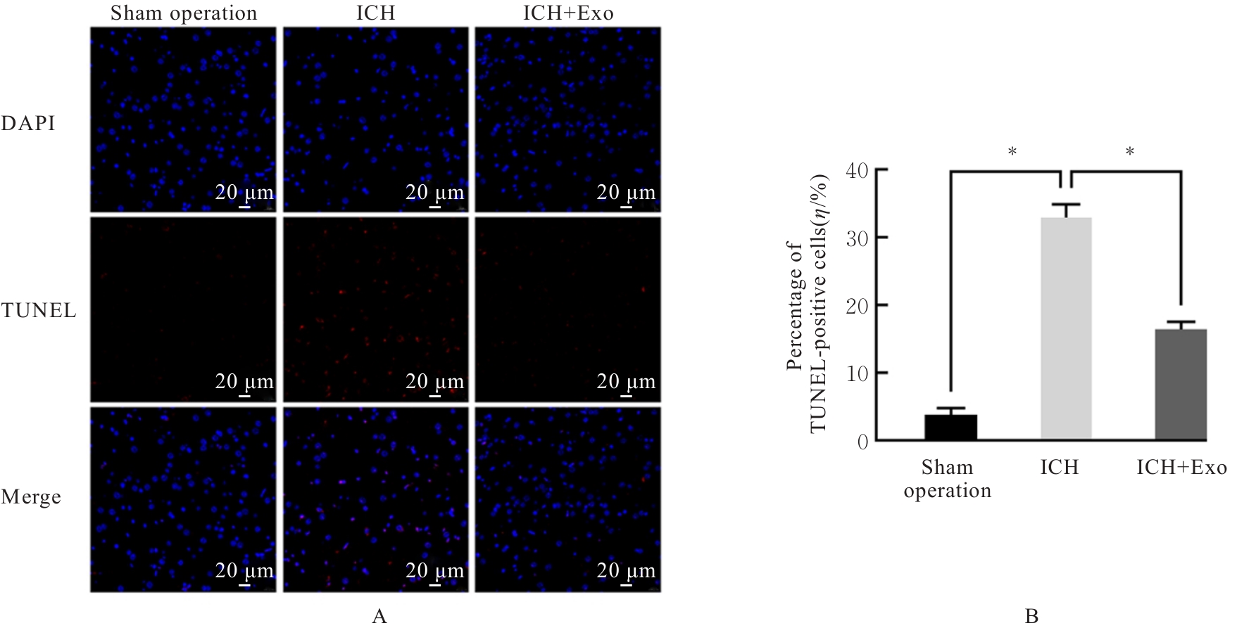
Fig. 4
Expressions of neuronal and glial cell markers in brain tissue of rats in various groups"
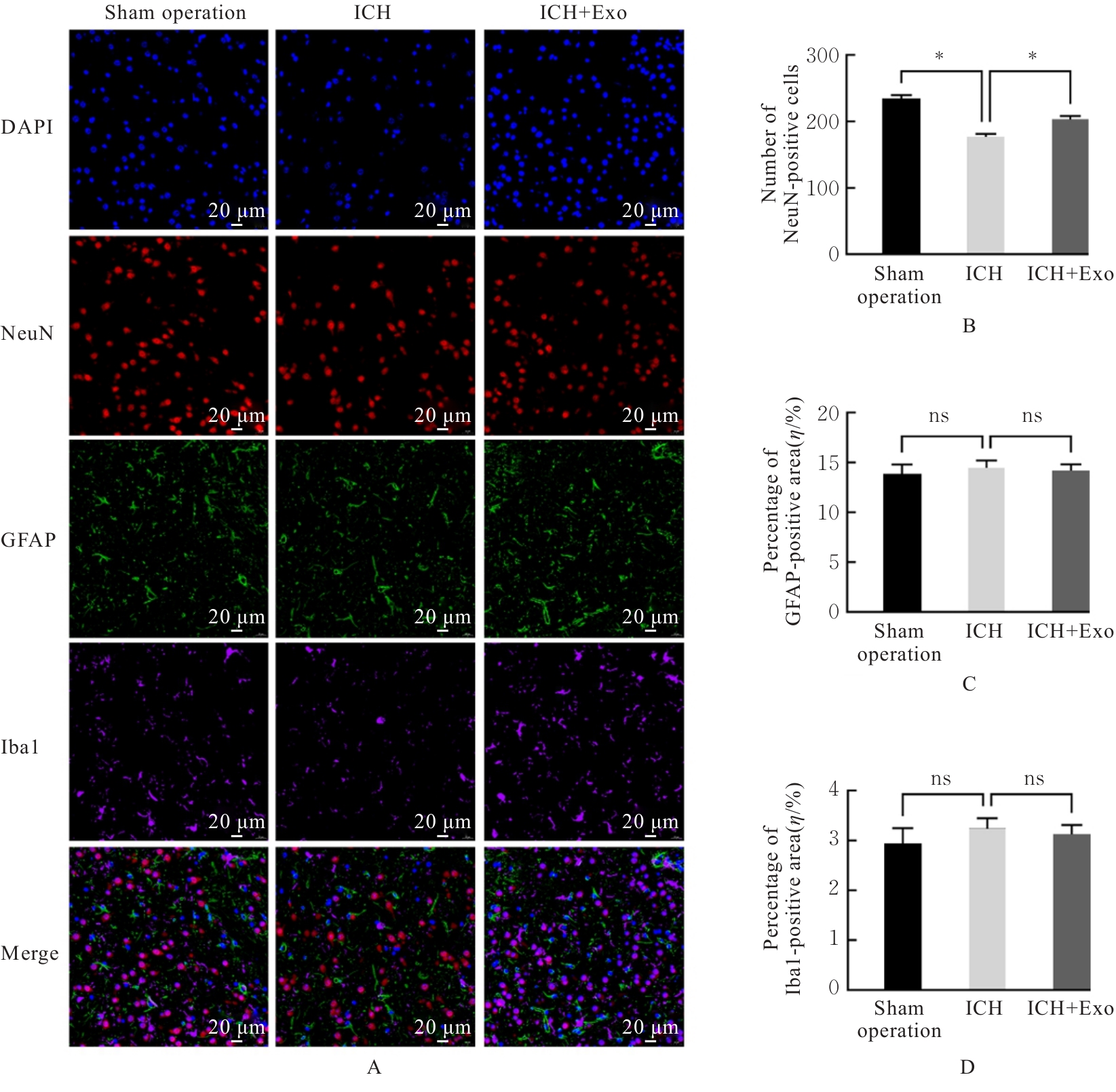
Fig. 5
Expressions of autophagy-related proteins in brain tissue of rats in various groups"
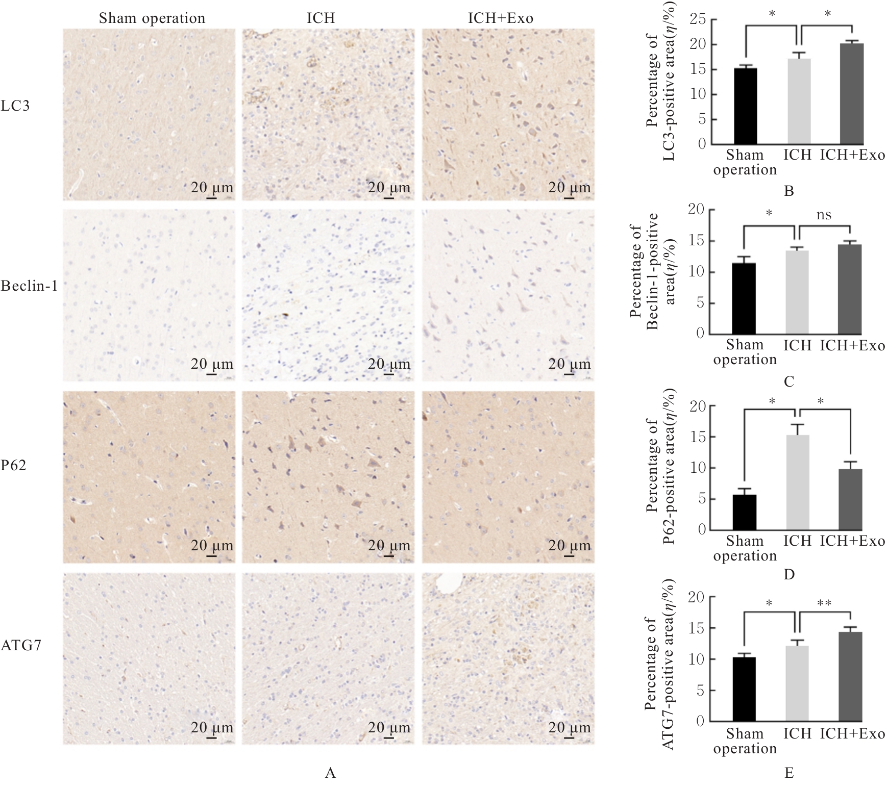
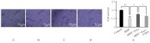
Fig. 6
Morphology of SH-SY5Y cells(A-D) and cell activities(E) in various groups"
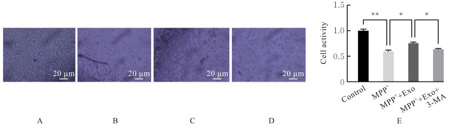
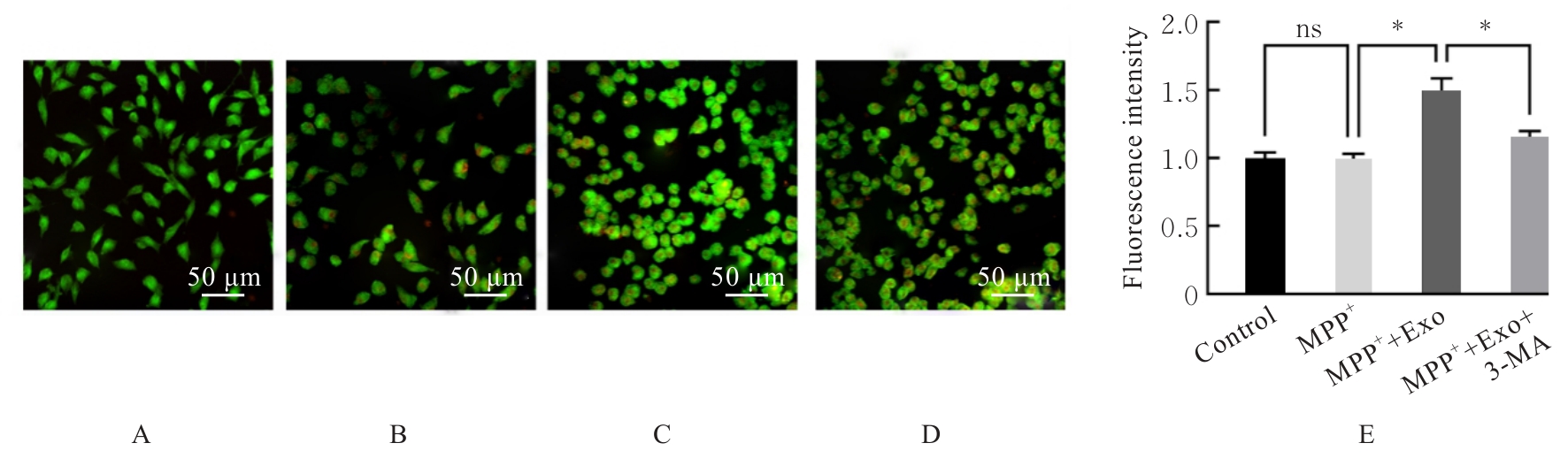
Fig. 7
Cell autophagy (A-D) and level of autophagy (E) in SH-SY5Y cells in various groups"
| [1] | MAGID-BERNSTEIN J, GIRARD R, POLSTER S, et al. Cerebral hemorrhage: pathophysiology, treatment, and future directions[J]. Circ Res, 2022, 130(8): 1204-1229. |
| [2] | SEIFFGE D J, FANDLER-HÖFLER S, DU Y, et al. Intracerebral haemorrhage: mechanisms, diagnosis and prospects for treatment and prevention[J]. Nat Rev Neurol, 2024, 20(12): 708-723. |
| [3] | GERMANS M R, BOOGAARTS H D, MACDONALD R L. Neuroprotection in critical care neurology[J]. Semin Neurol, 2016, 36(6): 642-648. |
| [4] | XIAO A Q, ZHANG Y Q, REN Y M, et al. GDF11 alleviates secondary brain injury after intracerebral hemorrhage via attenuating mitochondrial dynamic abnormality and dysfunction[J]. Sci Rep, 2021, 11(1): 3974. |
| [5] | 黄正旺, 张水诚, 庞杰荣. 磁敏感加权成像与动脉自旋标记成像在脑卒中溶栓后脑出血转化的评估价值[J]. 中国医学物理学杂志, 2024, 41(12): 1532-1536. |
| [6] | KO S B, CHOI H A, LEE K. Clinical syndromes and management of intracerebral hemorrhage[J]. Curr Atheroscler Rep, 2012, 14(4): 307-313. |
| [7] | HOSTETTLER I C, SEIFFGE D J, et al. Intracerebral hemorrhage: an update on diagnosis and treatment[J]. Expert Rev Neurother, 2019, 19(7): 679-694. |
| [8] | XU Q, ZHOU D Z, YU D. Bone marrow mesenchymal stem cells-derived exosomal long non-coding RNA KLF3 antisense RNA 1 enhances autophagy to protect against cerebral ischemia/reperfusion injury via ETS variant transcription factor 4/silent information regulator 1 axis[J]. Neuroscience, 2023, 521: 44-57. |
| [9] | SONG Z D, TAO Y, JIANG R, et al. Translational potential of mesenchymal stem cells in regenerative therapies for human diseases: challenges and opportunities[J]. Stem Cell Res Ther, 2024, 15(1): 266. |
| [10] | DJOUAD F, BOUFFI C, GHANNAM S, et al. Mesenchymal stem cells: innovative therapeutic tools for rheumatic diseases[J]. Nat Rev Rheumatol, 2009, 5(7): 392-399. |
| [11] | 鲁 霞, 王 凡. 间充质干细胞来源的外泌体在新生儿脑损伤治疗中的研究进展[J]. 解放军医学杂志, 2025, 50(8): 1038-1044. |
| [12] | ZHAO L Q, LI J W. Microglial uptake of hADSCs-Exo mitigates neuroinflammation in ICH[J]. Cell Signal, 2024, 119: 111146. |
| [13] | FANG X L, ZHOU D S, WANG X Y, et al. Exosomes: a cellular communication medium that has multiple effects on brain diseases[J]. Mol Neurobiol, 2024, 61(9): 6864-6892. |
| [14] | HAN Y X, SEYFRIED D, MENG Y L, et al. Multipotent mesenchymal stromal cell-derived exosomes improve functional recovery after experimental intracerebral hemorrhage in the rat[J]. J Neurosurg, 2019, 131(1): 290-300. |
| [15] | DUAN S R, WANG F, CAO J W, et al. Exosomes derived from microRNA-146a-5p-enriched bone marrow mesenchymal stem cells alleviate intracerebral hemorrhage by inhibiting neuronal apoptosis and microglial M1 polarization[J]. Drug Des Devel Ther, 2020, 14: 3143-3158. |
| [16] | LIU W, WANG Y X, GONG F Y, et al. Exosomes derived from bone mesenchymal stem cells repair traumatic spinal cord injury by suppressing the activation of A1 neurotoxic reactive astrocytes[J]. J Neurotrauma, 2019, 36(3): 469-484. |
| [17] | XU J X, ZHANG J, LIU Q Y, et al. Bone marrow mesenchymal stem cells-derived exosomes promote spinal cord injury repair through the miR-497-5p/TXNIP/NLRP3 axis[J]. J Mol Histol, 2024, 56(1): 16. |
| [18] | PARZYCH K R, KLIONSKY D J. An overview of autophagy: morphology, mechanism, and regulation[J]. Antioxid Redox Signal, 2014, 20(3): 460-473. |
| [19] | 唐 朝, 曹妍群, 陈超亮, 等. 红景天苷调节MAPK/ERK/mTOR信号通路对OGD/R诱导的海马神经元自噬和凋亡的影响[J]. 解放军医学杂志, 2025, 50(8): 1008-1014. |
| [20] | HE Y D, WAN S, HUA Y, et al. Autophagy after experimental intracerebral hemorrhage[J]. J Cereb Blood Flow Metab, 2008, 28(5): 897-905. |
| [21] | SHEN X, MA L, DONG W W, et al. Autophagy regulates intracerebral hemorrhage induced neural damage via apoptosis and NF-κB pathway [J]. Neurochem Int, 2016, 96: 100-112. |
| [22] | CANONICO B. The many faces of autophagy: balancing survival and cell death[J]. Biomolecules, 2024, 14(10): 1268. |
| [23] | 周 阳. hBM-MSCs-Exo激活自噬修复SH-SY5Y细胞氧化应激损伤机制研究[D]. 长春: 长春中医药大学, 2022. |
| [24] | 张 磊, 沈延春, 李 艳, 等. 采用Ⅶ型胶原酶构建大鼠脑出血模型[J]. 江汉大学学报(自然科学版), 2009, 37(2): 82-84. |
| [25] | WAN Y F, HOLSTE K G, HUA Y, et al. Brain edema formation and therapy after intracerebral hemorrhage[J]. Neurobiol Dis, 2023, 176: 105948. |
| [26] | CHEN Y H, CHEN S P, CHANG J B, et al. Perihematomal edema after intracerebral hemorrhage: an update on pathogenesis, risk factors, and therapeutic advances[J]. Front Immunol, 2021, 12: 740632. |
| [27] | GE Y, WU J J, ZHANG L, et al. A new strategy for the regulation of neuroinflammation: exosomes derived from mesenchymal stem cells[J]. Cell Mol Neurobiol, 2024, 44(1): 24. |
| [28] | HERIS R M, SHIRVALILOO M, ABBASPOUR-AGHDAM S, et al. The potential use of mesenchymal stem cells and their exosomes in Parkinson’s disease treatment[J]. Stem Cell Res Ther, 2022, 13(1): 371. |
| [29] | ZHAO L, LI H, WANG Y, et al. Autophagy deficiency leads to impaired antioxidant defense via p62-FOXO1/3 axis[J]. Oxid Med Cell Longev, 2019, 2019: 2526314. |
| [30] | FU K J, XU W L, LENAHAN C, et al. Autophagy regulates inflammation in intracerebral hemorrhage: Enemy or friend?[J]. Front Cell Neurosci, 2023, 16: 1036313. |
| [31] | CHEN H X, LIANG F C, GU P, et al. Exosomes derived from mesenchymal stem cells repair a Parkinson’s disease model by inducing autophagy[J]. Cell Death Dis, 2020, 11: 288. |
| [32] | HE S Z, WANG Q Q, CHEN L K, et al. miR-100a-5p-enriched exosomes derived from mesenchymal stem cells enhance the anti-oxidant effect in a Parkinson’s disease model via regulation of Nox4/ROS/Nrf2 signaling[J]. J Transl Med, 2023, 21(1): 747. |
| [33] | ZHANG Z X, ZHOU Y J, GU P, et al. Exosomes derived from human umbilical cord mesenchymal stem cells alleviate Parkinson’s disease and neuronal damage through inhibition of microglia[J]. Neural Regen Res, 2023, 18(10): 2291-2300. |
| [34] | ZENG H J, WU T H, LUO S, et al. MiRNA-loaded MSC exosomes restore autophagy flux for acute pancreatitis therapy[J]. Front Immunol, 2025, 16: 1613716. |
| [35] | XING H F, TAN J, MIAO Y Y, et al. Crosstalk between exosomes and autophagy: a review of molecular mechanisms and therapies[J]. J Cell Mol Med, 2021, 25(5): 2297-2308. |
| [36] | LI R Y, HU Q, SHI X, et al. Crosstalk between exosomes and autophagy in spinal cord injury: fresh positive target for therapeutic application[J]. Cell Tissue Res, 2023, 391(1): 1-17. |
| [1] | Yawen HU,Changhui FU,Yanmei YANG,Xianwei MENG,Pengli LI,Hongqi LIU,Na ZHANG,Bin GU. Promotive effect of osteogenic differentiation of mouse jaw bone marrow mesenchymal stem cells by CeO₂-loaded dendritic mesoporous silica nanoparticles and its mechanism [J]. Journal of Jilin University(Medicine Edition), 2026, 52(5): 1195-1205. |
| [2] | Yuqin AN,Ning CAO,Fanchao KONG,Huirong ZHANG. Effect of knockdown of NIPBL gene on osteogenic differentiation of mouse bone marrow mesenchymal stem cells through Shh signaling pathway [J]. Journal of Jilin University(Medicine Edition), 2026, 52(5): 1252-1262. |
| [3] | Yu ZHANG,Shuguang LI,Mingfu CAO,Di HUANG. Promotion effect of TXNDC9 gene knockdown on apoptosis and autophagy of colorectal cancer cells and its influence in xenograft tumor in nude mice [J]. Journal of Jilin University(Medicine Edition), 2026, 52(5): 1263-1272. |
| [4] | Lijia SUN,Chang SHEN,Libin WANG,Fanyu MENG,Tao LU,Xiaofei SUN,Anning LI,Guofeng XIA,Bo HONG,Chuanbo DING. Therapeutic effect of Mishitong capsule on kidney stones induced by lithogenic solution in mice and its mechanism [J]. Journal of Jilin University(Medicine Edition), 2026, 52(5): 1309-1321. |
| [5] | Yang JIANG,Boyuan WANG,Aizhuo SONG,Minghe LI. Promotive effect of exosome sustained-release system based on F127-DA hydrogel on cartilage regeneration in rats and its mechanism [J]. Journal of Jilin University(Medicine Edition), 2026, 52(4): 921-931. |
| [6] | Lina XU,Xiaoshuang HE,Wenyan XIN,Chao WU. Improvement effect of bone marrow mesenchymal stem cell-derived exosomes on airway resistance and lung tissue lesion in mice of asthmatic models and its mechanism [J]. Journal of Jilin University(Medicine Edition), 2026, 52(3): 602-611. |
| [7] | Jingxin WANG,Yuejuan GAO,Jingnan ZHANG,Jiawen NIU,Chunhua JIN. Effect of sesamin on learning and memory abilities of rats with Alzheimer’s disease model through regulating AMPK/SIRT1 autophagy pathway [J]. Journal of Jilin University(Medicine Edition), 2026, 52(3): 663-671. |
| [8] | Ling DENG,Shouqing LI,Chunxue NIU,Xiuying LIN,Xuguang MI,Jianhua FU. Effect of bisphenol AF on senescence of human endometrial stromal cells and inhibitory role of rapamycin [J]. Journal of Jilin University(Medicine Edition), 2026, 52(2): 340-347. |
| [9] | Di LI,Yang SU,Yong ZHANG,Jiaoqi WANG,Xiaofei ZHANG,Xuebing ZHAO. Expression levels of miR-328-3p and miR-671-5p in serum exosomes of Kawasaki disease model mice and their significance [J]. Journal of Jilin University(Medicine Edition), 2026, 52(2): 359-365. |
| [10] | Yao ZHENG,Mingxia FU,Weichen WANG,Weiwei CHEN,Yuchen HAN,Yu BAI,Jiajia AN. Effect of sodium selenite on biological behaviors of breast cancer doxorubicin-resistant MCF-7/ADR cells [J]. Journal of Jilin University(Medicine Edition), 2026, 52(2): 410-417. |
| [11] | Yu ZHAO,Xiaoshuang HE,Xiaoyin DONG,Fengyi GAO,Jiageng HE. Regulatory effect of lncRNA urothelial carcinoembryonic antigen 1 on anoikis in human gastrointestinal stromal tumor cells and its mechanism [J]. Journal of Jilin University(Medicine Edition), 2026, 52(2): 429-439. |
| [12] | Huiyan ZHU,Min CHEN,Jinxian LI,Chunli LI. Effect of enriched environment on neurofunctional damage in rats with ischemic stroke via transcription factor EB-mediated autophagy [J]. Journal of Jilin University(Medicine Edition), 2026, 52(1): 116-124. |
| [13] | Huiying YANG,Weihong LIANG,Xinru XU,Haodan SUN,Xin JIAO,Haiping WANG. Research progress in effect of bone marrow mesenchymal stem cells on differentiation induction of cardiomyocyte-like cells, protection of cardiomyocytes, and myocardial-related diseases [J]. Journal of Jilin University(Medicine Edition), 2026, 52(1): 281-289. |
| [14] | Xiaohan YAO,Mingchen YAO,Zhiqing WANG,Heyang LI,Yan YAN,Ningjing LEI. Effect of silencing GPR139 gene on proliferation, apoptosis and autophagy of breast cancer cells and its mechanism [J]. Journal of Jilin University(Medicine Edition), 2026, 52(1): 1-9. |
| [15] | Zhongzheng LIU,Shubo LIAN,Wenxuan FENG,Xin WEN,Hanyu LIU,Wei HE. Prometive effect of knockdown of KIF3B gene on autophagy in mouse embryonic palatal mesenchymal cells by inhibiting Shh signaling pathway [J]. Journal of Jilin University(Medicine Edition), 2025, 51(6): 1445-1451. |
|
||