Journal of Jilin University(Medicine Edition) ›› 2026, Vol. 52 ›› Issue (4): 987-998.doi: 10.13481/j.1671-587X.20260411
• Research in basic medicine • Previous Articles Next Articles
Promotion effect of exosome-regulated macrophage polarization on proliferation and metastasis of human esophageal squamous cell carcinoma cells and its mechanism
Jiangfen LI( ),Xinzhi FANG
- Department of Pathology,Fourth Affiliated Hospital,Xinjiang Medical University,Urumqi 830000,China
-
Received:2025-07-16Accepted:2025-11-10Online:2026-07-28Published:2026-07-27 -
Contact:Jiangfen LI E-mail:1427252351@qq.com

CLC Number:
- R735
Cite this article
Jiangfen LI,Xinzhi FANG. Promotion effect of exosome-regulated macrophage polarization on proliferation and metastasis of human esophageal squamous cell carcinoma cells and its mechanism[J].Journal of Jilin University(Medicine Edition), 2026, 52(4): 987-998.
share this article
Fig. 1
Relationship between BGN and M2 polarization of macrophages in ESCC-derived exosomes"
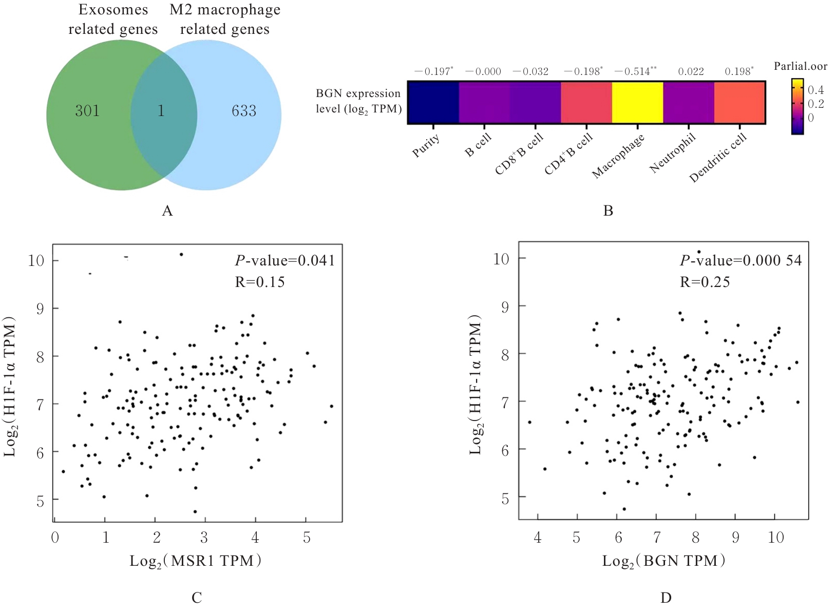
Fig. 2
Distribution differences and prognostic analysis on various subtypes of TAMs in esophageal cancer"
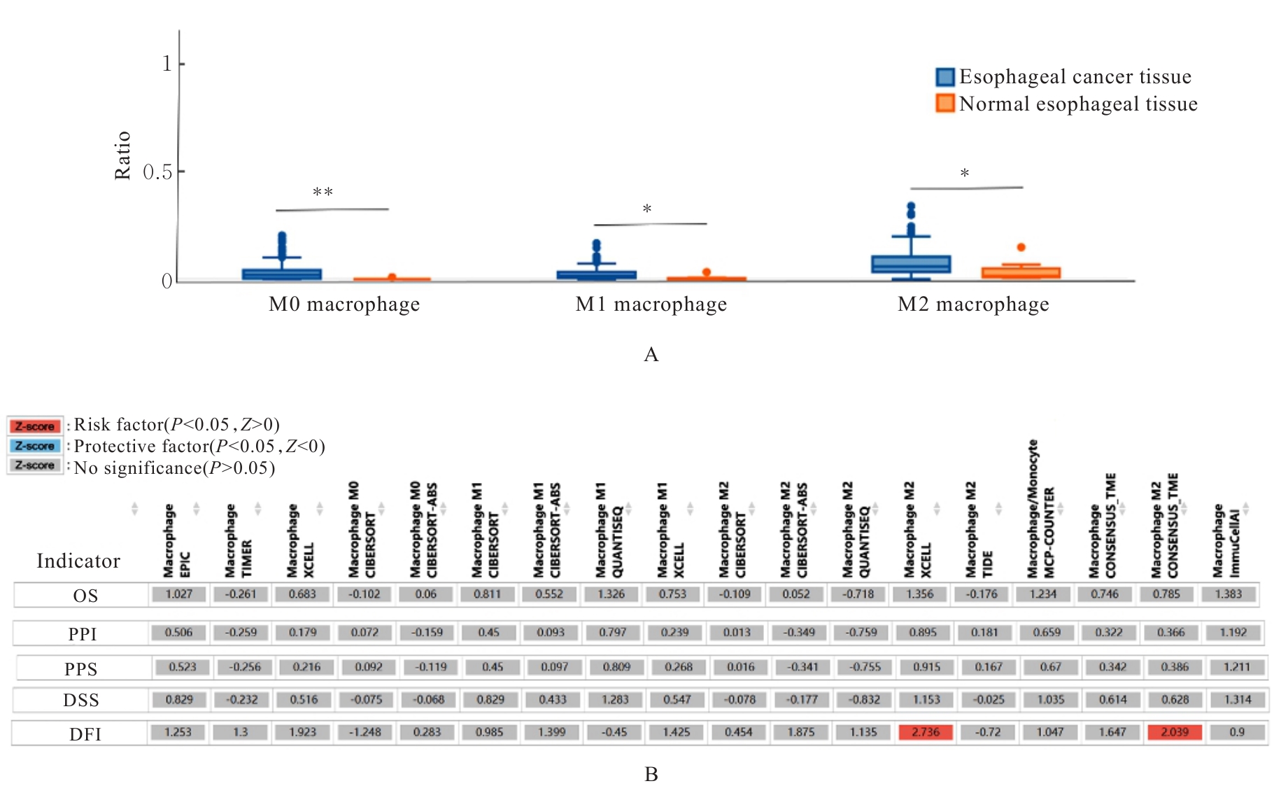
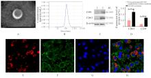
Fig. 3
Isolation and identification of exosomes derived from EC109 cells"
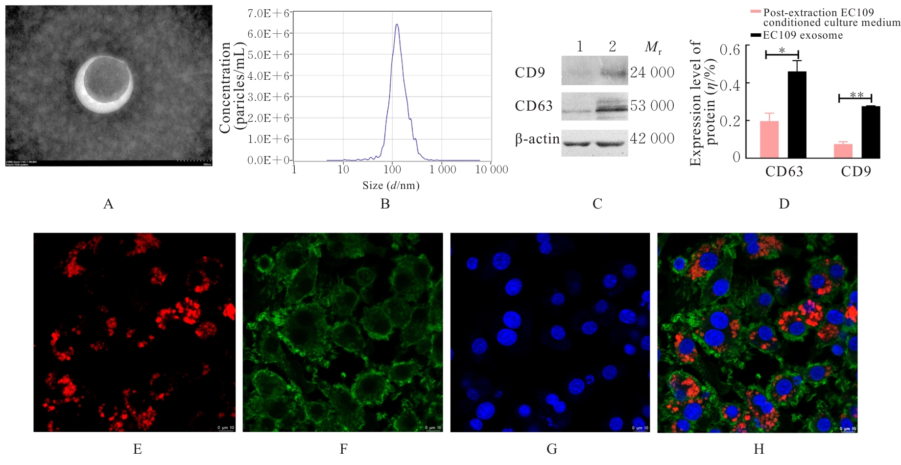
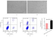
Fig. 4
Polarization status of macrophages after treated with exosomes"
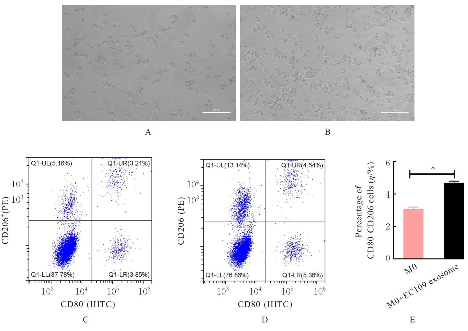
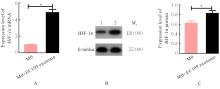
Fig. 5
Expressions of HIF-1α mRNA and protein in macrophages before and after treated with ESCC cell-derived exosomes"
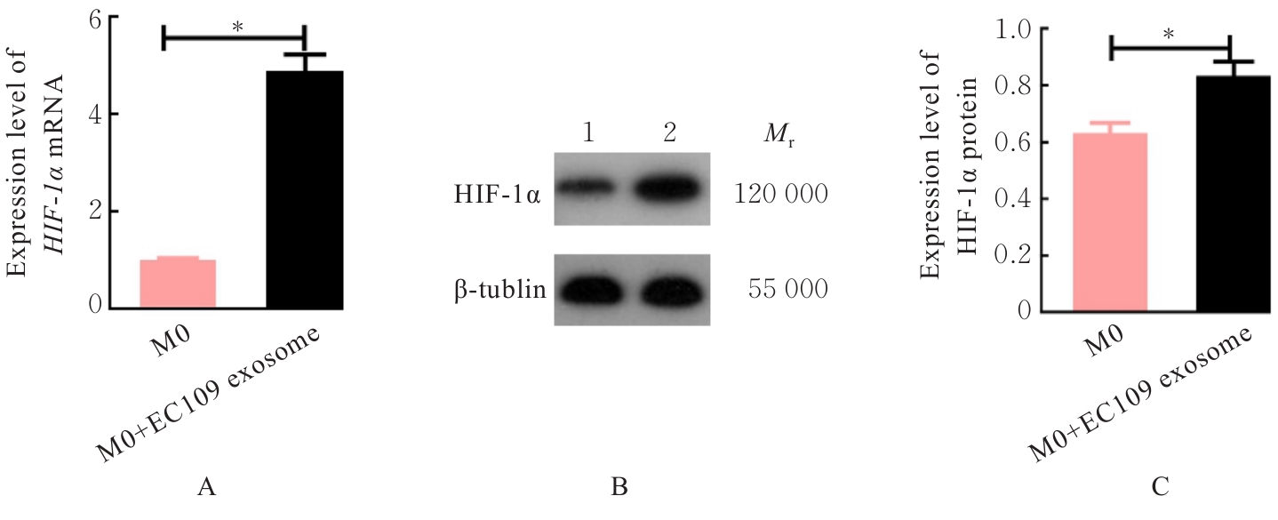
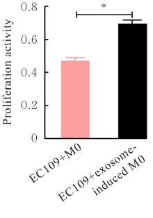
Fig. 6
Proliferation activities of macrophages before and after exosome induction"
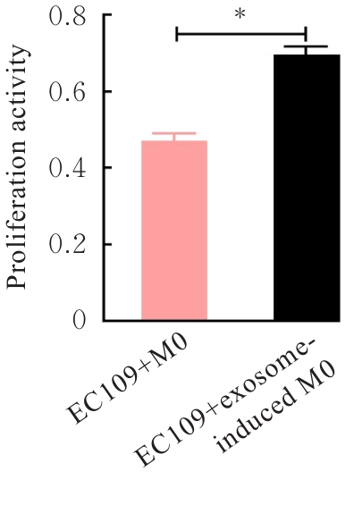
Fig. 7
Apoptosis of macrophages before and after exosome induction"
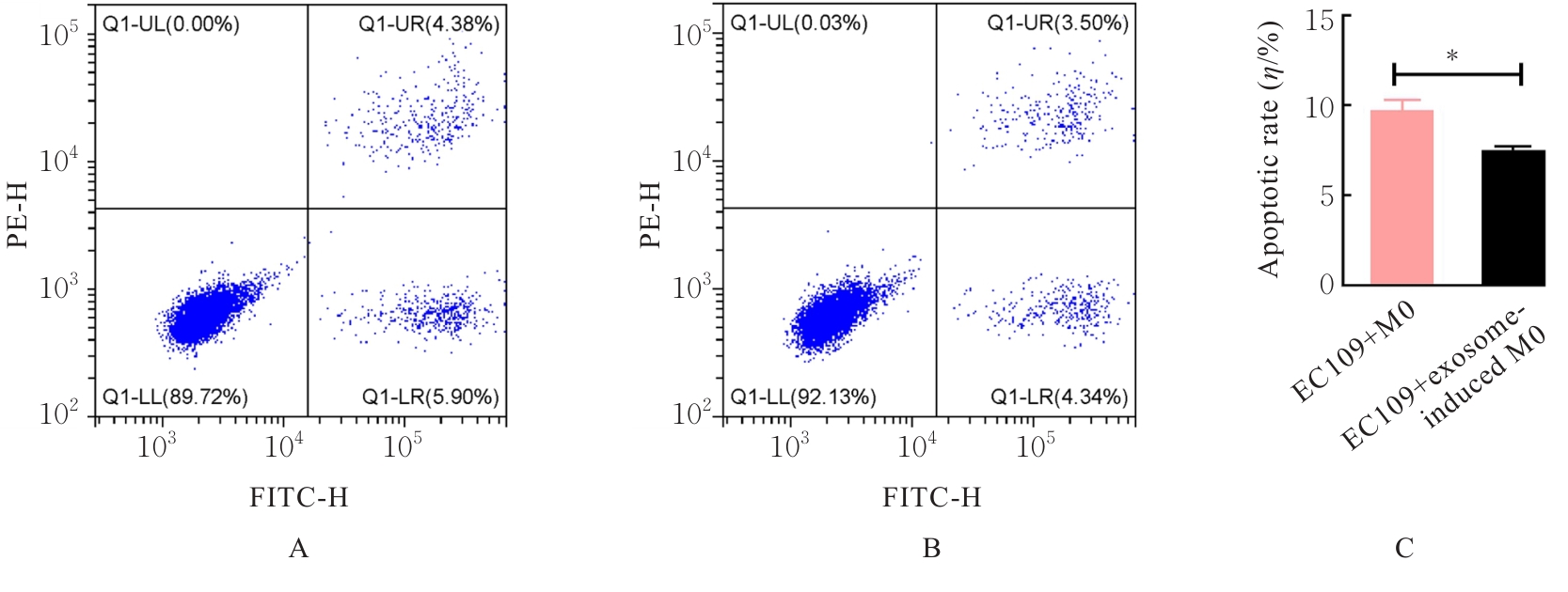
Fig. 8
Invasion of macrophages before and after exosome induction"
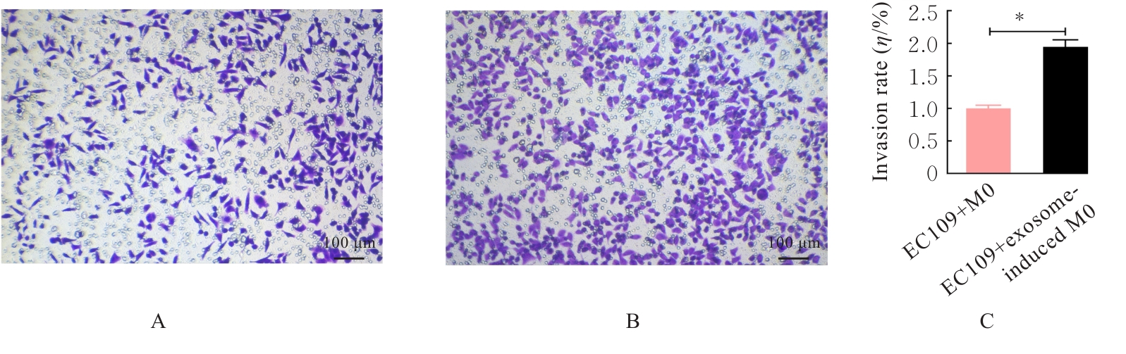
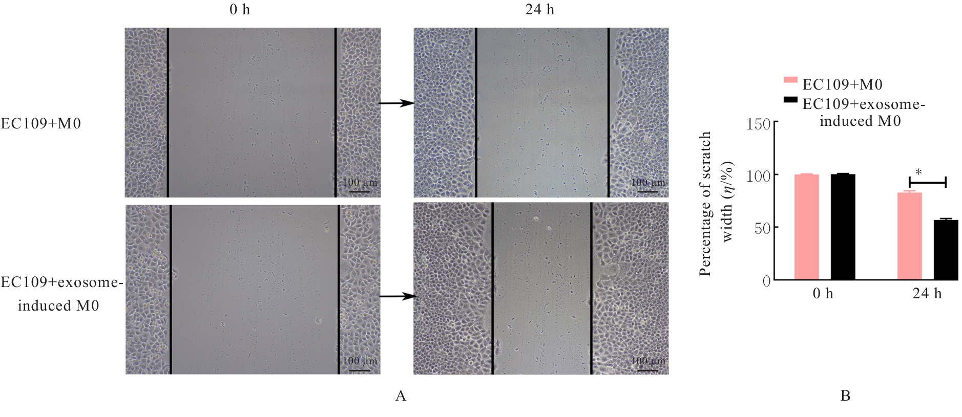
Fig. 9
Migration of macrophages before and after exosome induction"
| [1] | BRAY F, LAVERSANNE M, SUNG H, et al. Global cancer statistics 2022: GLOBOCAN estimates of incidence and mortality worldwide for 36 cancers in 185 countries[J]. CA Cancer J Clin, 2024, 74(3): 229-263. |
| [2] | JIANG W, ZHANG B, XU J Q, et al. Current status and perspectives of esophageal cancer: a comprehensive review[J]. Cancer Commun (Lond), 2025, 45(3): 281-331. |
| [3] | ZHENG Y X, CHEN Z Y, HAN Y C, et al. Immune suppressive landscape in the human esophageal squamous cell carcinoma microenvironment[J]. Nat Commun, 2020, 11(1): 6268. |
| [4] | LI Q, XU J W, HUA R J, et al. Nano-strategies for targeting tumor-associated macrophages in cancer immunotherapy[J]. J Cancer, 2025, 16(7): 2261-2274. |
| [5] | HU J M, LIU K, LIU J H, et al. CD163 as a marker of M2 macrophage, contribute to predicte aggressiveness and prognosis of Kazakh esophageal squamous cell carcinoma[J]. Oncotarget, 2017, 8(13): 21526-21538. |
| [6] | LI J F, XIE Y F, WANG X L, et al. Prognostic impact of tumor-associated macrophage infiltration in esophageal cancer: a meta-analysis[J]. Future Oncol, 2019, 15(19): 2303-2317. |
| [7] | DEB A, GUPTA S, MAZUMDER P B. Exosomes: a new horizon in modern medicine[J]. Life Sci, 2021, 264: 118623. |
| [8] | SONWANE S, TELRANDHE U, CHAMBHARE N, et al. Unraveling exosome-mediated cancer therapy resistance: pathways and therapeutic challenges[J]. J Egypt Natl Canc Inst, 2025, 37(1): 30. |
| [9] | TIAN L, YANG L, ZHENG W J, et al. RNA sequencing of exosomes revealed differentially expressed long noncoding RNAs in early-stage esophageal squamous cell carcinoma and benign esophagitis[J]. Epigenomics, 2020, 12(6): 525-541. |
| [10] | LI C W, TANG Z F, ZHANG W J, et al. GEPIA2021: integrating multiple deconvolution-based analysis into GEPIA[J]. Nucleic Acids Res, 2021, 49(W1): W242-W246. |
| [11] | LI T W, FAN J Y, WANG B B, et al. TIMER: a web server for comprehensive analysis of tumor-infiltrating immune cells[J]. Cancer Res, 2017, 77(21): e108-e110. |
| [12] | MURRAY P J, ALLEN J E, BISWAS S K, et al. Macrophage activation and polarization: nomenclature and experimental guidelines[J]. Immunity, 2014, 41(1): 14-20. |
| [13] | XU C C, CHEN J Y, TAN M, et al. The role of macrophage polarization in ovarian cancer: from molecular mechanism to therapeutic potentials[J]. Front Immunol, 2025, 16: 1543096. |
| [14] | LIAO L, WANG Y X, FAN S S, et al. The role and clinical significance of tumor-associated macrophages in the epithelial-mesenchymal transition of lung cancer[J]. Front Oncol, 2025, 15: 1571583. |
| [15] | CHEN Z P, ZHAO B Z. The role of tumor-associated macrophages in HPV induced cervical cancer[J]. Front Immunol, 2025, 16: 1586806. |
| [16] | REN B X, LIANG J N, LIU Y H, et al. Proguanil inhibits proliferation and migration in glioblastoma development through targeting CSF1R receptor[J]. Cell Signal, 2025, 127: 111550. |
| [17] | YU M J, WU Y M, LI Q F, et al. Colony-stimulating factor-1 receptor inhibition combined with paclitaxel exerts effective antitumor effects in the treatment of ovarian cancer[J]. Genes Dis, 2024, 11(3): 100989. |
| [18] | LI W C, WEI H F, LIU J J, et al. Exosomal Biglycan promotes gastric cancer progression via M2 polarization and CXCL10-mediated JAK/STAT1 activation[J]. Cancer Lett, 2025, 626: 217758. |
| [19] | EVENS A M, SEHN L H, FARINHA P, et al. Hypoxia-inducible factor-1 alpha expression predicts superior survival in patients with diffuse large B-cell lymphoma treated with R-CHOP[J]. J Clin Oncol, 2010, 28(6): 1017-1024. |
| [20] | HU L, ZANG M D, WANG H X, et al. Biglycan stimulates VEGF expression in endothelial cells by activating the TLR signaling pathway[J]. Mol Oncol, 2016, 10(9): 1473-1484. |
| [21] | GAO W D, JIN W W, LI Y N, et al. A highly bioactive bone extracellular matrix-biomimetic nanofibrous system with rapid angiogenesis promotes diabetic wound healing[J]. J Mater Chem B, 2017, 5(35): 7285-7296. |
| [22] | HUANG Y J, KANADA M, YE J X, et al. Exosome-mediated remodeling of the tumor microenvironment: From local to distant intercellular communication[J]. Cancer Lett, 2022, 543: 215796. |
| [23] | JAHAN S, MUKHERJEE S, ALI S, et al. Pioneer role of extracellular vesicles as modulators of cancer initiation in progression, drug therapy, and vaccine prospects[J]. Cells, 2022, 11(3): 490. |
| [24] | YONG T Y, WEI Z H, GAN L, et al. Extracellular-vesicle-based drug delivery systems for enhanced antitumor therapies through modulating the cancer-immunity cycle[J]. Adv Mater, 2022, 34(52): e2201054. |
| [25] | 赵 乐. HIF-1表达及CD206功能状态的相关性及其对肝移植术后肿瘤复发的影响[D]. 青岛: 青岛大学, 2019. |
| [26] | 刘苗苗, 邱 刚, 房保栓, 等. 肿瘤相关巨噬细胞和缺氧诱导因子-1α在宫颈鳞癌中表达及其临床意义[J]. 现代中西医结合杂志, 2018, 27(3): 264-266. |
| [27] | COLEGIO O R, CHU N Q, SZABO A L, et al. Functional polarization of tumour-associated macrophages by tumour-derived lactic acid[J]. Nature, 2014, 513(7519): 559-563. |
| [28] | LIANG J J, RAN Y Y, HU C B, et al. Inhibition of HIF-1α ameliorates pulmonary fibrosis by suppressing M2 macrophage polarization through PRMT1/STAT6 signals[J]. Int Immunopharmacol, 2025, 146: 113931. |
| [29] | LIANG Y H, LI J Y, YUAN Y L, et al. Exosomal miR-106a-5p from highly metastatic colorectal cancer cells drives liver metastasis by inducing macrophage M2 polarization in the tumor microenvironment[J]. J Exp Clin Cancer Res, 2024, 43(1): 281. |
| [30] | 王子奇, 张晓菊. 早期肺癌新型分子诊断的意义及评价[J]. 中国实用内科杂志, 2025, 45(6): 449-453. |
| [31] | JIAOJIAO X, ZHE L, QINLI T, et al. CD133+PD-L1+ cancer cells confer resistance to adoptively transferred engineered macrophage-based therapy in melanoma [J]. Nat Commun, 2025, 16(1): 895. |
| [1] | Rui LI,Xiaoyan HAO,Liu YANG,Jiayun LIU,Mu HE. Construction and validation of machine learning-based prediction models for esophageal cancer using routine clinical laboratory data [J]. Journal of Jilin University(Medicine Edition), 2026, 52(4): 1086-1095. |
| [2] | Yang JIANG,Boyuan WANG,Aizhuo SONG,Minghe LI. Promotive effect of exosome sustained-release system based on F127-DA hydrogel on cartilage regeneration in rats and its mechanism [J]. Journal of Jilin University(Medicine Edition), 2026, 52(4): 921-931. |
| [3] | Chunrong XIANG,Yong HE,Nuo JIANG,Shouqi HUANG,Li HONG. Ameliorative effect of Pueraria lobata-derived exosome-like nanovesicles on D-galactose-induced senescence in L929 mouse fibroblasts and its mechanism [J]. Journal of Jilin University(Medicine Edition), 2026, 52(3): 590-601. |
| [4] | Lina XU,Xiaoshuang HE,Wenyan XIN,Chao WU. Improvement effect of bone marrow mesenchymal stem cell-derived exosomes on airway resistance and lung tissue lesion in mice of asthmatic models and its mechanism [J]. Journal of Jilin University(Medicine Edition), 2026, 52(3): 602-611. |
| [5] | Wei ZHOU,Yangyang HE,Youde WANG,Wenzhuo MA,Heng KANG. Effect of histone deacetylase 6 inhibitor ACY-241 on macrophage polarization and biological behaviors of esophageal cancer cells and its mechanism [J]. Journal of Jilin University(Medicine Edition), 2026, 52(3): 621-632. |
| [6] | Ning MA,Shuang LIU,Chengyao WANG,Jiajun LU,Linyu CHEN,Ziyi WANG,Weiqin ZHAO,Weitong WANG,Pengcheng CHE,Hong SUN. Improvement effect of topical application of exosome loaded hydrogel DCC on peritoneal adhesion in mice and its mechanism [J]. Journal of Jilin University(Medicine Edition), 2026, 52(2): 318-329. |
| [7] | Di LI,Yang SU,Yong ZHANG,Jiaoqi WANG,Xiaofei ZHANG,Xuebing ZHAO. Expression levels of miR-328-3p and miR-671-5p in serum exosomes of Kawasaki disease model mice and their significance [J]. Journal of Jilin University(Medicine Edition), 2026, 52(2): 359-365. |
| [8] | Xiqian ZHANG,Zhibo DANG,Yunan DU,Pei WU,Hang XIE,Gaofeng TAN. Inhibitory effect of Chaiqi Yigan formula on malignant biological behaviors of liver cancer HepG2 cells by regulating macrophage polarization [J]. Journal of Jilin University(Medicine Edition), 2026, 52(2): 398-409. |
| [9] | Ruihan GE,Chen LI,Shengpeng WANG,Yang LU,Caixia TAN,Haotian CUI,Xinmin WANG,Le ZHANG. Bioinformatic analysis on regulatory mechanism of MAPK-Mcl-1 signaling pathway and macrophage polarization during Bacillus Calmette-Guérin infection and its experimental validation [J]. Journal of Jilin University(Medicine Edition), 2026, 52(2): 440-450. |
| [10] | Tian LI,Jiafeng WANG,Zhimin ZHANG. Research progress in application of exosomes in diagnosis and treatment of oral diseases [J]. Journal of Jilin University(Medicine Edition), 2026, 52(2): 543-550. |
| [11] | Huiying YANG,Weihong LIANG,Xinru XU,Haodan SUN,Xin JIAO,Haiping WANG. Research progress in effect of bone marrow mesenchymal stem cells on differentiation induction of cardiomyocyte-like cells, protection of cardiomyocytes, and myocardial-related diseases [J]. Journal of Jilin University(Medicine Edition), 2026, 52(1): 281-289. |
| [12] | Xiaoqian TANG,Shengcong WEN,Zhenya DONG,Jingyi CHEN,Yu CAO,Yunhua ZHANG. Improvement effect of engineered exosomes delivering ANGPTL6 mRNA on liver fibrosis in mice [J]. Journal of Jilin University(Medicine Edition), 2025, 51(6): 1452-1463. |
| [13] | Jing LIU,Yan WANG,Xu HUANG. Promoting effect of overexpressed circRNAs-modified dental pulp stem cell-derived exosomes on angiogenesis of human umbilical vein endothelial cells [J]. Journal of Jilin University(Medicine Edition), 2025, 51(6): 1561-1570. |
| [14] | Meng XIANG,Bing XU,Peisha WANG,Siqi LIU,Shaohua ZHANG. Effect of exosomes loaded with miR-520a-5p on pregnancy outcomes in fetal mice with intrauterine growth restriction and its mechanism [J]. Journal of Jilin University(Medicine Edition), 2025, 51(5): 1230-1239. |
| [15] | Kun YANG,Qianyao FU,Yongqiang SUN,Kun YANG,Jun MENG. Protective effect of dexmedetomidine on intestinal mucosal injury in rats with enterogenous sepsis and its mechanism [J]. Journal of Jilin University(Medicine Edition), 2025, 51(4): 855-865. |
|
||