吉林大学学报(医学版) ›› 2025, Vol. 51 ›› Issue (6): 1584-1594.doi: 10.13481/j.1671-587X.20250614

• 基础研究 • 上一篇    

甲型流感病毒PR8感染小鼠早期记忆T淋巴细胞的改变及其意义

徐昆1,田晶2()   

  1. 1.锦州医科大学附属第一医院心脏康复中心,辽宁 锦州 121000
    2.锦州医科大学基础医学院免疫 教研室,辽宁 锦州 121000
  • 收稿日期:2025-01-08 接受日期:2025-02-12 出版日期:2025-11-28 发布日期:2025-12-15
  • 通讯作者: 田晶 E-mail:tianjing@jzmu.edu.cn
  • 作者简介:徐 昆(1982-),女,辽宁省大连市人,主治医师,医学硕士,主要从事呼吸道病毒对心肌损伤方面的研究。
  • 基金资助:
    辽宁省教育厅基本科研面上项目(LJKMZ20221239);辽宁省科技厅联合计划面上项目(2025-MSLH-250)

Changes of early memory T lymphocytes in mice infected with influenza A virus PR8 and its significance

Kun XU1,Jing TIAN2()   

  1. 1.Cardiac Rehabilitation Center,First Affiliated Hospital,Jinzhou Medical University,Jinzhou 121000,China
    2.Department of Immunology,School of Basic Medical Sciences,Jinzhou Medical University,Jinzhou 121000,China
  • Received:2025-01-08 Accepted:2025-02-12 Online:2025-11-28 Published:2025-12-15
  • Contact: Jing TIAN E-mail:tianjing@jzmu.edu.cn

摘要:

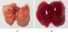
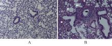
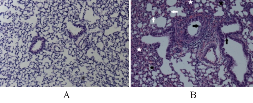
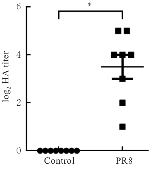
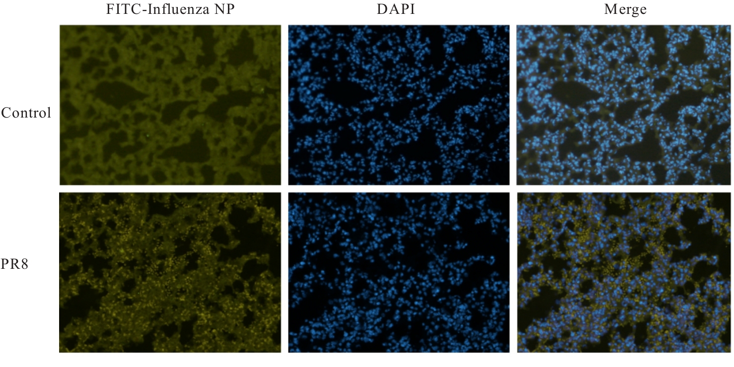
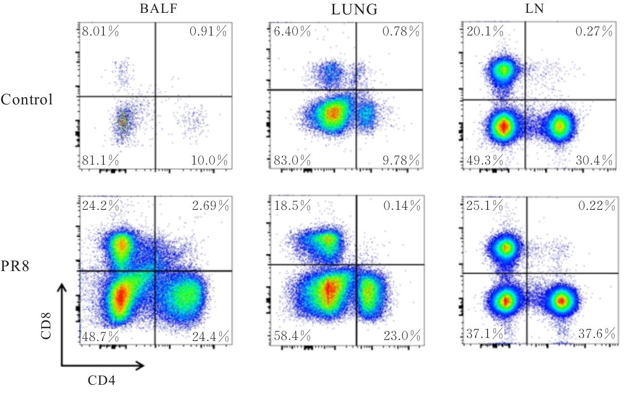
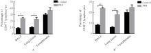
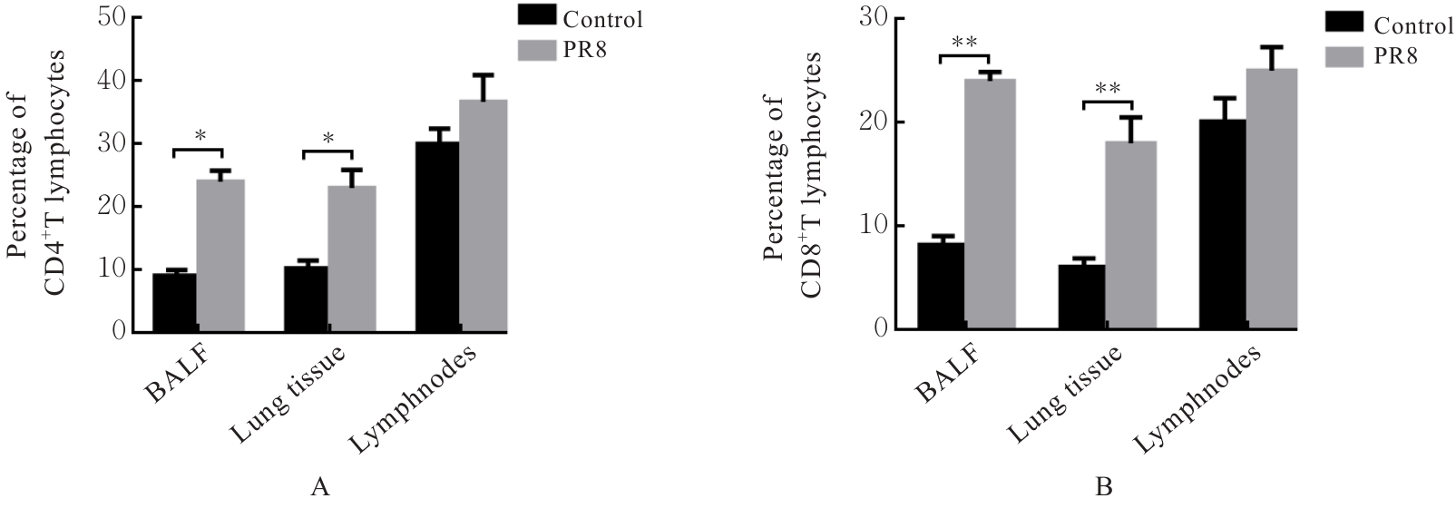
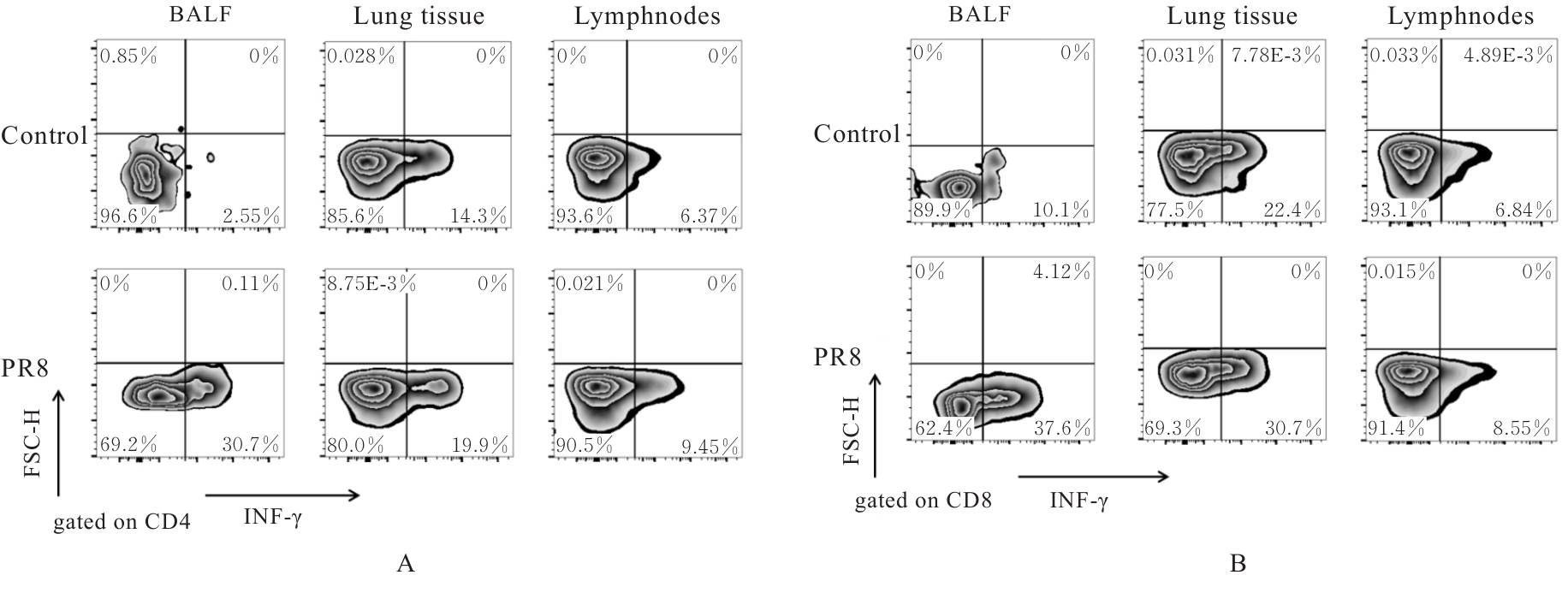
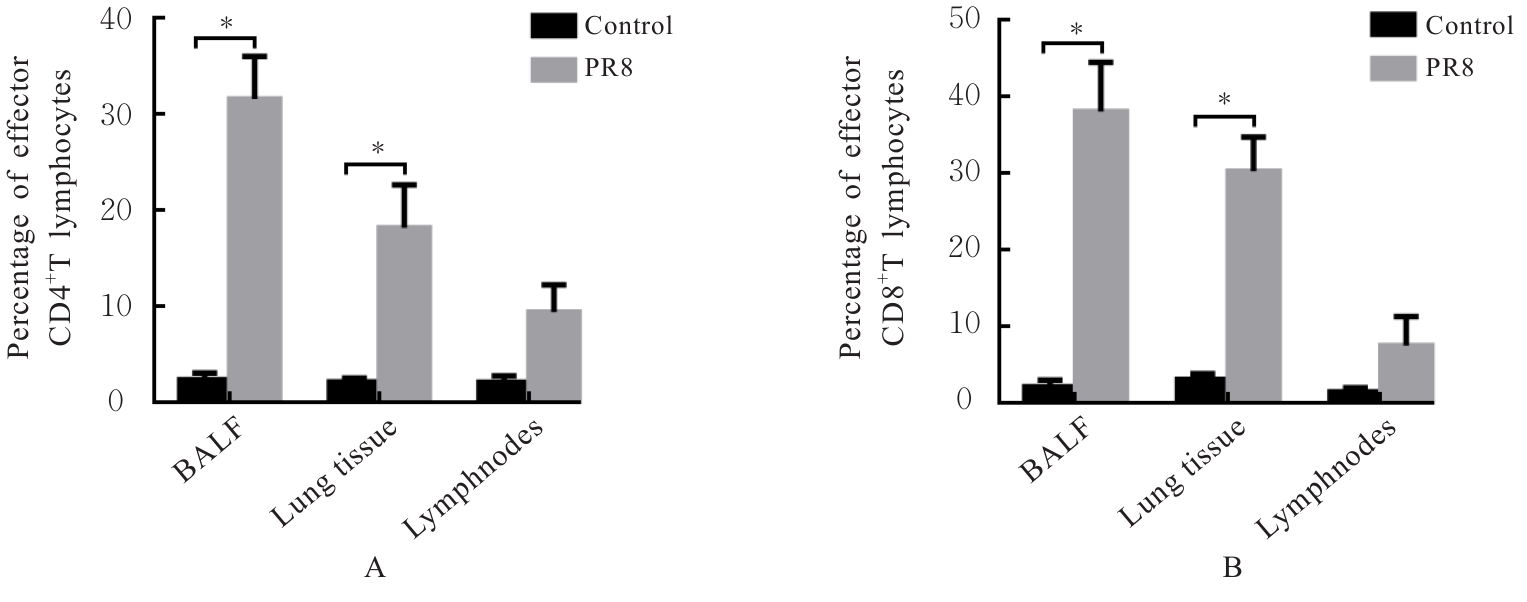
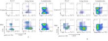
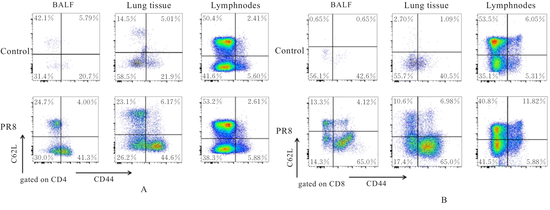
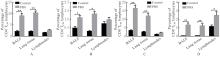
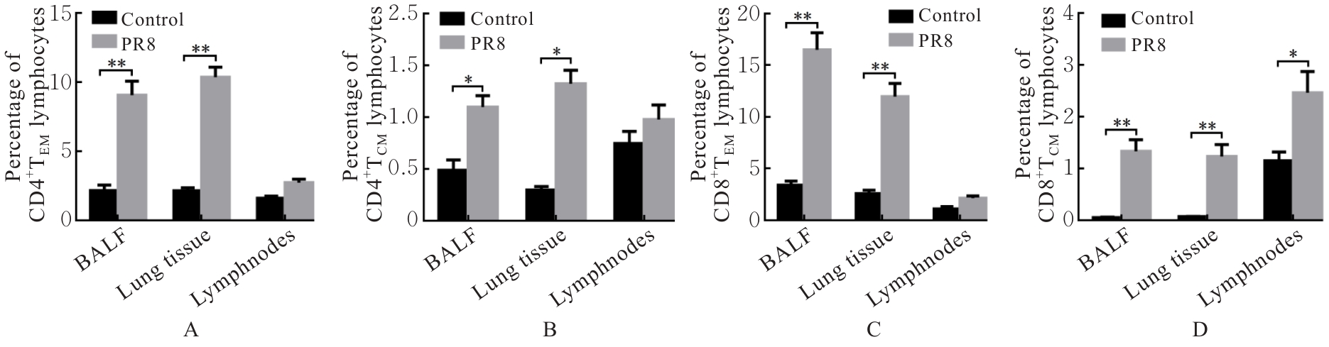
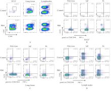
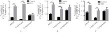
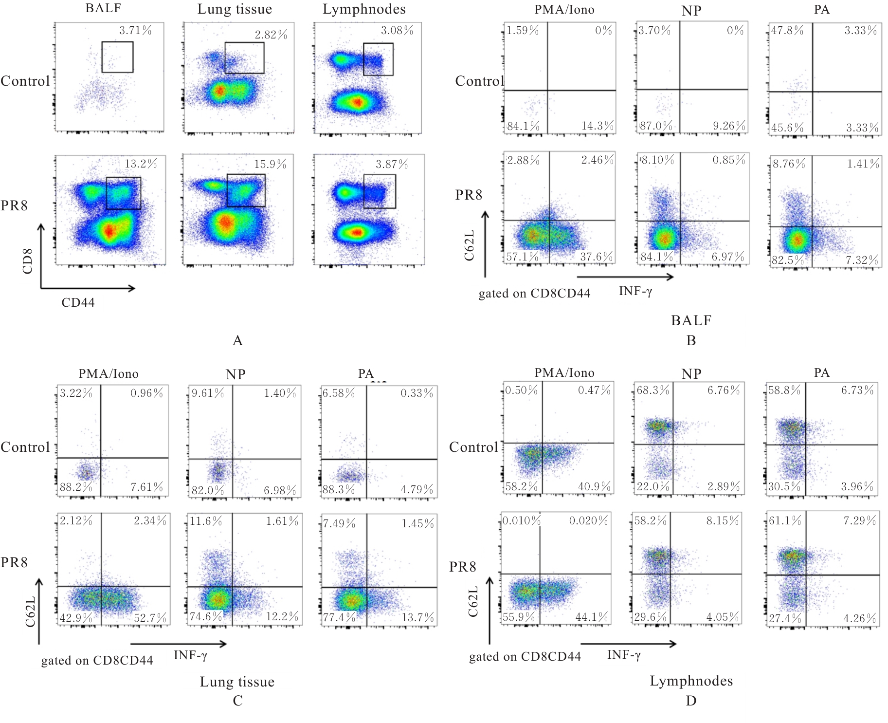
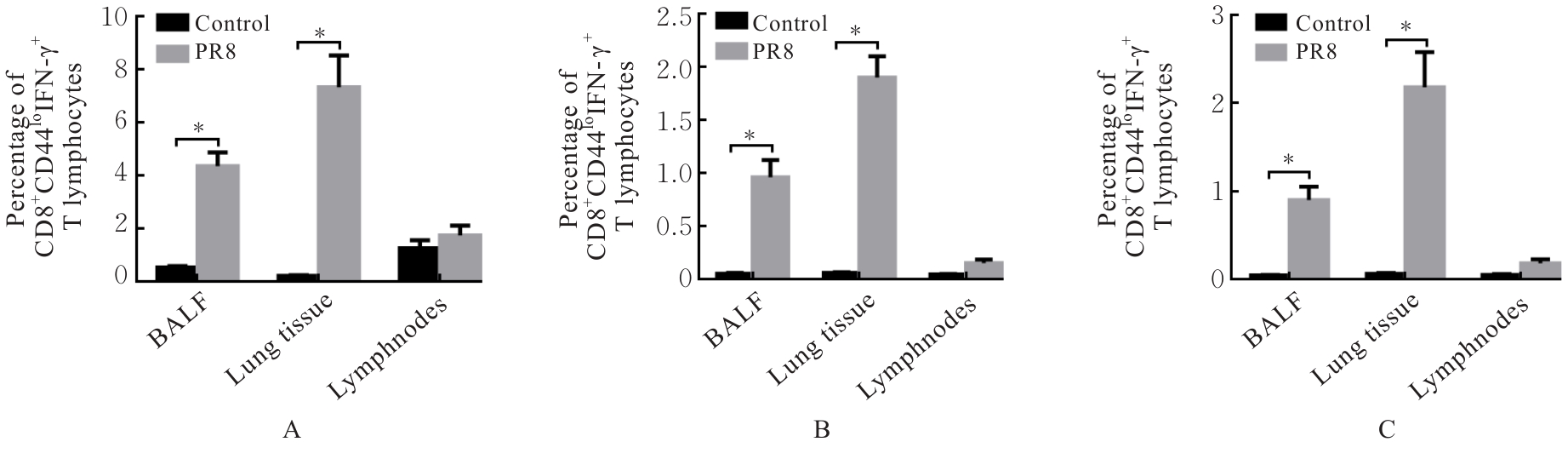
目的 探讨早期记忆T淋巴细胞(TM)在甲型流感病毒(H1N1)PR8感染小鼠后的变化情况,并阐明其意义。 方法 将32只C57BL/6小鼠随机分为对照组和流感病毒(PR8)组,每组16只。对照组小鼠鼻滴30 μL无菌磷酸盐缓冲液(PBS),PR8组小鼠给予鼻滴2 LD50 PR8病毒液。于第10天处死小鼠,收集支气管肺泡灌洗液(BALF)、肺组织和淋巴结用于后续实验。HE染色观察2组小鼠肺组织病理形态表现,血凝试验、实时荧光定量PCR(RT-qPCR)法和免疫荧光法检测小鼠肺组织病毒感染情况,流式细胞术检测2组小鼠BALF、肺组织和淋巴结中T淋巴细胞、效应性T淋巴细胞、中央型TM淋巴细胞(TCM)和效应性TM淋巴细胞(TEM)及核蛋白(NP)/聚合酶酸性蛋白(PA)特异性TEM淋巴细胞百分率。 结果 与对照组比较,PR8组小鼠肺组织出现明显的病理损伤,且肺组织病毒效价和NP蛋白表达水平均明显升高(P<0.01)。与对照组比较,PR8组小鼠BALF和肺组织中CD4+T淋巴细胞、CD8+T淋巴细胞、效应性CD4+T淋巴细胞、效应性CD8+T淋巴细胞、CD4+TEM淋巴细胞和CD8+TEM淋巴细胞百分率均明显升高(P<0.01)。与对照组比较,PR8组小鼠BALF和肺组织中CD4+TCM、CD8+TCM、CD4+TEM、CD8+TEM及NP/PA特异性CD4+TEM和CD8+TEM淋巴细胞百分率均明显升高(P<0.05或P<0.01),淋巴结中CD8+TCM淋巴细胞百分率明显升高(P<0.05)。 结论 流感病毒感染可诱导机体T淋巴细胞增殖分化为效应性T淋巴细胞,在呼吸道黏膜局部早期建立保守表位NP/PA的特异性TM淋巴细胞。

关键词: 流感病毒, 记忆T淋巴细胞, 中央型记忆T淋巴细胞, 效应型记忆T淋巴细胞, 保守表位

Abstract:

Objective To discuss the changes of early memory T lymphocytes (TM) in the mice infected with influenza A virus (H1N1) PR8, and to clarify its significance. Methods A total of 32 C57BL/6 mice were randomly divided into control group and influenza virus (PR8) group, with 16 mice in each group. The mice in control group were intranasally administered 30 μL of sterile phosphate buffered saline (PBS), and the mice in PR8 group were intranasally administered 2 LD50 PR8 virus solution. The mice were sacrificed on the 10 th day, and bronchoalveolar lavage fluid (BALF), lung tissue, and lymphnodes were collected for subsequent experiments. HE staining was used to observe the pathomorphology of lung tissue in the mice in two groups; hemagglutination assay, real-time fluorescence quantitative PCR (RT-qPCR) method, and immunofluorescence method were used to detect the viral infection in lung tissue of the mice; flow cytometry was used to detect the percentages of T lymphocytes, effector T lymphocytes, central memory T lymphocytes (TCM), effector memory T lymphocytes (TEM), and nuclear protein(NP)/pdymerase acidic protein(PA)-specific TEM lymphocytes in BALF, lung tissue, and lymphnodes of the mice in two groups. Results Compared with control group, the lung tissue of the mice in PR8 group showed significant pathological damage, and the lung tissue virus titer and NP protein expression level were significantly increased (P<0.01). Compared with control group, the percentages of CD4+ T lymphocytes, CD8+ T lymphocytes, effector CD4+ T lymphocytes, effector CD8+ T lymphocytes, CD4+ TEM lymphocytes, and CD8+ TEM lymphocytes in BALF and lung tissue of the mice in PR8 group were significantly increased (P<0.01). Compared with control group, the positive percentages of CD4+ TCM, CD8+ TCM, CD4+ TEM, CD8+ TEM, and NP/PA-specific CD4+ TEM, CD8+ TEM lymphocytes in BALF and lung tissue of the mice in PR8 group were significantly increased (P<0.05 or P<0.01), and the percentage of CD8+ TCM lymphocytes in the lymph nodes was significantly increased (P<0.05). Conclusion Influenza virus infection can induce the proliferation and differentiation of T lymphocytes into effector T lymphocytes, leading to the early establishment of virus-specific TM lymphocytes targeting the conserved NP/PA epitopes in the respiratory mucosa.

Key words: Influenza virus, Memory T lymphocyte, Central memory T lymphocyte, Effector memory T lymphocyte, Conserved epitope

中图分类号: 

  • R392