吉林大学学报(医学版) ›› 2022, Vol. 48 ›› Issue (3): 648-656.doi: 10.13481/j.1671-587X.20220313

• 基础研究 • 上一篇    

脂肪间充质干细胞对卵巢早衰模型大鼠的治疗作用及其机制

颜培玉1,张爱臣1,章宏2,李杨2,张萌萌2,洛梦泽1,潘颖1()   

  1. 1.吉林大学中日联谊医院妇产科, 吉林 长春 130033
    2.吉林大学基础医学院生理学系, 吉林 长春 130021
  • 收稿日期:2021-09-26 出版日期:2022-05-28 发布日期:2022-06-21
  • 通讯作者: 潘颖 E-mail:panying424@163.com
  • 作者简介:颜培玉(1994-),女,吉林省和龙市人,在读硕士研究生,主要从事妇科肿瘤方面的研究。
  • 基金资助:
    吉林省科技厅科研项目(192499YY01026)

Therapeutic effect of adipose-derived mesenchymal stem cells on premature ovarian failure model rats and its mechanism

Peiyu YAN1,Aichen ZHANG1,Hong ZHANG2,Yang LI2,Mengmeng ZHANG2,Mengze LUO1,Ying PAN1()   

  1. 1.Department of Obstetrics and Gynecology,China-Japan Union Hospital,Jilin University,Changchun 130033,China
    2.Department of Physiology,School of Basic Medical Sciences,Jilin University,Changchun 130021,China
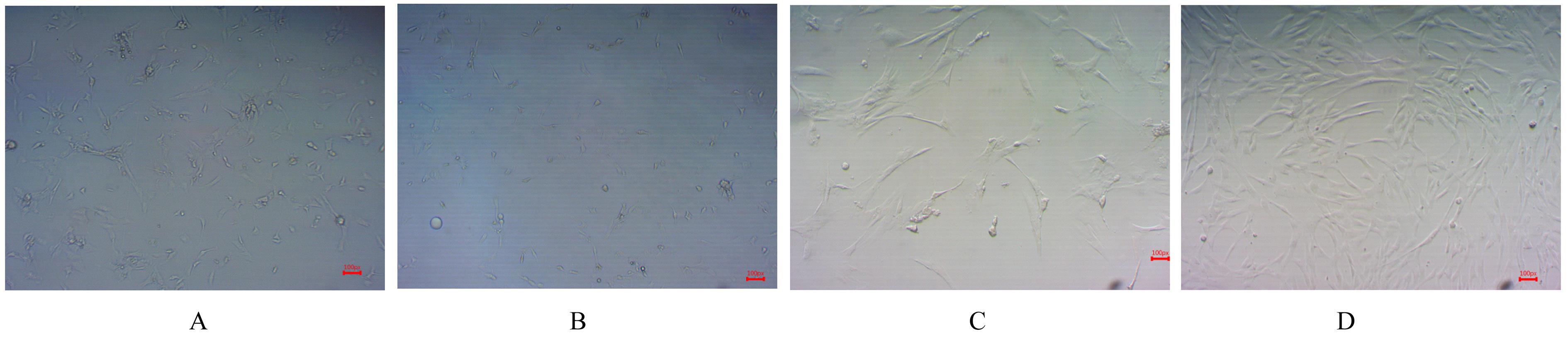
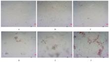
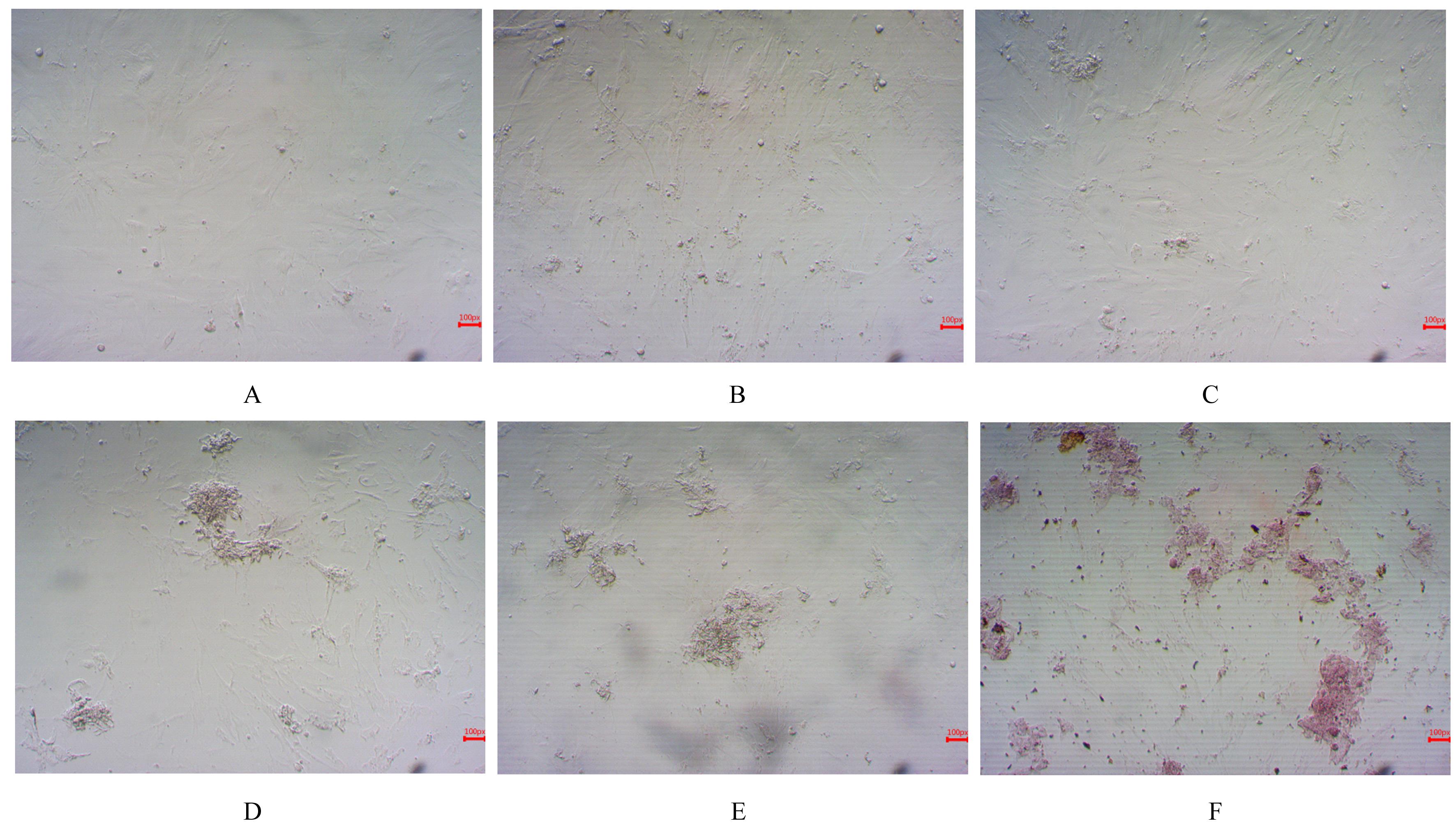
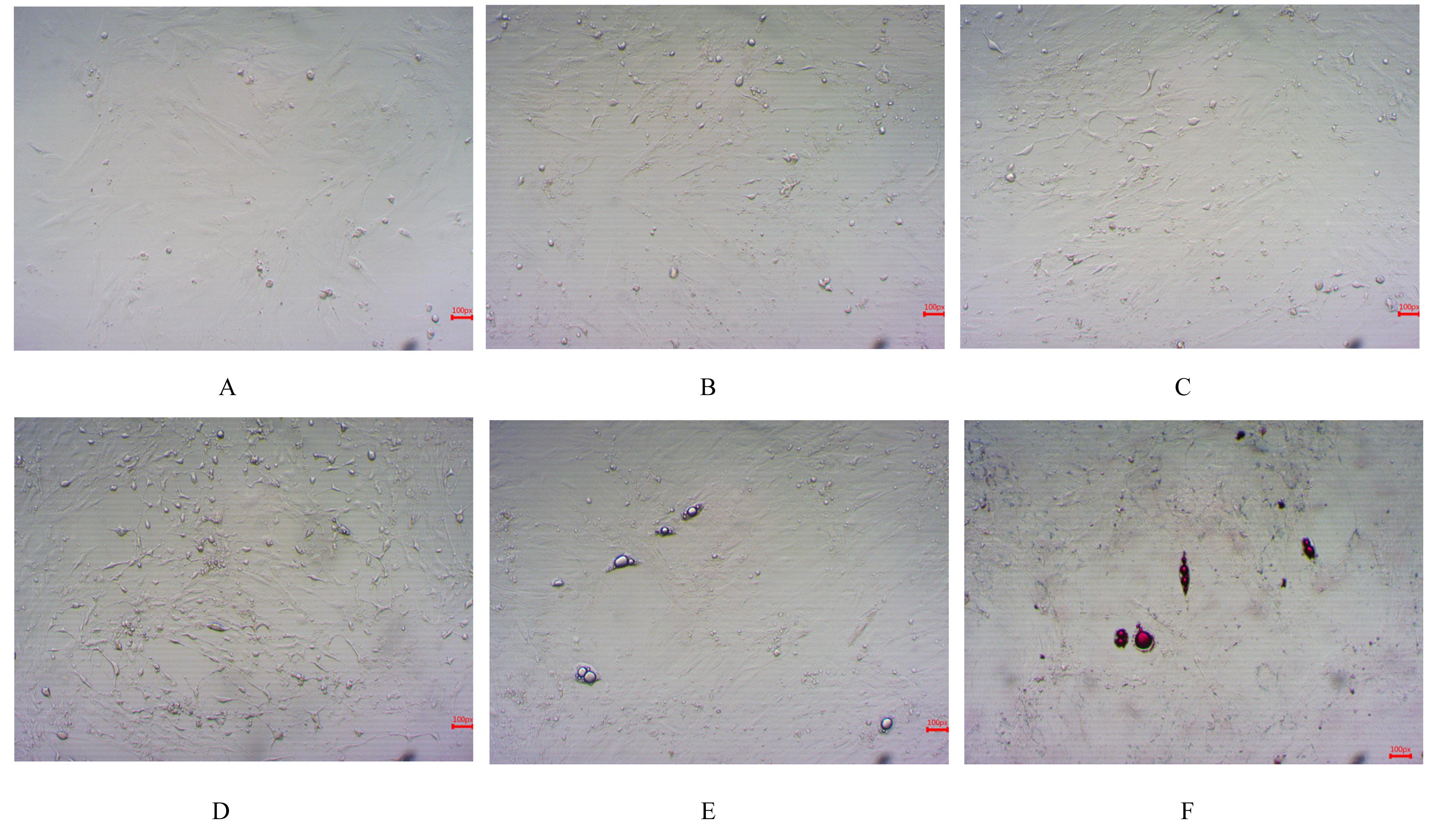
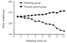
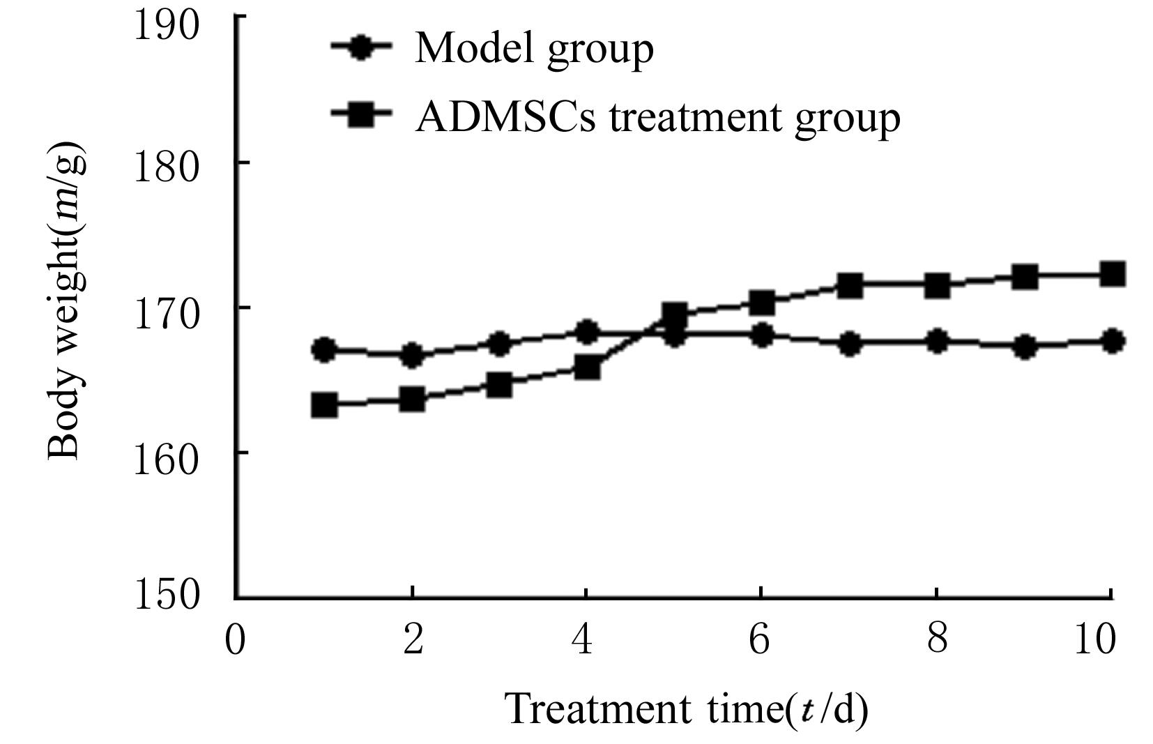
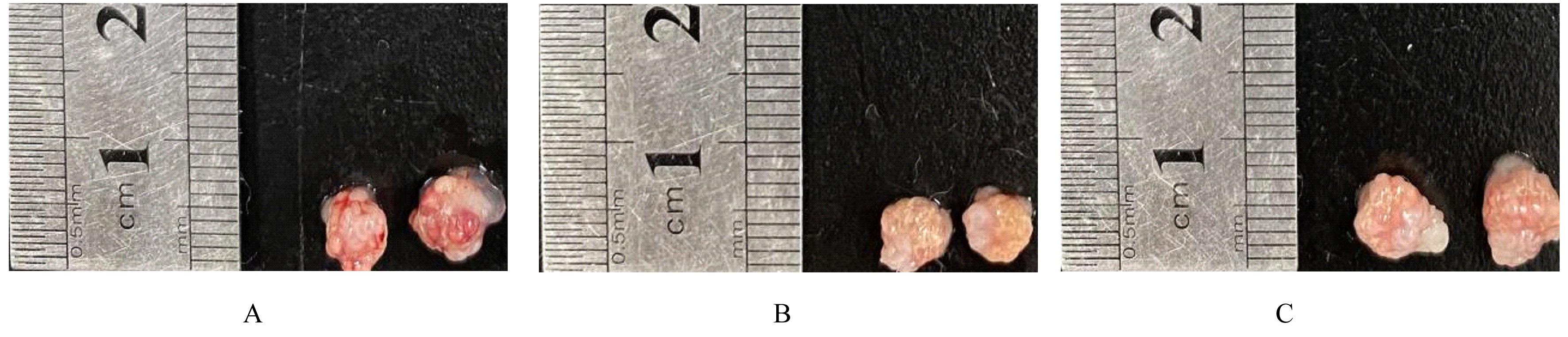
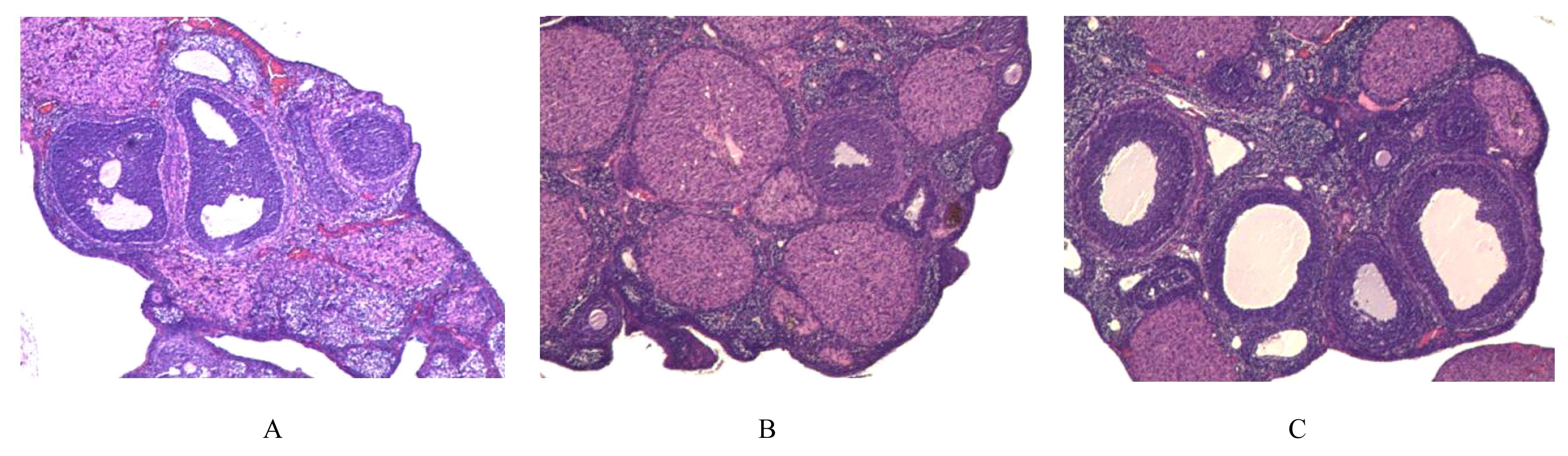
  • Received:2021-09-26 Online:2022-05-28 Published:2022-06-21
  • Contact: Ying PAN E-mail:panying424@163.com

摘要: 目的

探讨脂肪间充质干细胞(ADMSCs)的生物学特性,阐明ADMSCs对卵巢早衰(POF)模型大鼠的治疗作用及其机制。

方法

分离培养SD大鼠的ADMSCs,光学显微镜观察细胞形态表现,成骨和成脂细胞诱导试剂诱导ADMSCs成骨及成脂分化,流式细胞术检测ADMSCs表面抗原标记物CD44、CD90和CD45的表达。CCK-8法检测ADMSCs的生长状态并绘制生长曲线。通过腹腔注射顺铂建立大鼠POF模型。30只SD大鼠分为正常对照组(10只)和造模组(20只),造模组大鼠再随机分为模型组和ADMSCs治疗组,每组10只。ADMSCs治疗组大鼠每3 d经尾静脉注射异体ADMSCs;正常对照组和模型组大鼠每3 d经尾静脉注射生理盐水。记录各组大鼠体质量,ELISA法检测各组大鼠血清雌二醇(E2)、卵泡刺激素(FSH)和黄体生成素(LH)水平,HE染色观察各组大鼠卵巢组织病理形态表现。

结果

ADMSCs为贴壁生长细胞,生长旺盛,可向成骨和成脂分化,ADMSCs高表达CD44和CD90,表达率分别为75.85%及59.71%;低表达CD45,表达率为39.12%。与正常对照组比较,造模组大鼠体质量明显降低(P<0.05),血清E2水平明显降低(P<0.05),FSH和LH水平明显升高(P<0.05),缺乏各级卵泡,闭锁卵泡数目增多。与模型组比较,ADMSCs治疗组大鼠血清E2水平升高(P<0.05),FSH和LH水平降低(P<0.05),各级卵泡数目增多,闭锁卵泡数目减少。

结论

ADMSCs能够在体外生长繁殖,并具有较强生长增殖能力和多向分化能力;ADMSCs能够改善POF模型大鼠血清E2水平及卵巢功能,对POF模型大鼠具有一定的治疗作用。

关键词: 脂肪间充质干细胞, 细胞分化, 细胞增殖, 卵巢早衰, 卵巢功能

Abstract: Objective

To explore the biological characteristics of adipose-derived mesenchymal stem cells (ADMSCs), and to clarify the therapeutic effect of ADMSCs in the premature ovarian failure (POF) model rats.

Methods

The ADMSCs from SD rats were isolated and cultured, and the morphology of cells was observed under optical microscope. The osteogenic and adipogenic differentiations of ADMSCs were induced by osteogenic and adipogenic cell induction reagents. The expressions of surface antigen markers CD44, CD90,and CD45 of ADMSCs were detected by flow cytometry.The growth of ADMSCs was determined by CCK-8 assay and the growth curves were drawn. The models of PDF were established by intraperitoneal injection of cisplatin.Thirty SD rats were randomly divided into normal control group (n=10) and modeling group (n=20). The rats in modeling group were randomly divided into model group and ADMSCs treatment group, and there were 10 rats in each group.The rats in ADMSCs treatment group were injected with allogeneic ADMSCs through the tail vein every 3 d; the rats in normal control group and model group were injected with normal saline every 3 d through the tail vein. The body weights of the rats in various groups were detected, and the levels of estradiol (E2), follicle stimulating hormone (FSH), and luteinizing hormone (LH) in serum of the rats in various groups were detected by ELISA mehod. The pathomorphology of ovary tissue of the rats in various groups was observed by HE staining.

Results

The ADMSCs were adherent growth cells, which grew vigorously and could differentiate into the osteogenic and adipogenic cells. The expressions of CD44 and CD90 in the ADMSCs were highly, and the expression rates were 75.85% and 59.71%; the expression of CD45 was low, and the expression rate of 39.12%. Compared with normal control group, the body weight of the rats in model group was significantly decreased (P<0.05); the serum E2 level was significantly decreased (P<0.05), and the levels of FSH and LH were significantly increased (P<0.05), lacking of follicles at all levels, and the number of atretic follicles was increased. Compared with model group, the serum E2 level of the rats in ADMSCs treatment group was increased (P<0.05), the FSH and LH levels were decreased (P<0.05); the number of follicles was increased,and the number of atretic follicles was decreased.

Conclusion

ADMSCs can grow and reproduce in vitro, and have strong growth and proliferation abilities and multi-directional differentiation abilities; ADMSCs can improve the serum E2 level and ovarian function of the POF model rats, and have a certain therapeutic effect in the POF model rats.

Key words: Adipose-derived mesenchymal stem cells, Cell differentiation, Cell proliferation, Premature ovarian failure, Ovarian function

中图分类号: 

  • R711