吉林大学学报(医学版) ›› 2023, Vol. 49 ›› Issue (4): 858-866.doi: 10.13481/j.1671-587X.20230405

• 基础研究 • 上一篇    下一篇

包虫抗原B通过肿瘤坏死因子受体2调控巨噬细胞极化对小鼠免疫性血小板减少症的改善作用

宋传龙,焦红杰,海力其古丽·努日丁null,岳迎宾,严媚()   

  1. 新疆医科大学第一附属医院儿科中心,新疆 乌鲁木齐 830054
  • 收稿日期:2022-09-21 出版日期:2023-07-28 发布日期:2023-07-26
  • 通讯作者: 严媚 E-mail:yan1omei25@163.com
  • 作者简介:宋传龙(1988―),男,山东省临沂市人,住院医师,在读博士研究生,主要从事小儿急危重症医学及儿童血液系统疾病基础和临床方面的研究。
  • 基金资助:
    国家自然科学基金项目(82160031);省部共建中亚高发病成因与防治国家重点实验室包虫病研究专项项目(SKL-HIDCA-2020-BC1);新疆维吾尔自治区科技厅天山创新团队计划项目(2020D14027)

Improvement effect of hydatid antigen B on immune thrombocytopenia in mice by regulating macrophage polarization through tumor necrosis factor receptor 2

Chuanlong SONG,Hongjie JIAO, HAILIQIGULI·Nuriding,Yingbin YUE,Mei YAN()   

  1. Pediatric Center,First Affiliated Hospital,Xinjiang Medical University,Urumqi 830054,China
  • Received:2022-09-21 Online:2023-07-28 Published:2023-07-26
  • Contact: Mei YAN E-mail:yan1omei25@163.com

摘要:

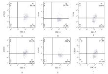
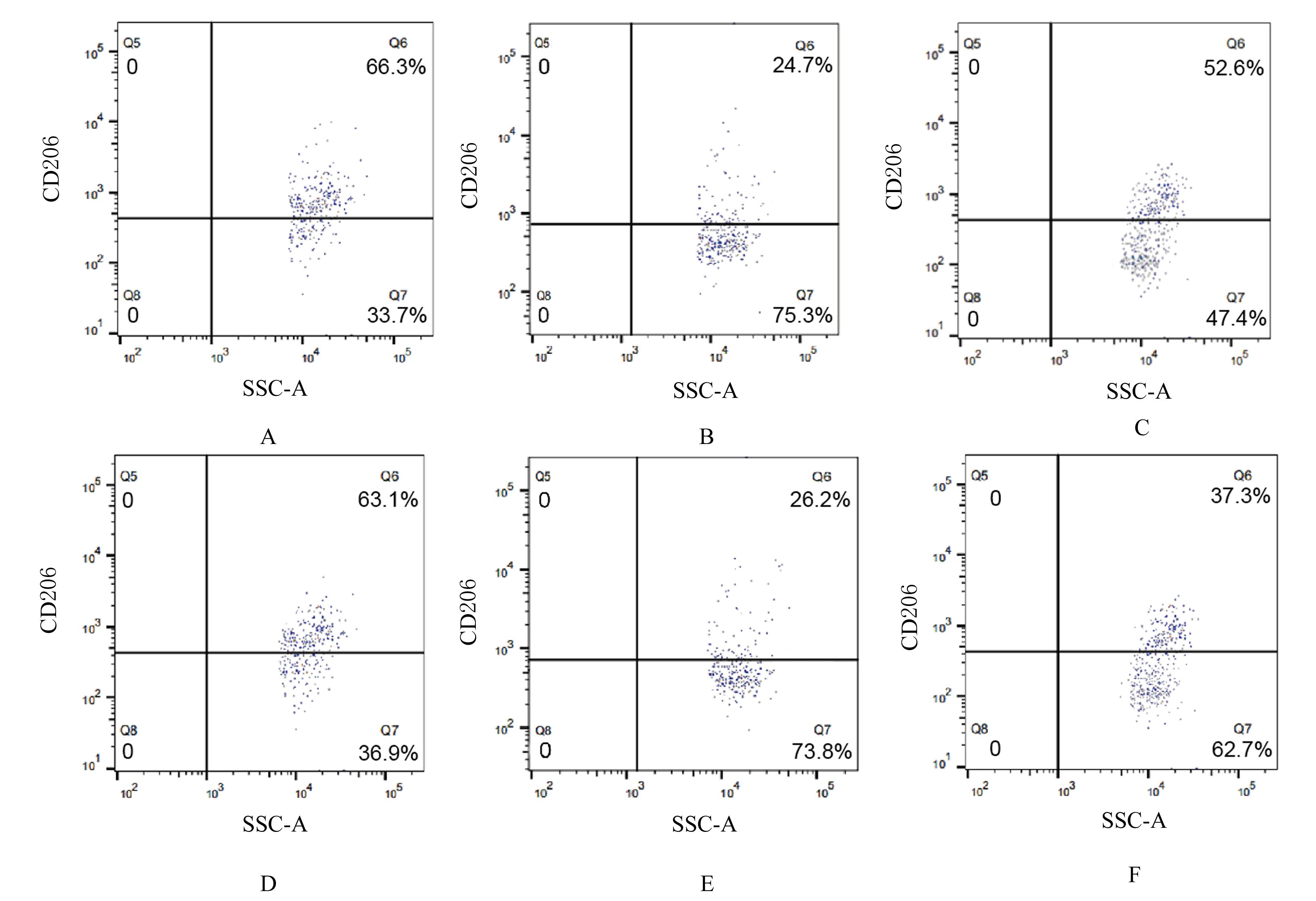
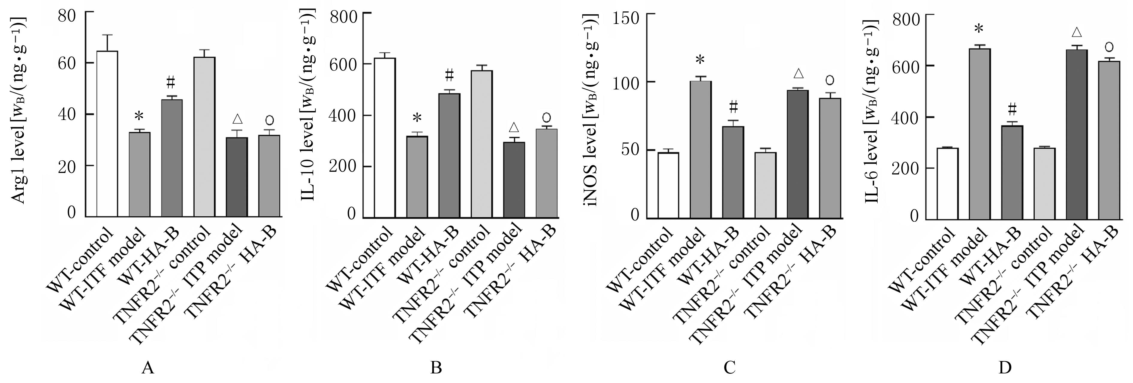
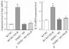
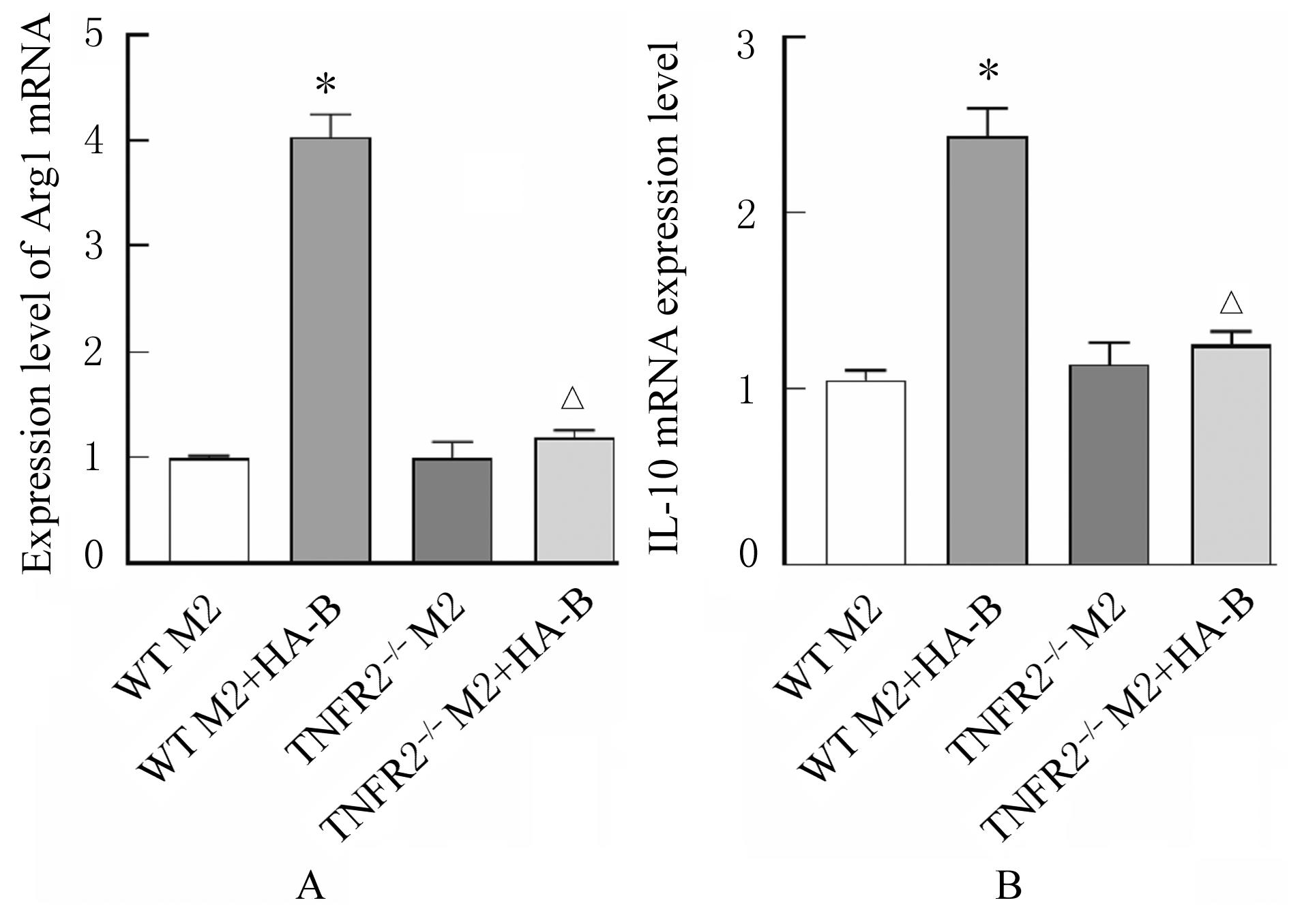
目的 探讨包虫抗原B(HA-B)对小鼠免疫性血小板减少症(ITP)的改善作用,阐明其相关作用机制。 方法 将野生型(WT)和肿瘤坏死因子受体2(TNFR2)基因敲除(TNFR2-/-)的C57/B6小鼠分为WT对照组、WT-ITP模型组、WT-HA-B组、TNFR2-/-对照组、TNFR2-/- ITP模型组和TNFR2-/- HA-B组,检测各组小鼠体质量、脏器指数和血常规指标,采用流式细胞术检测各组小鼠外周血中M2巨噬细胞百分率,酶联免疫吸附试验(ELISA)法检测各组小鼠血清中精氨酸酶1(Arg1)、白细胞介素10(IL-10)、诱导型一氧化氮合成酶(iNOS)和白细胞介素6(IL-6)水平,采用实时荧光定量PCR(RT-qPCR)法检测各组小鼠骨髓来源巨噬细胞(BMDM)中Arg1、IL-10、iNOS和IL-6表达水平。分别将WT对照组和TNFR2-/-对照组小鼠BMDM诱导为M2巨噬细胞(WT M2组和TNFR2-/- M2组),加入HA-B(WT M2+HA-B组和TNFR2-/-M2+HA-B组),采用RT-qPCR法检测各组细胞中Arg1和IL-10 mRNA表达水平。 结果 分别与WT对照组和TNFR2-/-对照组比较,WT-ITP模型组和TNFR2-/- ITP模型组小鼠体质量降低(P<0.05),脾脏和胸腺指数升高(P<0.05),血小板和红细胞数量减少(P<0.05),血红蛋白水平降低(P<0.05),白细胞数量增加(P<0.05),凝血时间延长(P<0.05);外周血中M2巨噬细胞百分率降低(P<0.05),血清中Arg1和IL-10水平降低(P<0.05),iNOS和IL-6水平升高(P<0.05);BMDM中Arg1和IL-10 mRNA表达水平降低(P<0.05),iNOS和IL-6 mRNA表达水平升高(P<0.05)。与WT-ITP模型组比较,WT-HA-B组小鼠体质量增加(P<0.05),脾脏和胸腺指数降低(P<0.05),血小板和红细胞数量增加(P<0.05),血红蛋白水平升高(P<0.05),白细胞数量减少(P<0.05),凝血时间缩短(P<0.05);外周血中M2巨噬细胞百分率升高(P<0.05),血清中Arg1和IL-10水平升高(P<0.05),iNOS和IL-6水平降低(P<0.05);BMDM中Arg1和IL-10 mRNA表达水平升高(P<0.05),iNOS和IL-6 mRNA表达水平降低(P<0.05)。与WT-HA-B组比较,TNFR2-/- HA-B组小鼠体质量降低(P<0.05),脾脏和胸腺指数升高(P<0.05),血小板和红细胞数量减少(P<0.05),血红蛋白水平降低(P<0.05),白细胞数量增加(P<0.05),凝血时间延长(P<0.05);外周血中M2巨噬细胞百分率降低(P<0.05),血清中Arg1和IL-10水平降低(P<0.05),iNOS和IL-6水平升高(P<0.05);BMDM中Arg1和IL-10 mRNA表达水平降低(P<0.05),iNOS和IL-6 mRNA表达水平升高(P<0.05)。与WT M2组比较,WT M2+HA-B组M2巨噬细胞中Arg1和IL-10 mRNA表达水平升高(P<0.05);与WT M2+HA-B组比较,TNFR2-/- M2+HA-B组M2巨噬细胞中Arg1和IL-10 mRNA表达水平降低(P<0.05)。 结论 HA-B可通过TNFR2促进巨噬细胞M2极化,进而发挥治疗ITP的作用。

关键词: 包虫抗原B, 巨噬细胞M2极化, 免疫性血小板减少症, 肿瘤坏死因子受体2

Abstract:

Objective To discuss the improvement effect of hydatid antigen B (HA-B) on the immune thrombocytopenia (ITP) of the mice, and to clarify its related mechanism. Methods The C57/B6 mice with wild-type (WT) and tumor necrosis factor receptor 2 (TNFR2) gene knockout (TNFR2-/-) were divided into WT control group, WT-ITP model group, WT-HA-B group, TNFR2-/-control group, TNFR2-/- ITP model group, and TNFR2-/- HA-B group. The body weights, organ indexes, and blood routine indexes of mice in various groups were detected. The percentages of M2 macrophages in peripheral blood of mice in various groups were detected by flow cytometry; enzyme-linked immunosorbent assay (ELISA) method was used to detect the levels of Arginase 1 (Arg1), interleukin-10 (IL-10),inductible nitric oxide synthase (iNOS),and interleukin-6 (IL-6) in serum of the mice in various groups; real-time fluorescence quantitative PCR (RT-qPCR) method was used to detect the expression levels of Arg1, IL-10, iNOS and IL-6 in bone marrow derived macrophages (BMDM) of the mice in various groups. The BMDM of the mice in WT control group and TNFR2-/- control group were induced into the M2 macrophages (WT M2 group and TNFR2-/- M2 group). After adding HA-B (WT M2+HA-B group and TNFR2-/- M2+HA-B group), the expression levels of Arg1 and IL-10 mRNA in the cells in various groups were detected by RT-qPCR method. Results Compared with WT control group and TNFR2-/- control group, the body weights of the rats in WT-ITP model group and TNFR2-/- ITP model group were decreased (P<0.05), the spleen and thymus indexes were increased (P<0.05), the platelet counts and red blood cell counts were decreased (P<0.05), the hemoglobin levels were decreased (P<0.05), the white blood cell counts were increased (P<0.05), and the coagulation time were increased(P<0.05); the percentages of M2 macrophages in the peripheral blood were decreased (P<0.05), the levels of Arg1 and IL-10 in serum were decreased (P<0.05), and the levels of iNOS and IL-6 were increased (P<0.05); the expression levels of Arg1 and IL-10 mRNA in the BMDM were decreased (P<0.05), while the expression levels of iNOS and IL-6 mRNA were increased (P<0.05). Compared with WT-ITP model group, the body weight of the mice in WT-HA-B group was increased (P<0.05), the spleen and thymus indexes were decrease (P<0.05), the platelet count and red blood cell count were increased (P<0.05), the hemoglobin level was increased (P<0.05), the white blood cell count was decrease (P<0.05), and the coagulation time was decreased (P<0.05); the percentage of M2 macrophages in the peripheral blood was increased (P<0.05), the levels of Arg1 and IL-10 in the serum were increased (P<0.05), and the levels of iNOS and IL-6 in serum were decreased (P<0.05);the expression levels of Arg1 and IL-10 mRNA in the BMDM were increased(P<0.05), while the expression levels of iNOS and IL-6 mRNA were decreased (P<0.05). Compared with WT-HA-B group, the body weight of the mice in TNFR2-/- HA-B group was decreased (P<0.05), the spleen and thymus indexes were increased (P<0.05), the platelet count and red blood cell count were decreased (P<0.05), the hemoglobin level was decreased (P<0.05), the white blood cell count was increased (P<0.05), and the coagulation time was increased (P<0.05); the percentage of M2 macrophages in the peripheral blood was decreased (P<0.05), the levels of Arg1 and IL-10 in the serum were decreased (P<0.05), and the levels of iNOS and IL-6 were increased (P<0.05); the expression levels of Arg1 and IL-10 mRNA in the BMDM were decreased (P<0.05), while the expression levels of iNOS and IL-6 mRNA were increased (P<0.05). Compared with WT M2 group, the expression levels of Arg1 and IL-10 mRNA in the macrophages in WT M2+HA-B group were increased (P<0.05); compared with WT M2+HA-B group, the expression levels of Arg1 and IL-10 mRNA in the M2 macrophages in TNFR2-/-M2+HA-B group were decreased (P<0.05). Conclusion HA-B can promote the macrophage M2 polarization through TNFR2, thereby exerting the therapeutic effect on ITP.

Key words: Hydatid antigen B, Macrophages M2 polarization, Immune thrombocytopenia, Tumor necrosis factor receptor 2

中图分类号: 

  • R558.2