吉林大学学报(医学版) ›› 2024, Vol. 50 ›› Issue (6): 1606-1613.doi: 10.13481/j.1671-587X.20240614

• 基础研究 • 上一篇    

内侧前额叶皮质区核氧化还原蛋白对卒中后抑郁小鼠抑郁样行为的影响及其机制

赵丹(),史博,魏志玄,崔群建   

  1. 南阳医学高等专科学校第一附属医院神经外科,河南 南阳 473058
  • 收稿日期:2023-12-06 出版日期:2024-11-28 发布日期:2024-12-10
  • 通讯作者: 赵丹 E-mail:zhaod820518@163.com
  • 作者简介:赵 丹(1982-),男,河南省南阳市人,主治医师,医学硕士,主要从事脑血管疾病基础和临床方面的研究。
  • 基金资助:
    河南省卫健委医科科技攻关计划联合共建项目(LHGJ20210983)

Effect of nucleoredoxin in medial prefrontal cortex on depression-like behavior in mice with post-stroke depression and its mechanism

Dan ZHAO(),Bo SHI,Zhixuan WEI,Qunjian CUI   

  1. Department of Neurosurgery,First Affiliated Hospital,Nanyang Medical College,Nanyang 473058,China
  • Received:2023-12-06 Online:2024-11-28 Published:2024-12-10
  • Contact: Dan ZHAO E-mail:zhaod820518@163.com

摘要:

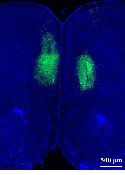
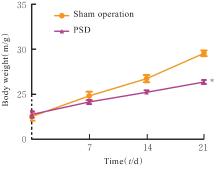
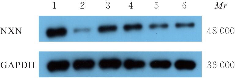
目的 探讨内侧前额叶皮质(mPFC)区核氧化还原蛋白(NXN)对小鼠卒中后抑郁(PSD)的影响,并阐明其可能的作用机制。 方法 80只C57BL/6小鼠,随机选取42只分为NXN过表达腺相关病毒感染组(AAV-NXN-OE组,n=21)和阴性对照腺相关病毒感染组(AAV-NC组,n=21),剩余小鼠分为假手术组(n=20)和PSD组(n=18),注射NXN过表达腺相关病毒后,将AAV-NXN-OE组和AAV-NC组剩余小鼠分为PSD+AAV-NC组(n=18)和PSD+AAV-NXN-OE组(n=18)。术前3周,采用脑立体定位法向小鼠脑组织mPFC区注射NXN过表达腺相关病毒,显微镜下观察病毒在小鼠脑组织mPFC区表达情况,Western blotting法检测小鼠脑组织中NXN蛋白表达水平。采用线栓法建立大脑中动脉闭塞(MCAO)卒中模型,术后1周使用慢性不可预见的中等应激(CUMS)结合孤养法持续干预3周,构建PSD模型小鼠。造模期间监测小鼠体质量变化,造模结束后采用糖水偏好实验、悬尾实验和强迫游泳实验观察各组小鼠抑郁样行为学表现,生化法检测各组小鼠脑组织mPFC区中丙二醛(MDA)和还原型谷胱甘肽(GSH)水平及超氧化物歧化酶(SOD)活性,2',7'-二氯荧光素二乙酸酯(DCFH-DA)荧光探针标记法检测各组小鼠脑组织mPFC区中活性氧(ROS)水平,Western blotting法检测各组小鼠脑组织mPFC区、杏仁核区和海马组织中NXN蛋白表达水平。 结果 PSD小鼠脑组织mPFC区有大量绿色荧光,表明携带ZsGreen绿色荧光蛋白标签的AAVs病毒在PSD小鼠脑组织mPFC区成功感染并表达。与AAV-NC组比较,AAV-NXN-OE组小鼠脑组织mPFC区中NXN蛋白表达水平明显升高(P<0.05)。与假手术组比较,PSD组小鼠体质量增长缓慢(P<0.05),糖水偏好率明显降低(P<0.05),小鼠在悬尾实验和强迫游泳实验中不动时间均明显增加(P<0.05);与假手术组比较,PSD+AAV-NC组小鼠糖水偏好率明显降低(P<0.05),小鼠在悬尾实验和强迫游泳实验中不动时间均明显增加(P<0.05);与PSD+AAV-NC组比较,PSD+AAV-NXN-OE组小鼠糖水偏好率明显升高(P<0.05),小鼠在悬尾实验和强迫游泳实验中不动时间均明显减少(P<0.05)。与假手术组比较,PSD+AAV-NC组小鼠脑组织mPFC区中MDA和ROS水平均明显升高(P<0.05),GSH水平和SOD活性均明显降低(P<0.05);与PSD+AAV-NC组比较,PSD+AAV-NXN-OE组小鼠脑组织mPFC区中MDA和ROS水平均明显降低(P<0.05),GSH水平和SOD活性均明显升高(P<0.05)。与假手术组比较,PSD组小鼠脑组织mPFC区中NXN蛋白表达水平明显降低(P<0.05),杏仁核区和海马组织中NXN蛋白表达水平差异无统计学意义(P>0.05)。 结论 小鼠脑组织mPFC区中NXN过表达可改善PSD小鼠抑郁样行为,其作用机制可能与调节氧化还原平衡有关。

关键词: 卒中后抑郁, 核氧化还原蛋白, 内侧前额叶皮质, 氧化还原平衡

Abstract:

Objective To discuss the effect of nucleoredoxin (NXN) in medial prefrontal cortex (mPFC) region of the mice with post-stroke depression (PSD), and to clarify its possible mechanism. Methods A total of 42 mice among 80 C57BL/6 mice were randomly divided into NXN over-expression adeno-associated virus infection group (AAV-NXN-OE group, n=21) and negative control adeno-associated virus infection group (AAV-NC group, n=21). The remaining mice were divided into sham operation group (n=20) and PSD group (n=18). After injectied with NXN over-expression adeno-associated virus, the remaining mice in AAV-NXN-OE group and AAV-NC group were further divided into PSD+AAV-NC group (n=18) and PSD+AAV-NXN-OE group (n=18). Three weeks before surgery, NXN over-expression adeno-associated virus was injected into the mPFC region of brain tissue of the mice by stereotaxic method, and the expression of the virus in mPFC region of the mice was observed under microscope. Western blotting method was used to detect the expression levels of NXN protein in brain tissue of the mice in various groups; middle cerebral artery occlusion (MCAO) model was established by thread embolism method, followed one week post-surgery by three weeks of chronic unpredictable moderate stress (CUMS) combined with isolation feeding to construct the PSD mice model. During modeling, the body weight changes of the mice were monitored. After modeling, sucrose preference test, tail suspension test, and forced swim test were used to observe the depressive-like behavioral changes of the mice in various groups; biochemical method was used to detect the levels of malondialdehyde (MDA) and reduced glutathione (GSH), and superoxide dismutase (SOD) activities in mPFC region of brain tissue of the mice in various groups; DCFH-DA fluorescence probe labeling method was used to detect the reactive oxygen species (ROS) levels in mPFC region of brain tissue of the mice in various groups; Western blotting method was used to detect expression levels of NXN protein in mPFC region, amygdala, and hippocampus tissues of the mice in various groups. Results A large amount of green fluorescence was observed in the mPFC region in brain tissue of the PSD mice, indicating successful infection and expression of AAVs virus labeled with ZsGreen green fluorescent protein in mPFC region in brain tissue of the PSD mice. Compared with AAV-NC group, the expression level of NXN protein in mPFC region in brain tissue of the mice in AAV-NXN-OE group was significantly increased (P<0.05). Compared with sham operation group, the body weight of the mice in PSD group was increased slowly (P<0.05), the sucrose preference rate was significantly decreased (P<0.05), and the immobility time of the mice in the tail suspension test and forced swim test was significantly increased (P<0.05). Compared with sham operation group, the sucrose preference rate of the mice in PSD+AAV-NC group was significantly decreased (P<0.05), and the immobility time in tail suspension test and forced swim test was significantly increased (P<0.05). Compared with PSD+AAV-NC group, the sucrose preference rate of the mice in PSD+AAV-NXN-OE group was significantly increased (P<0.05), and the immobility time of the mice in tail suspension test and forced swim test was significantly decreased (P<0.05). Compared with sham operation group, the MDA and ROS levels in mPFC region in brain tissue of the mice in PSD+AAV-NC group were significantly increased (P<0.05), while the GSH level and SOD activity were significantly decreased (P<0.05). Compared with PSD+AAV-NC group, the levels of MDA and ROS in mPFC region in brain tissue of the mice in PSD+AAV-NXN-OE group were significantly decreased (P<0.05), while the GSH level and SOD activity were significantly increased (P<0.05). Compared with sham operation group, the expression level of NXN protein in the mPFC region of brain tissue of the mice in PSD group was significantly decreased (P<0.05), the expression levels of NXN protein in amygdala and hippocampus tissue had no statistically significant difference (P>0.05). Conclusion Over-expression of NXN in mPFC region of brain tissue of the mice can improve the depressive-like behavior in the PSD mice, and its mechanism is possibly related to regulating the redox balance.

Key words: Post-stroke depression, Nucleoredoxin, Medial prefrontal cortex, Redox balance

中图分类号: 

  • R749.13