吉林大学学报(医学版) ›› 2021, Vol. 47 ›› Issue (3): 630-636.doi: 10.13481/j.1671-587X.20210312

• 基础研究 • 上一篇    下一篇

miR-106b靶向调控TGF-β/Smad通路对结肠癌细胞侵袭和迁移的促进作用

马博(),李建刚,王俊,候军丽,李亮   

  1. 新疆医科大学第二附属医院普外科,新疆 乌鲁木齐 830063
  • 收稿日期:2020-10-10 出版日期:2021-05-28 发布日期:2021-05-28
  • 通讯作者: 马博 E-mail:1401596054@qq.com
  • 作者简介:马 博(1968-),男,新疆维吾尔自治区乌鲁木齐市人,主任医师,医学硕士,主要从事胃肠外科基础和临床方面的研究。
  • 基金资助:
    新疆维吾尔自治区科技厅自然科学基金项目(2017D01C241)

Promotion effect of miR-106b on invasion and migration of colon cancer cells through targeting TGF-β/Smad pathway

Bo MA(),Jiangang LI,Jun WANG,Junli HOU,Liang LI   

  1. Department of General Surgery,Second Affiliated Hospital,Xinjiang Medical University,Urumqi 830063,China
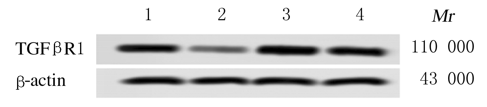
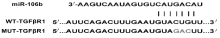
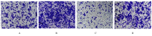
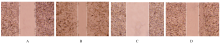
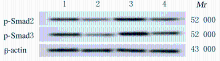
  • Received:2020-10-10 Online:2021-05-28 Published:2021-05-28
  • Contact: Bo MA E-mail:1401596054@qq.com

摘要: 目的

探讨miR-106b靶向转化生长因子β受体1(TGF-βR1)对结肠癌细胞侵袭和迁移的影响,阐明miR-106b与TGF-βR1基因的靶向关系及其可能作用机制。

方法

选取30例结肠癌患者癌组织和癌旁正常结肠组织、结肠癌SW-480细胞及正常结肠上皮NCM460细胞为研究对象;采用实时荧光定量PCR(RT-qPCR)法检测各种组织和细胞中miR-106b表达水平及细胞中TGF-βR1 mRNA表达水平。将结肠癌SW-480细胞分为空质粒组(转染miR-NC空质粒)、miR-106b组(转染miR-106b-mimics)、pGL3-TGF-βR1组(转染pGL3-TGF-βR1)和miR-106b-mimics+pGL3-TGF-βR1组(同时转染miR-106b-mimics和pGL3-TGF-βR1);生物信息分析法预测miR-106b与TGF-βR1的靶向作用关系,荧光素酶报告实验检测各组SW-480细胞中荧光素酶活性。Transwell小室实验检测各组SW-480细胞侵袭能力,细胞划痕实验检测各组SW-480细胞划痕愈合率,Western blotting法检测各组SW-480细胞中TGF-βR1、磷酸化Smad同源物2(p-Smad2)和磷酸化Smad同源物3(p-Smad3)蛋白表达水平。

结果

结肠癌组织中miR-106b表达水平高于正常结肠组织,结肠癌SW-480细胞中miR-106b表达水平高于正常结肠上皮NCM460细胞(P<0.01)。双荧光素酶报告基因检测结果提示TGF-βR1为miR-106b的靶基因。与空质粒组比较,miR-106b组SW-480细胞中TGF-βR1 mRNA和蛋白表达水平明显降低(P<0.01),侵袭细胞数和细胞划痕愈合率明显升高(P<0.01),细胞中p-Smad2和p-Smad3蛋白表达水平明显降低(P<0.01)。与miR-106b组比较,miR-106b-mimics+pGL3-TGF-βR1组SW-480细胞中TGF-βR1 mRNA和蛋白表达表达水平明显升高(P<0.01),侵袭细胞数和细胞划痕愈合率明显降低(P<0.01),细胞中p-Smad2和p-Smad3蛋白表达水平明显升高(P<0.01)。

结论

miR-106b过表达可能通过抑制TGF-β/Smad通路促进结肠癌细胞的侵袭和迁移。

关键词: miR-106b, 转化生长因子β受体 1, 结肠肿瘤, 侵袭, 转移

Abstract: Objective

To investigate the effect of miR-106b targeting transforming growth factor-β receptor1(TGF-βR1)on the invasion and migration of colon cancer cells, and to clarify the targeting relationship between miR-106b and TGF-βR1 and its possible mechanism.

Methods

A total of 30 cases of colon cancer tissue and adjacent normal colon tissue,the colon cancer SW-480 cells and normal colon epithelial NCM460 cells were selected. The Real-time fluorescence quantitative PCR(RT-qPCR)method was used to detect the expression levels of miR-106b and TGF-βR1 mRNA in different kinds of tissues and cells.The colon cancer SW-480 cells were transfected and divided into empty vector group (transfected with miR-NC),miR-106b group (transfected with miR-106b-mimics),pGL3-TGF-βR1 group (transfected with pGL3-TGF-βR1), and miR-106b-mimics+pGL3-TGF-βR1 group (transfected with miR-106b-mimics and pGL3-TGF-βR1). The level of miR-106b in different kinds of tissues and cells,and the expression levels of TGF-βR1 mRNA in different kinds of cells were detected by RT-qPCR method;the targarc relationship between miR-106b and TGF-βR1 was predicted by bio-information analysis method.The luciferase activities of SW-480 cells in various groups were detected by luciferase reporter assay;the invasion abilities of the colon cancer SW-480 cells in various groups were detected by Transwell chamber test; the wound healing rates of the colon cancer SW-480 cells in various groups were detected by cell scratch test; the expression levels of TGF-βR1, phosphorylated Smad family member 2 (p-Smad2) and phosphorylated Smad family member 3(p-Smad3) proteins in colon cancer SW-480 cells in various groups were detected by Western blotting method.

Results

The level of miR-106b in the colon cancer tissue and colon cancer SW-480 cells were lower than those in normal colon tissue and normal colon epithelial NCM460 cells (P<0.01).The results of double luciferase reporter gene analysis showed that TGFβR1 was the target gene of miR-106b.Compared with empty vector group,the expression levels of TGF-βR1 mRNA and protein in the colon cancer SW-480 cells in miR-106b-mimics+pGL3-TGF-βR1 group were significantly decreased(P<0.01);the number of invasion cells and wound healing rate in the colon cancer SW-480 cells were significantly increased (P<0.01),and the expression levels of p-Smad2 and p-Smad3 proteins in the colon cancer SW-480 cells were decreased (P<0.01).Compared with miR-106b group,the expression levels of TGF-βR1 mRNA and protein in the SW-480 cells in miR-106b-minics+PGL3-TGF-βR1 group were significantly increased(P<0.01),the number of invasion cells and the scrath healing rate were decreased(P<0.01),and the expression levels of p-Smad2 and p-Smad3 proteins in the cells were significantly increased(P<0.01).

Conclusion

Over-expression of miR-106b may promote the invasion and migration of the colon cancer cells expression by inhibiting the TGF-β / Smad pathway.

Key words: miR-106b, transforming growth factor β receptor 1, colon neoplasms, cell invasion, cell migration

中图分类号: 

  • R735.35