吉林大学学报(医学版) ›› 2022, Vol. 48 ›› Issue (2): 299-307.doi: 10.13481/j.1671-587X.20220205

• 基础研究 • 上一篇    下一篇

商陆皂苷甲对哮喘小鼠气道炎症的缓解作用及对肺组织中IL-6和STAT3表达水平的影响

王静1,2,徐畅1,3,宋艺兰1,3,王重阳1,3,姜京植1,3,李良昌1,3,延光海1,3,苏立明1,2()   

  1. 1.延边大学 吉林省过敏性常见疾病免疫与靶向研究重点实验室,吉林 延吉 133002
    2.延边大学 附属医院儿科,吉林 延吉 133002
    3.延边大学医学院解剖学教研室,吉林 延吉 133002
  • 收稿日期:2021-09-08 出版日期:2022-03-28 发布日期:2022-05-10
  • 通讯作者: 苏立明 E-mail:suliming1997@163.com
  • 作者简介:王 静(1994-),女,山东省菏泽市人,在读硕士研究生,主要从事儿童哮喘和炎症性疾病方面的研究。
  • 基金资助:
    国家自然科学基金项目(81860729)

Alleviation of esculentoside A on airway inflammation of asthmatic mice and its effect on expression levels of IL-6 and STAT3 in lung tissue

Jing WANG1,2,Chang XU1,3,Yilan SONG1,3,Chongyang WANG1,3,Jingzhi JIANG1,3,Liangchang LI1,3,Guanghai YAN1,3,Liming SU1,2()   

  1. 1.Key Laboratory of Immunization and Targeting Research on Common Allergic Diseases of Jilin Province,Yanbian University,Yanji 133002,China
    2.Department of Pediatrics,Affiliated Hospital,Yanbian University,Yanji 133002 China
    3.Department of Anatomy,School of Medicine,Yanbian University,Yanji 133002,China
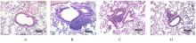
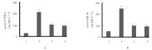
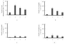
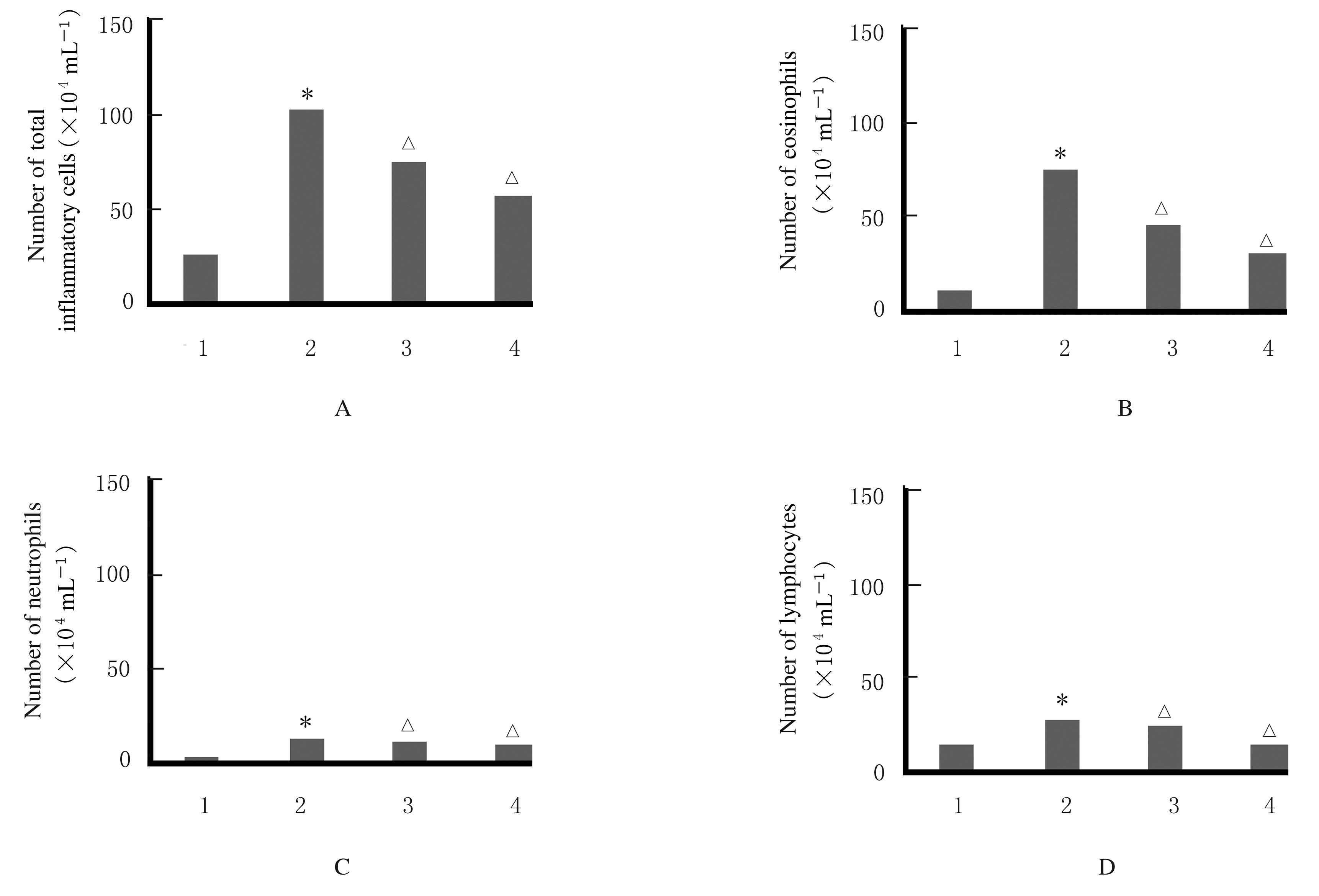
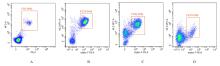
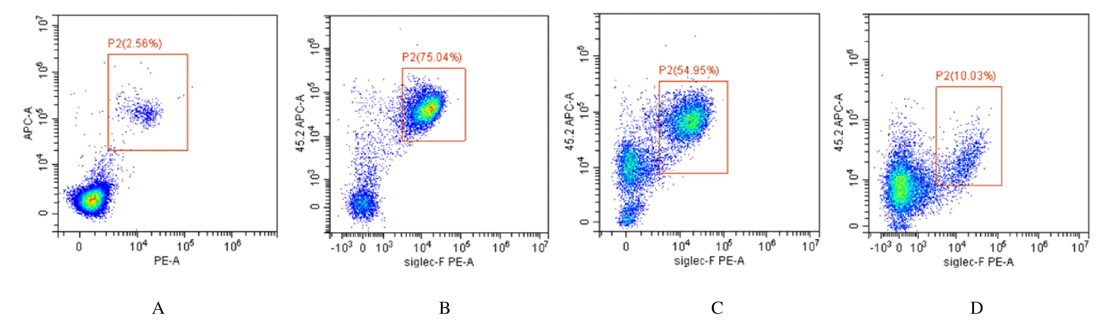
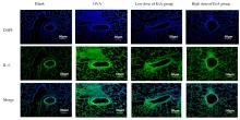
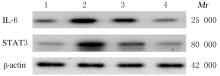
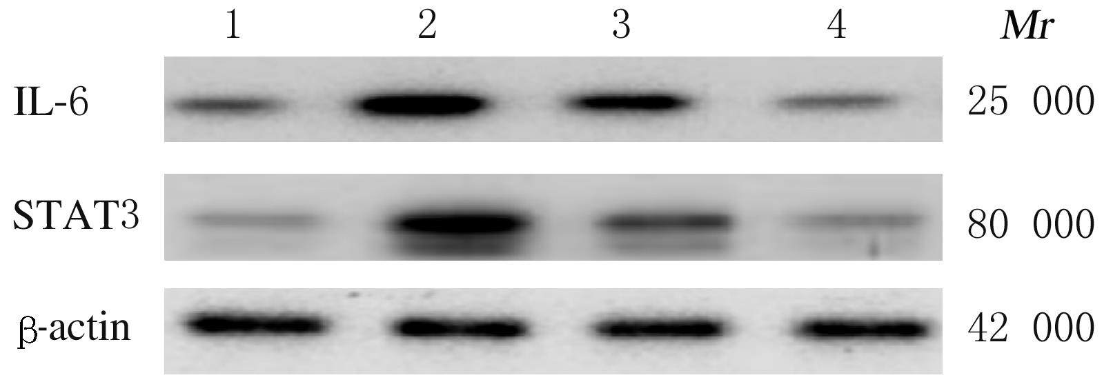
  • Received:2021-09-08 Online:2022-03-28 Published:2022-05-10
  • Contact: Liming SU E-mail:suliming1997@163.com

摘要: 目的

探讨商陆皂苷甲(EsA)对卵清蛋白(OVA)诱导哮喘小鼠气道炎症的药理作用,并阐明其作用机制。

方法

40 只清洁级 BALB/c雌性小鼠随机分为空白对照组、OVA组、低剂量EsA组和高剂量EsA组,每组 10 只;除空白对照组外,其他组小鼠用腹腔注射致敏,分别在第1、7和14天致敏,从第21天开始采用1% OVA激发,每 1 d为 1 个激发周期,每个激发周期激发1次,共进行3次;空白对照组和OVA 组小鼠应用生理盐水 0.2 mL 灌胃给药,低剂量EsA组和高剂量EsA组小鼠分别按照 10和20 mg·kg-1 灌胃给药,从第 17天开始连续给药7 d,每天1次。采用HE染色法观察各组小鼠肺组织形态表现,直接计数法计算各组小鼠肺泡灌洗液 (BALF)中总炎症细胞、嗜酸性粒细胞、中性粒细胞和淋巴细胞数量,流式细胞术检测 BALF 中嗜酸性粒细胞、白细胞介素4(IL-4)和干扰素γ(IFN-γ)水平,酶联免疫吸附测定(ELISA)法检测各组小鼠 BALF 中白细胞介素1β(IL-1β)和肿瘤坏死因子α(TNF-α)水平,Western blotting 法检测各组小鼠肺组织中白细胞介素6(IL-6)和信号传导及转录激活蛋白-3(STAT3)蛋白表达水平,免疫组织化学法和免疫荧光法检测 各组小鼠肺组织中IL-6 和 STAT3 蛋白表达量。

结果

与空白对照组比较,OVA 组小鼠肺组织支气管和血管周围炎性细胞浸润,支气管部分损伤;与OVA 组比较,低和高剂量EsA组小鼠肺组织中炎性细胞浸润明显减轻,气道损伤缓解。与空白对照组比较,OVA 组小鼠 BALF中总炎症细胞、嗜酸性粒细胞、中性粒细胞和淋巴细胞数量增加(P<0.05),IL-4、IL-1β和 TNF-α水平升高(P<0.05或P<0.01),而 IFN-γ水平降低(P<0.05);与 OVA 组比较,低和高剂量EsA组小鼠 BALF中总炎症细胞、嗜酸性粒细胞、中性粒细胞和淋巴细胞数量降低(P<0.05),IL-4、IL-1β和 TNF-α水平降低(P<0.05或P<0.01),而 IFN-γ水平升高(P<0.05)。与空白对照组比较,OVA组小鼠肺组织中IL-6和STAT3蛋白表达水平明显升高(P<0.05);与 OVA 组比较,低和高剂量EsA组小鼠肺组织中IL-6和STAT3蛋白表达水平降低(P<0.05)。

结论

EsA 可缓解哮喘小鼠气道炎症,其作用机制与调节IL-6和STAT3信号通路有关。

关键词: 商陆皂苷甲, 哮喘, 白细胞介素6, 信号传导及转录激活蛋白-3

Abstract: Objective

To explore the pharmacological effect of esculentoside A (EsA) on the ovalbumin (OVA)-induced airway inflammation in the asthmatic mice, and to clarify its mechanism of action.

Methods

Forty clean BALB/c female mice were randomly divided into blank control group,OVA group, low dose of EsA group and high dose of EsA group,with 10 mice in each group. They were sensitized by intraperitoneal injection and sensitization occurred on days 1, 7,and 14, respectively. Starting from the 21st day with 1% OVA, and one excitation period was perfomed every day,with each excitation period being excited once for three times.The mice in blank control group and OVA group were given 0.2 mL of normal saline by gavage. The mice in low dose of EsA group and high dose of EsA group received 10 and 20 mg·kg-1 EsA by gavage, starting from the 17th day, for 7 consecutive days,once a day. HE staining method was used to observe the morphological performance of lung tissue of the mice in various groups, and direct counting method was used to calculate the number of total inflammatory cells, eosinophils, neutrophils and lymphocytes in BALF of the mice in various groups. Cytometry method was used to detect the expressions of eosinophils,IL-4 and IFN-γ in BALF, and ELISA method was used to detect the levels of IL-1β and TNF-α; Western blotting method was used to determine the expression levels of IL-6 and STAT3 proteins in lung tissue of the mice in various groups;immunohistochemistry and immunofluorescence methods were used to detect the expressions of IL-6 and STAT3 proteins in lung tissue of the mice in various groups.

Results

Compared with blank control group, the bronchi and perivascular inflammatory cells in lung tissue of the mice in OVA group were infiltrated with partial bronchial damage; compared with OVA group, the infiltration of inflammatory cells in lung tissue of the mice in low and high doses of EsA groups were reduced and airway injury was alleviated; compared with blank control group, the number of total inflammatory cells, eosinophils, neutrophils and lymphocytes in BALF of the mice in OVA group was increased(P<0.05),the expression levels of IL-4, IL-1βand TNF-α were increased (P<0.01), while the expression level of IFN-γ was decreased (P<0.05); compared with OVA group, the number of total inflammatory cells, eosinophils, neutrophils and lymphocytes in BALF of the mice in low and high doses of EsA groups were decreased(P<0.05),the levels of IL-4, IL-1β and TNF-α were decreased (P<0.01), and the levels of IFN-γ were increased (P<0.05).Compared with blank control group, the expression levels of IL-6 and STAT3 proteins in lung tissue of the mice in OVA group were significantly increased (P<0.05); compared with OVA group, the expression levels of IL-6 and STAT3 proteins in lung tissue of the mice in low and high doses of EsA groups were decreased (P<0.05).

Conclusions

EsA may alleviate the airway inflammation in the asthmatic mice, and its mechanism may be related to regulating the IL-6 and STAT3 signaling pathway.

Key words: Esculentoside A, Asthma, Interleukin-6, Signal transduction and activator of transcription-3

中图分类号: 

  • R562.25