吉林大学学报(医学版) ›› 2022, Vol. 48 ›› Issue (3): 561-567.doi: 10.13481/j.1671-587X.20220302

• 基础研究 • 上一篇    

丹酚酸B对小鼠动脉粥样硬化病变和巨噬细胞胞葬作用的影响及其机制

张一凡,丁洁,杜敏,冯骁腾,刘萍()   

  1. 上海中医药大学附属龙华医院心病科,上海 200032
  • 收稿日期:2021-09-28 出版日期:2022-05-28 发布日期:2022-06-21
  • 通讯作者: 刘萍 E-mail:liuping0207@126.com
  • 作者简介:张一凡(1993-),女,河北省唐山市,在读博士研究生,主要从事中医药防治心血管疾病方面的研究。
  • 基金资助:
    国家自然科学基金项目(81873117)

Effects of salvianolic acid B on atherosclerosis and efferocytosis of macrophages of mice and their mechanisms

Yifan ZHANG,Jie DING,Min DU,Xiaoteng FENG,Ping LIU()   

  1. Department of Cardiology,Longhua Hospital,Shanghai University of Traditional Chinese Medicine,Shanghai 200032,China
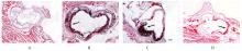
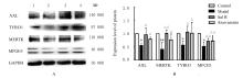
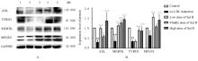
  • Received:2021-09-28 Online:2022-05-28 Published:2022-06-21
  • Contact: Ping LIU E-mail:liuping0207@126.com

摘要: 目的

探讨丹酚酸B(Sal B)对小鼠动脉粥样硬化病变和氧化型低密度脂蛋白(ox-LDL)诱导的小鼠巨噬细胞胞葬作用的影响,阐明Sal B抗动脉粥样硬化的作用机制。

方法

将32只雄性低密度脂蛋白受体基因敲除(LDLR-/-)小鼠随机分为对照组、模型组、Sal B组和阿托伐他汀组,每组8只。对照组小鼠给予普通饲料喂养,其他各组小鼠给予高脂饲料喂养12周。对照组和模型组小鼠每日给予生理盐水腹腔注射,Sal B组小鼠给予Sal B溶液腹腔注射,阿托伐他汀组小鼠给予阿托伐他汀溶液灌胃。检测各组小鼠血清总胆固醇(TC)、甘油三酯(TG)和低密度脂蛋白胆固醇(LDL-c)水平,油红O染色法观察小鼠主动脉粥样硬化斑块面积百分率。RAW264.7细胞分为对照组、ox-LDL组(采用40 mg·L-1 ox-LDL诱导24 h),低、中和高剂量Sal B组(给予含1.25、2.50和5.00 mg·L-1 Sal B的培养基)。实时荧光定量PCR(RT-qPCR)和Western blotting法检测各组小鼠主动脉组织及RAW264.7细胞中AXL、MERTK、TYRO3和乳脂球表皮生长因子8 (MFGE8) mRNA及蛋白表达水平。

结果

与对照组比较,模型组小鼠血清TC、TG和LDL-c水平升高(P<0.05),小鼠主动脉粥样硬化斑块面积百分率升高(P<0.05),小鼠主动脉组织中AXL、MERTK、TYRO3和MFGE8 mRNA及蛋白表达水平降低(P<0.05或P<0.01);与模型组比较,Sal B和阿托伐他汀组小鼠血清TC、TG和LDL-c水平降低(P<0.01),小鼠主动脉粥样硬化斑块面积百分率降低(P<0.01),小鼠主动脉组织中AXL、MERTK、TYRO3和MFGE8 mRNA及蛋白表达水平升高(P<0.05或P<0.01)。与对照组比较,ox-LDL诱导组RAW264.7细胞中AXL、MERTK、TYRO3和MFGE8 mRNA及蛋白表达水平明显降低(P<0.05);与ox-LDL诱导组比较,Sal B组RAW264.7细胞中AXL、MERTK、TYRO3、MFGE8 mRNA及蛋白表达水平升高(P<0.05或P<0.01)。

结论

Sal B通过上调胞葬相关分子表达,促进LDLR-/-小鼠及RAW264.7细胞的胞葬作用,从而发挥抗动脉粥样硬化的作用。

关键词: 丹酚酸B, 低密度脂蛋白受体, 动脉粥样硬化, RAW264.7细胞, 胞葬作用

Abstract: Objective

To investigate the effects of salvianolic arid B (Sal B) on atherosclerosis of the mice and oxidized low-density lipoprotein (ox-LDL)-induced efferocytosis of macrophages of the mice,and to clarify the mechanism of anti-atherosclerosis of Sal B.

Methods

Thirty-two male low-density lipoprotein receptor gene knockout(LDLR-/-) mice were randomly divided into control group, model group, Sal B group, and atorvastatin group,and there were 8 mice in each group. Except for control group (fed with ordinary feed), the mice in the other groups were fed with high-fat feed for 12 weeks.The mice in control group and model group were injected intraperitoneally with normal saline daily, the mice in Sal B group were injected intraperitoneally with Sal B solution, and the mice in atorvastatin group were administered with atorvastatin solution by gavage. The serum levels of total cholesterol(TC), triglyceride(TG),and low density lipoprotein-cholesterol(LDL-c)of the mice in various groups were detected. Oil red O staining method was used to observe the percentages of aortic atherosclerotic plaque area of the mice in various groups. The RAW 264.7 cells were divided into control group,ox-LDL induction group (induced by 40 mg·L-1 ox-LDL for 24 h), low, middle, and high doses of Sal B groups(given 1.25, 2.50,and 5.00 mg·L-1 Sal B). Real-time fluorescence quantitative PCR(RT-qPCR)and Western blotting methods were used to detect the expression levels of AXL, MERTK, TYRO3,and milk fat globule epidermal growth factor 8(MFGE8)mRNA and proteins in arterial tissue of the mice and RAW264.7 cells in various groups.

Results

Compared with control group, the serum levels of TC, TG, and LDL-C of the mice in model group were increased (P<0.01), and the percentage of aortic atherosclerotic plaque area was increased (P<0.01),the expression levels of AXL, MERTK, TYRO3, and MFGE8 mRNA and proteins in arterial tissue were significantly decreased(P<0.05 or P<0.01). Compared with model group, the serum levels of TC, TG, LDL-C of the mice in Sal B and atorvastatin groups were decreased (P<0.01), and the percentages of aortic atherosclerotic plaque areas were decreased (P<0.01); the expression levels of AXL, MERTK, TYRO3 and MFGE8 mRNA and proteins in arterial tissue of the mice were increased (P<0.05 or P<0.01). Compared with control group, the expression levels of AXL, MERTK, TYRO3, MFGE8 mRNA and proteins in the RAW264.7 cells in ox-LDL group were decreased (P<0.05); compared with ox-LDL induction group, the expression levels of AXL, MERTK, TYRO3,and MFGE8 mRNA and proteins in the RAW264.7 cells in Sal B groups were increased (P<0.05 or P<0.01).

Conclusion

Sal B promotes the efferocytosis of the LDLR-/- mice and RAW264.7 cells by up-regulating the expressions of efferocytosis-related molecules, thereby exerting an anti-atherosclerotic effect.

Key words: Salvianolic acid B, Low-density lipoprotein receptor, Atherosclerosis, RAW264.7 cells, Efferocytosis

中图分类号: 

  • R543.5