吉林大学学报(医学版) ›› 2022, Vol. 48 ›› Issue (4): 866-874.doi: 10.13481/j.1671-587X.20220404

• 基础研究 • 上一篇    下一篇

整合素β1和层黏连蛋白受体1在小鼠牙发育不同阶段牙胚组织中的表达及其意义

郑赫南1,2,周怡君3,王爽爽3,任飞龙2,4,范心怡2,4,史册2,4(),刘红1,2()   

  1. 1.吉林大学口腔医院综合治疗科, 吉林 长春 130021
    2.吉林省牙发育及颌骨重塑与再生重点实验室, 吉林 长春 130021
    3.中国医科大学口腔医学院·附属口腔医院口腔病理科, 辽宁 沈阳 110002
    4.吉林大学口腔医院病理科, 吉林 长春 130021
  • 收稿日期:2021-10-29 出版日期:2022-07-28 发布日期:2022-07-26
  • 通讯作者: 史册,刘红 E-mail:ceshi@jlu.edu.cn;l_h99@jlu.edu.cn
  • 作者简介:郑赫南(1996-),男,河北省廊坊市人,在读硕士研究生,主要从事牙发育方面的研究。
  • 基金资助:
    国家自然科学基金项目(81970903);吉林省科技厅自然科学基金项目(20200201527JC);吉林省卫健委青年科技骨干培养计划项目(2019Q013);吉林省财政厅科技项目(JCSZ2019378-6);中国博士后科学基金项目(2017M621219)

Expressions of integrin β1 and laminin receptor 1 in mouse tooth germs at different stages of tooth development and their significances

Henan ZHENG1,2,Yijun ZHOU3,Shuangshuang WANG3,Feilong REN2,4,Xinyi FAN2,4,Ce SHI2,4(),Hong LIU1,2()   

  1. 1.Department of Oral Comprehensive Therapy,Stomatology Hospital,Jilin University,Changchun 130021,China
    2.Jilin Provincial Key Laboratory of Tooth Development and Jaw Bone Remodeling and Regeneration,Changchun 130021,China
    3.Department of Oral Pathology,Stomatology Hospital,School of Stomatology,China Medical University,Shenyang 110002,China
    4.Department of Oral Pathology,Stomatology Hospital,Jilin University,Changchun 130021,China
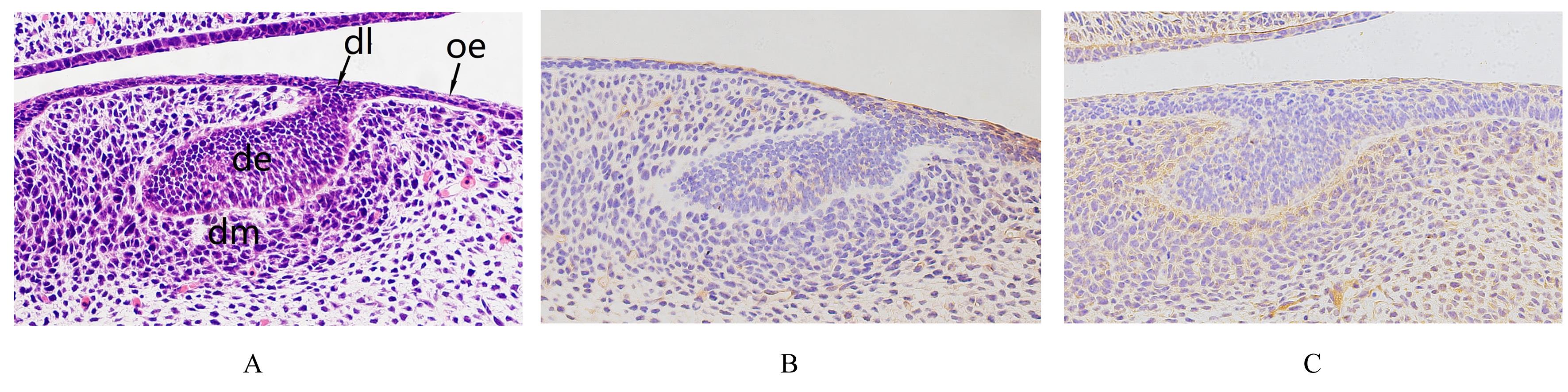
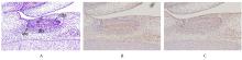
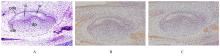
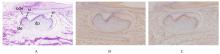
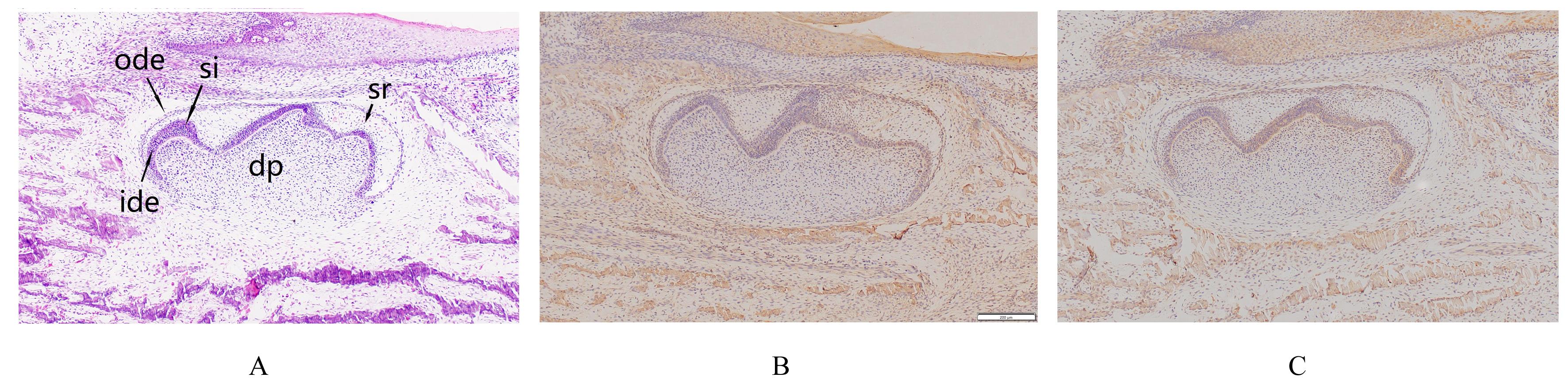
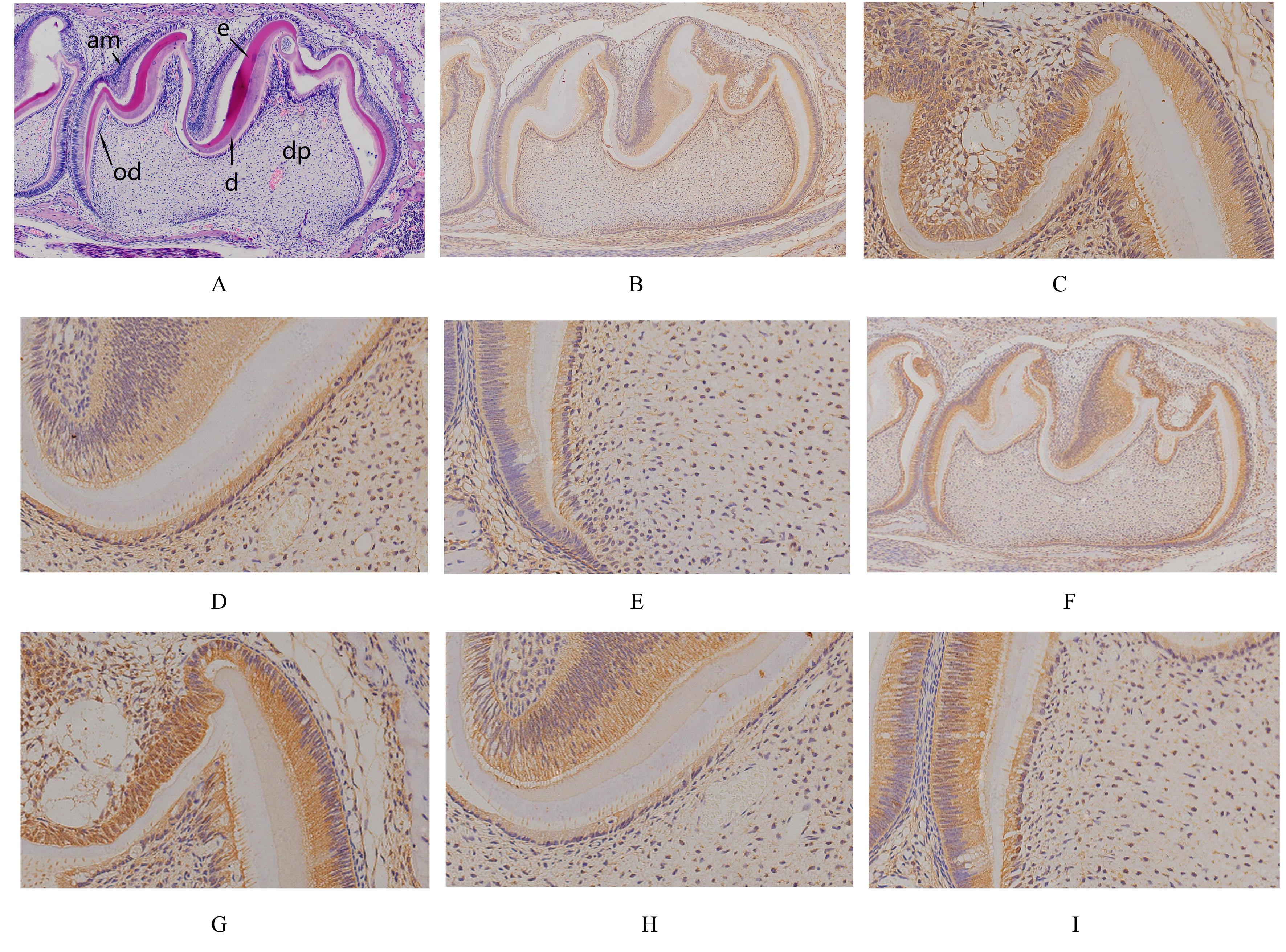
  • Received:2021-10-29 Online:2022-07-28 Published:2022-07-26
  • Contact: Ce SHI,Hong LIU E-mail:ceshi@jlu.edu.cn;l_h99@jlu.edu.cn

摘要: 目的

研究小鼠磨牙发育不同阶段牙胚组织中整合素β1和层黏连蛋白受体1(LAMR1)的表达模式,探讨二者在小鼠磨牙牙胚组织发育过程中可能的作用机制及其意义。

方法

取胚胎期第13.5天(E13.5)、E14.5、E16.5、E18.5和出生后5 d(PN5)的小鼠,分别作为E13.5、E14.5、E16.5、E18.5和PN5组,每组5只,分离小鼠头部,固定、脱钙、脱水、包埋和切片,HE染色观察各阶段小鼠牙胚组织形态表现,免疫组织化学染色观察小鼠磨牙发育不同阶段牙胚组织中整合素β1和LAMR1蛋白的表达分布情况和表达水平。

结果

HE染色,E13.5组小鼠牙胚组织发育处于蕾状期,上皮细胞突入到外胚间充质中,形成上皮芽,状如花蕾;E14.5组小鼠牙胚组织发育处于帽状期,上皮芽体积增大,称为帽状期成釉器,成釉器周围外胚间充质细胞密度增加,形成牙乳头;E16.5组小鼠牙胚组织发育处于钟状早期,成釉器进一步长大,形似吊钟,初步具有牙尖形态;E18.5组小鼠牙胚组织发育处于钟状晚期,成釉器进一步发育,內釉上皮细胞向前成釉细胞分化,形态由立方状变为高柱状;PN5组小鼠牙冠发育完成,形成颈环、上皮根鞘和上皮隔等结构,成釉细胞和成牙本质细胞分化成熟,呈高柱状整齐排列,已有釉质和牙本质形成。免疫组织化学染色,整合素β1和LAMR1蛋白在小鼠磨牙发育不同阶段牙胚组织中的上皮和牙乳头均有表达,且在各部位的表达强度随细胞分化的进程整体呈逐渐增强的趋势。PN5组小鼠牙胚组织成釉细胞中整合素β1蛋白表达水平高于E14.5、E16.5和E18.5组小鼠牙胚组织内釉上皮细胞/前成釉细胞(P<0.05);PN5组小鼠牙胚组织成牙本质细胞中整合素β1蛋白表达水平高于E14.5、E16.5和E18.5组小鼠牙胚组织前成牙本质细胞(P<0.05);E16.5和E18.5组小鼠牙胚组织内釉上皮细胞/前成釉细胞中LAMR1蛋白表达水平高于E14.5组小鼠牙胚组织内釉上皮细胞(P<0.05),且低于PN5组小鼠牙胚组织的成釉细胞(P<0.05);E18.5组小鼠牙胚组织前成牙本质细胞中LAMR1蛋白表达水平高于E16.5组小鼠牙胚组织前成牙本质细胞(P<0.05),且低于PN5组小鼠牙胚组织成牙本质细胞(P<0.05)。PN5组小鼠牙胚组织牙尖处较为成熟的成牙本质细胞中整合素β1和LAMR1蛋白表达水平高于牙尖之间和牙颈部尚未完全成熟的成牙本质细胞(P<0.05)。

结论

整合素β1和LAMR1表达于小鼠磨牙发育不同阶段牙胚组织的基底膜、(前)成釉细胞和(前)成牙本质细胞中,二者可能参与细胞外基质信号的转导,并在成釉细胞和成牙本质细胞的分化和极性形成过程中起促进作用。

关键词: 整合素β1, 层黏连蛋白受体1, 成牙本质细胞, 成釉细胞, 牙胚发育

Abstract: Objective

To study the expression patterns of integrin β1 and laminin receptor 1 (LAMR1) in the mouse molar germs at different stages of molar development, and to explore the possible mechanism and significance of integrin β1 and LAMR1 in mouse molar germ development.

Methods

The mice on the embry on days 13.5 (E13.5), E14.5, E16.5, E18.5 and postnatal days 5 (PN5) were obtained, and used as E13.5,E14.5,E16.5E18.5,and PN5 groups,with 5 mice in each group;their heads were separated, immobilized, decalcified, dehydrated, embedded and sliced, and the morphology of tooth embryo tissue at different stages was observed by HE staining. The expression levels of integrin β1 and LAMR1 in the molar germ tissue at different stages of mouse molar development were observed by immunohistochemical staining.

Results

The HE staining results showed that the tooth germ tissue development of the mice in E13.5 group corresponded to the bud stage, the epithelial cells protruded into the ectodermal mesenchyma and formed epithelial buds like flower buds;the tooth germ tissue development of the mice in E14.5 group corresponded to the cap stage, the epithelial bud volume was increased, which was called the enamel organ at the cap stage, and the density of ectomesenchymal cells around the enamel organ was increased, forming the dental papilla; the tooth germ tissue development of the mice in E16.5 group corresponded to the bell shaped early stage, the enamel organ further grew, resembled a hanging bell, and initially had the shape of the cusp; the tooth germ tissue development of the mice in E18.5 group corresponded to the late bell shaped stage, the ameloblast further developed, the inner ameloblast differentiated forward, and the shape changed from cubic to high columnar; the tooth crown of the mice in PN5 group developed into cervical loop, epithelial root sheath, epithelial septum and other structures, the ameloblasts and odontoblasts differentiated and matured and arranged neatly in high columnar shape, and enamel and dentin were formed. The results of immunohistochemical staining showed that integrin β1 and LAMR1 were expressed in the epithelium and dental papilla of mouse molars at different stages of molar development, and the expression intensities of integrin β1 and LAMR1 in different parts gradually were increased with the process of cell differentiation. The expression levels of integrin β1 protein in the ameloblasts of the mice in PN5 group was higher than those in the inner ameloblasts / preameloblasts in germ tissue of the mice in E14.5, E16.5 and E18.5 groups(P<0.05); the expression levels of integrin β1 protein in the odontoblasts in tooth germ tissue of the mice in PN5 group were higher than those in the preodontoblasts in dental germ of the mice in E14.5, E16.5 and E18.5 groups(P<0.05). The expression levels of LAMR1 protein in the inner ameloblasts / preameloblasts in E16.5 and E18.5 groups were higher than that in the inner ameloblasts in E14.5 group(P<0.05), and were lower than that in the ameloblasts in dental germ of the mice in PN5 group(P<0.05); the expression level of LAMR1 protein in the preodontoblasts in E18.5 group was higher than that in the preodontoblasts in E16.5 group(P<0.05), and was lower than that in the odontoblasts in dental germ of the mice in PN5 group(P<0.05). In PN5 group, the expression levels of integrin β1 and LAMR1 proteins in the mature odontoblasts at the cusp were higher than those in the immature odontoblasts between the apical and the neck (P<0.05).

Conclusion

Integrin β1 and LAMR1 are expressed in the basement membranes, (pre)ameloblasts and (pre)odontoblasts during molars development in the mice. They may be involved in signal transduction of extracellular matrix and play a promoting role in the differentiation and polarity formation of the ameloblasts and odontoblasts.

Key words: Integrin β1, LAMR1, Odontoblasts, Ameloblasts, Tooth germ development

中图分类号: 

  • R780.2