吉林大学学报(医学版) ›› 2022, Vol. 48 ›› Issue (4): 905-914.doi: 10.13481/j.1671-587X.20220409

• 基础研究 • 上一篇    下一篇

丹参酮ⅡA对缺氧缺血性脑病新生大鼠海马组织中miR-132表达的调节作用及其机制

张洋(),陈美月,崔莹,刘娜   

  1. 吉林大学第一医院二部儿科,吉林 长春 130031
  • 收稿日期:2021-10-17 出版日期:2022-07-28 发布日期:2022-07-26
  • 通讯作者: 张洋 E-mail:513396998@qq.com
  • 作者简介:张 洋(1990-),女,吉林省长春市人,主治医师,医学硕士,主要从事新生儿缺氧缺血性脑病方面的研究。
  • 基金资助:
    吉林省科技厅卫生科研人才计划项目(2019SCZ045)

Effects of tanshinone ⅡA on miR-132 expression in hippocampus tissue of neonatal rats with hypoxic-ischemic encephalopathy and its mechanism

Yang ZHANG(),Meiyue CHEN,Ying CUI,Na LIU   

  1. Department of Pediatrics,Second Department of First Hospital,Jilin University,Changchun 130031,China
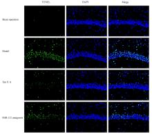
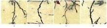
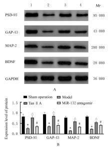
  • Received:2021-10-17 Online:2022-07-28 Published:2022-07-26
  • Contact: Yang ZHANG E-mail:513396998@qq.com

摘要: 目的

探讨丹参酮 ⅡA(Tan ⅡA)对新生大鼠缺氧缺血性脑病的干预作用,并阐明其作用机制。

方法

40只SD大鼠随机分为假手术组、模型组、Tan ⅡA组和miR-132抑制剂(miR-132 antagomir)组,每组10只。大鼠双重结扎颈总动脉并放入缺氧舱建立缺血缺氧性脑病模型。Tan ⅡA组大鼠造模后腹腔注射Tan ⅡA注射液(24 mg·kg-1),双侧脑室注射2 μg miR-132阴性对照质粒(NC);假手术组和模型组大鼠腹腔注射等量生理盐水;miR-132 antagomir组大鼠造模后腹腔注射Tan ⅡA注射液(24 mg·kg-1),双侧脑室注射2 μg miR-132 antagomir;各组大鼠连续注射7 d。实时荧光定量PCR(RT-qPCR)法和荧光原位杂交检测各组大鼠海马组织中miR-132表达水平和表达强度,采用改良神经功能缺失评分评价各组大鼠神经功能,HE染色观察各组大鼠海马组织病理形态表现, TUNEL法检测各组大鼠海马组织中神经元细胞凋亡率,酶联免疫吸附测定(ELISA)法检测各组大鼠海马组织中单胺类神经递质水平,高尔基染色检测各组大鼠海马组织中树突棘密度,Western blotting法检测各组大鼠海马组织中突触后致密物95(PSD-95)、生长相关蛋白43(GAP-43)、微管相关蛋白2(MAP-2)和脑源性神经营养因子(BDNF)蛋白表达水平。

结果

与假手术组比较,模型组大鼠海马组织中miR-132表达水平和表达强度明显降低(P<0.05);与模型组比较,Tan ⅡA组大鼠海马组织中miR-132表达水平和表达强度明显升高(P<0.05);与Tan ⅡA组比较,miR-132 antagomir组大鼠海马组织中miR-132表达水平和表达强度明显降低(P<0.05)。与假手术组比较,模型组大鼠神经功能缺失评分明显升高(P<0.05);与模型组比较,Tan ⅡA组大鼠神经功能缺失评分明显降低(P<0.05);与Tan ⅡA组比较,miR-132 antagomir组大鼠神经功能缺失评分明显升高(P<0.05)。HE染色,假手术组大鼠海马组织无损伤;与假手术组比较,模型组大鼠海马组织损伤严重;与模型组比较,Tan ⅡA组大鼠海马组织损伤明显改善;与Tan ⅡA组比较,miR-132 antagomir组大鼠海马组织损伤严重。与假手术组比较,模型组大鼠海马组织神经元凋亡率明显升高(P<0.05);与模型组比较,Tan ⅡA组大鼠海马组织神经元凋亡率明显降低(P<0.05);与Tan ⅡA组比较,miR-132 antagomir组大鼠海马组织神经元凋亡率明显升高(P<0.05)。与假手术组比较,模型组大鼠海马组织中去甲肾上腺素、多巴胺和5-羟色胺水平明显降低(P<0.05);与模型组比较,Tan ⅡA组大鼠海马组织中去甲肾上腺素、多巴胺和5-羟色胺水平明显升高(P<0.05);与Tan ⅡA组比较,miR-132 antagomir组大鼠海马组织中去甲肾上腺素、多巴胺和5-羟色胺水平明显降低(P<0.05)。与假手术组比较,模型组大鼠海马组织中树突棘密度明显降低(P<0.05);与模型组比较,Tan ⅡA组大鼠海马组织中树突棘密度明显升高(P<0.05);与Tan ⅡA组比较,miR-132 antagomir组大鼠海马组织中树突棘密度明显降低(P<0.05)。与假手术组比较,模型组大鼠海马组织中PSD-95、GAP-43、MAP-2和BDNF蛋白表达水平明显降低(P<0.05);与模型组比较,Tan ⅡA组大鼠海马组织中PSD-95、GAP-43、MAP-2和BDNF蛋白表达水平明显升高(P<0.05);与Tan ⅡA组比较,miR-132 antagomir组大鼠海马组织中PSD-95、GAP-43、MAP-2和BDNF蛋白表达水平明显降低(P<0.05)。

结论

Tan ⅡA可上调缺氧缺血性脑病新生大鼠海马组织中miR-132表达,改善新生大鼠神经功能和海马组织病理损伤,其机制可能与抑制海马神经元凋亡以及增加海马神经递质水平、树突棘密度、突触前蛋白、MAP-2和BDNF蛋白表达有关。

关键词: 丹参酮ⅡA磺酸钠, 缺氧缺血性脑病, 微小RNA-132, 树突棘密度, 突触前蛋白

Abstract: Objective

To investigate the intervention effect of tanshinone ⅡA(Tan ⅡA) in the neonatal rats with hypoxic-ischemic encephalopathy, and to clarify its mechanism.

Methods

Forty SD rats were randomly divided into sham operation group, model group, Tan ⅡA group and miR-132 antagomir group, with 10 rats in each group. The rat models of hypoxic-ischemic encephalopathy were established by double ligation of the common carotid artery and being put into anoxic chamber. The rats in TanⅡA group were intrapeitoneally injected with Tan ⅡA injection (24 mg·kg-1), and 2 μg miR-132 negative control (NC) was injected into the bilateral ventricles. The rats in sham operation group and model group were intraperitoneally injected with the same amount of normal saline. After modeling, the rats in miR-132 antagomir group were intraperitoneally injected with tanshinone ⅡA injection (24 mg·kg-1), and 2 μg miR-132 antagomir was injected into the bilateral ventricles; the tars in various groups were injected for 7 d. The expression levels and fluorescence intensities of miR-132 in hippocampus tissue of the rats in various groups were detected by real-time fluorescence quantitative PCR(RT-qPCR) and fluorescence in situ hybridization. Modified neurological deficit score was used to evaluate the neural function of the rats in various groups. HE staining was used to observe the pathomorphology of hippocampus tissue of the rats in various groups. TUNEL assay was used to detect the apoptotic rates of neurons in hippocampus tissue of the rats in various groups. The levels of monoamine neurotransmitters in hippocampus tissue of the rats in various groups were detected by enzyme-linked immunosorbent assay (ELISA). The densities of dendritic spines in hippocampus tissues of the rats in various groups were detected by Golgi staining. The protein expression levels of postsynaptic density-95(PSD-95),growth associated protein-43(GAP-43),microtubule-associated protein-2 (MAP)-2 and brain-derived neurotrophic factor (BDNF) in hippocampus tissue of the rats in various groups were detected by Western blotting method.

Results

Compared with sham operation group, the expression level of miR-132 in the hippocampus tissue and the fluorescence intensity of the rats in model group were significantly decreased (P<0.05); compared with model group, the expression level of miR-132 in hippocampus tissue and the fluorescence intensity of the rats in Tan ⅡA group were significantly increased (P<0.05); compared with Tan ⅡA group, the miR-132 expression level in hippocampus tissue and the fluorescence intensity of the rats in miR-132 antagomir group were significantly decreased (P<0.05). Compared with sham operation group, neurological deficit score of the rats in model group were significantly increased (P<0.05); compared with model group, the neurological deficit score of the rats in Tan ⅡA group was significantly decreased (P<0.05); compared with Tan ⅡA group, the neurological deficit score of the rats in miR-132 antagomir group was significantly increased (P<0.05).The HE staining results showed that the hippocampus tissue of the rats in sham operation group was not damaged;compared with sham operation group, the hippocampus tissue of the rats in model group was seriously damaged;compared with model group, the damage of hippocampus tissue of the rats in Tan ⅡA group was improved;compared with Tan ⅡA group, the hippocampus tissue of the rats in miR-132 antagomir group was seriously damaged. Compared with sham operation group, the apoptotic rate of cells of hippocampal tissue of the rats in model group was significantly increased (P<0.05); compared with model group, the apoptotic rate of cells of hippocampus tissue in the Tan ⅡA group was significantly decreased (P<0.05); compared with Tan ⅡA group, the apoptotic rate of cells of hippocampus tissue in miR-132 antagomir group was significantly increased (P<0.05). Compared with sham operation group, the levels of norepinephrine, dopamine and 5-hydroxytryptamine in hippocampus tissue of the rats in model group were significantly decreased (P<0.05); compared with model group, the levels of norepinephrine, dopamine and 5-hydroxytryptamine in hippocampus tissue of the rats in Tan ⅡA group were significantly increased (P<0.05); compared with Tan ⅡA group, the levels of norepinephrine, dopamine and 5-hydroxytryptamine in hippocampus tissue of the rats in miR-132 antagomir group were significantly decreased (P<0.05). Compared with sham operation group, the density of dendritic spines in hippocampus tissue of the rats in model group was significantly decreased (P<0.05); compared with model group, the density of dendritic spines in hippocampus tissue of the rats in Tan ⅡA group was significantly increased (P<0.05); compared with Tan ⅡA group, the density of dendritic spines in hippocampus tissue of the rats in miR-132 antagomir group was significantly decreased (P<0.05). Compared with sham operation group, the expression levels of PSD-95, GAP-43, MAP-2 and BDNF proteins in hippocampus tissue of the rats in model group were significantly decreased (P<0.05); compared with model group, the expression levels of PSD-95, GAP-43, MAP-2 and BDNF proteins in hippocampus tissue of the rats in Tan ⅡA group were significantly increased (P<0.05); compared with Tan ⅡA group, the expression levels of PSD-95, GAP-43, MAP-2 and BDNF proteins in hippocampus tissue of the rats in miR-132 antagomir group were significantly decreased (P<0.05).

Conclusion

Tan ⅡA can up-regulate the expression of miR-132 in the hippocampus tissue of the neonatal rats with hypoxic-ischemic encephalopathy, and improve the neurological function and pathological damage of hippocampus tissue; its mechanism may be related to inhibiting the apoptosis of hippocampal neurons, increasing the level of neurotransmitters, the density of dendrite spines, preprotrusional proteins and the expressions of MAP-2 and BDNF proteins.

Key words: Tanshinone ⅡA sulfonate sodium, Hypoxic-ischemic encephalopathy, MicroRNA-132, Dendritic spine density, Preprotrusional proteins

中图分类号: 

  • R-332