吉林大学学报(医学版) ›› 2023, Vol. 49 ›› Issue (3): 647-655.doi: 10.13481/j.1671-587X.20230313

• 基础研究 • 上一篇    下一篇

PAX3基因沉默对P19细胞向心肌样细胞分化的影响及其机制

万朝辉1,曾良1,李晶3,雷长城3()   

  1. 1.南华大学衡阳医学院附属第二医院急诊科,湖南 衡阳 421001
    2.南华大学衡阳医学院附属第三医院急诊科,湖南 衡阳 421900
    3.南华大学衡阳医学院附属第二医院心血管内科,湖南 衡阳 421001
  • 收稿日期:2022-06-20 出版日期:2023-05-28 发布日期:2023-06-20
  • 通讯作者: 雷长城 E-mail:qtfcb6547@163.com
  • 作者简介:万朝辉(1978-),男,湖南省衡阳市人,主治医师,医学硕士,主要从事急危重症相关诊治方面的研究。
  • 基金资助:
    湖南省教育厅科学研究项目(19C1618);湖南省卫健委一般资助项目(20200881);湖南省衡阳市科技局指导性项目(2020jh042800)

Effect of PAX3 gene silencing on differentiation of P19 cells into cardiomyocyte-like cells and its mechanism

Zhaohui WAN1,Liang ZENG1,Jin LI3,Changcheng LEI3()   

  1. 1.Department of Emergency,Second Affiliated Hospital,Hengyang Medical School,University of South China,Hengyang 421001,China
    2.Department of Emergency,Third Affiliated Hospital,Hengyang Medical School,University of South China,Hengyang 421001,China
    3.Department of Cardiovascular Medicine,Second Affiliated Hospital,Hengyang Medical School,University of South China,Hengyang 421001,China
  • Received:2022-06-20 Online:2023-05-28 Published:2023-06-20
  • Contact: Changcheng LEI E-mail:qtfcb6547@163.com

摘要:

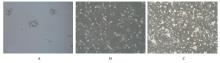
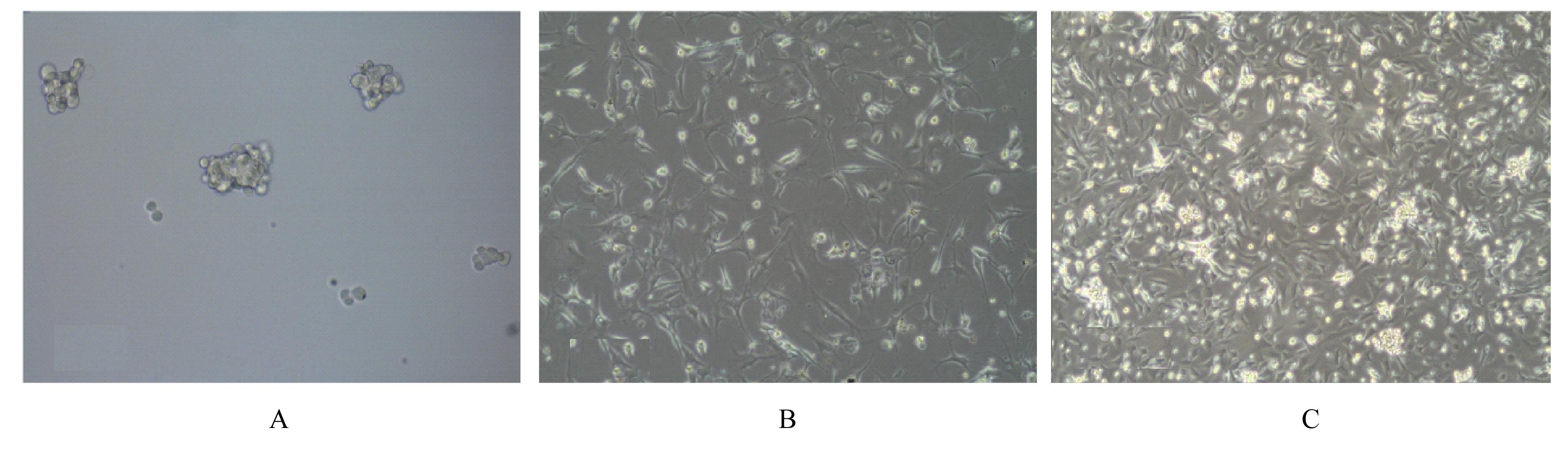
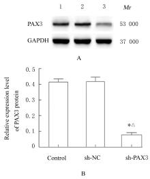
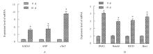
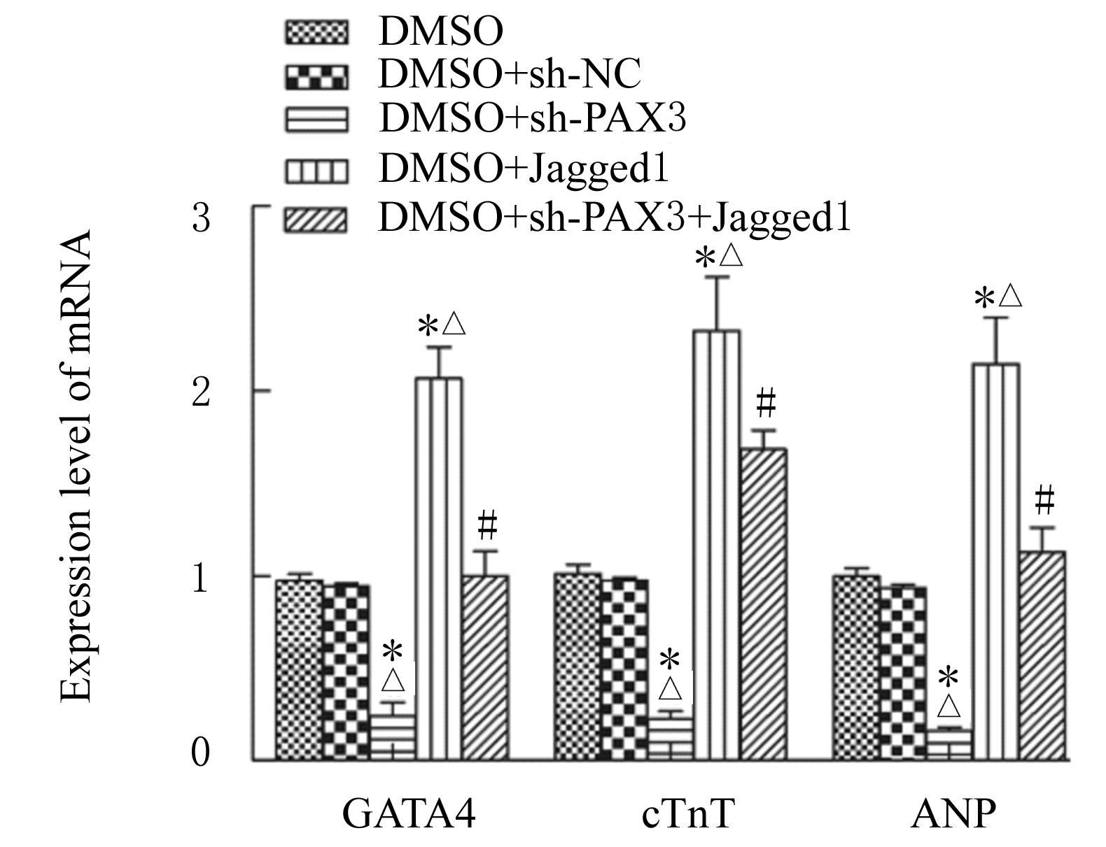
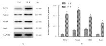
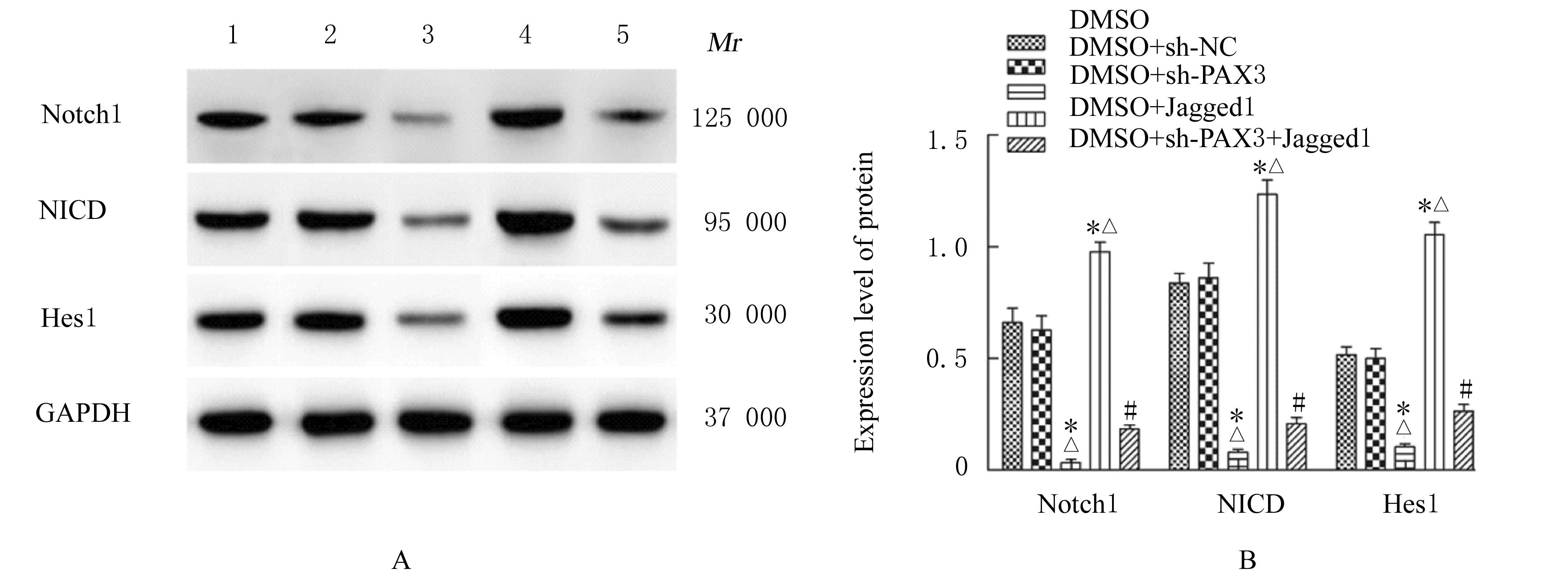
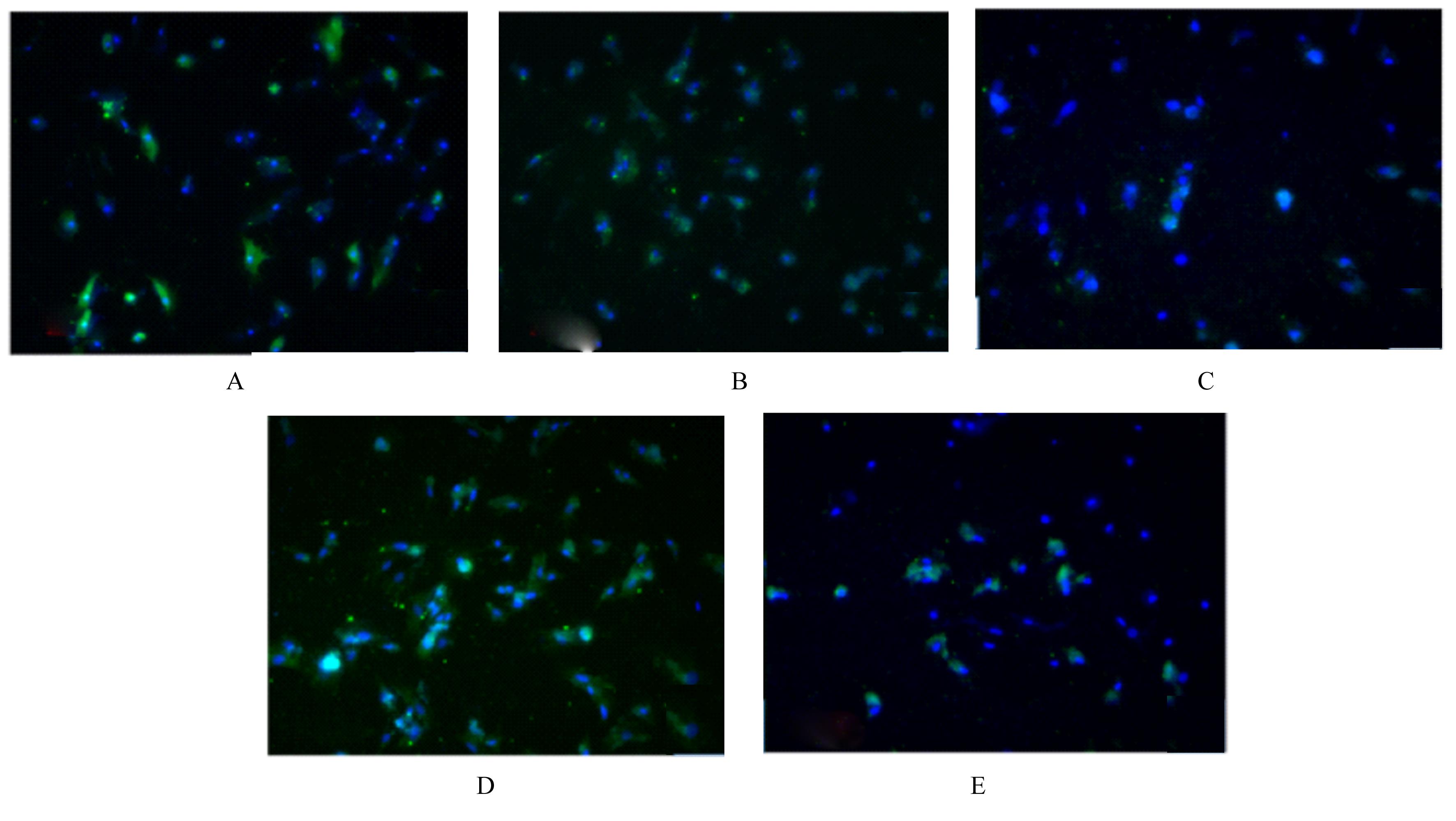
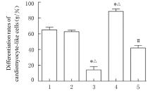
目的 探讨配对盒转录因子3(PAX3)基因沉默对P19细胞向心肌样细胞分化的影响,并阐明其可能作用机制。 方法 采用二甲基亚砜(DMSO)诱导P19细胞向心肌样细胞分化,观察诱导分化过程中细胞形态表现,并收集诱导分化第0天(0 d组)和第10天(10 d组)的细胞。采用sh-PAX3慢病毒感染P19细胞,将细胞分为对照组、sh-NC组(阴性对照)和sh-PAX3组(PAX3干扰),实时荧光定量PCR(RT-qPCR)法和Western blotting法检测PAX3基因沉默的P19细胞构建情况。采用Notch信号激动剂Jagged1处理慢病毒感染后的P19细胞,并采用DMSO诱导分化10 d,将细胞分为DMSO组、DMSO+sh-NC组、DMSO+sh-PAX3组、DMSO+Jagged1组和DMSO+sh-PAX3+Jagged1组。RT-qPCR法检测各组细胞中GATA结合蛋白4(GATA4)、心房利钠尿多肽(ANP)、心肌肌钙蛋白(cTn)T、PAX3、Notch1、Notch胞内结构域(NICD)及Hes 家族bHLH 转录因子1(Hes1)mRNA表达水平。Western blotting法检测各组细胞中PAX3、Notch1、NICD和Hes1蛋白表达水平。免疫荧光染色法检测各组细胞中cTnI阳性表达情况和心肌样细胞分化率。 结果 诱导分化第10天,可观察到自发性节律收缩的心肌样细胞簇。RT-qPCR和Western blotting法检测,慢病毒感染后,与对照组和sh-NC组比较,sh-PAX3组P19细胞中PAX3 mRNA和蛋白表达水平均降低(P<0.05),表明PAX3基因沉默的P19细胞构建成功。RT-qPCR法检测,与0 d组比较,10 d组细胞中GATA4、ANP和cTnT mRNA表达水平均升高(P<0.05),PAX3、Notch1、NICD和Hes1 mRNA表达水平均升高(P<0.05);与DMSO组和DMSO+sh-NC组比较,DMSO+sh-PAX3组细胞中GATA4、ANP和cTnT mRNA表达水平均降低(P<0.05),DMSO+Jagged1组细胞中GATA4、ANP和cTnT mRNA表达水平均升高(P<0.05);与DMSO+sh-PAX3组比较,DMSO+sh-PAX3+Jagged1组细胞中GATA4、ANP和cTnT mRNA表达水平均升高(P<0.05)。Western blotting法检测,与0 d组比较,10 d组细胞中PAX3、Notch1、NICD和Hes1蛋白表达水平均升高(P<0.05);与DMSO组和DMSO+sh-NC组比较,DMSO+sh-PAX3组细胞中Notch1、NICD和Hes1蛋白表达水平均降低(P<0.05),DMSO+Jagged1组细胞中Notch1、NICD和Hes1蛋白表达水平均升高(P<0.05);与DMSO+sh-PAX3组比较,DMSO+sh-PAX3+Jagged1组细胞中Notch1、NICD和Hes1蛋白表达水平均升高(P<0.05)。免疫荧光法检测,慢病毒感染后,各组细胞中均有cTnI蛋白阳性表达,与DMSO组和DMSO+sh-NC组比较,DMSO+ sh-PAX3组细胞中cTnI蛋白阳性表达减少,心肌样细胞分化率降低(P<0.05);与DMSO组和DMSO+sh-NC组比较,DMSO+Jagged1组细胞中cTnI蛋白阳性表达增加,心肌样细胞分化率升高(P<0.05);与DMSO+sh-PAX3组比较,DMSO+sh-PAX3+ Jagged1组细胞中cTnI蛋白阳性表达增加,心肌样细胞分化率升高(P<0.05)。 结论 PAX3基因沉默可抑制P19细胞向心肌样细胞分化,其机制可能是通过降低PAX3基因表达抑制Notch信号通路活化实现的。

关键词: P19细胞, 配对盒转录因子3, Notch信号通路, 心肌样细胞

Abstract:

Objective To investigate the effect of paired box transcription factor 3 (PAX3) gene silencing on the differentiation of the P19 cells into the cardiomyocyte-like cells,and to clarify its possible mechanism. Methods Dimethylene maple (DMSO) was used to induce the differentiation of the P19 cells into cardiomyocyte-like cells, and the morphology of the cells during the induction process was observed. The cells were collected on the 0th day (0 d group) and 10th day (10 d group) after induction and differentiation.The P19 cells were infected with sh-PAX3 Lentivirus.The cells were divided into control group,sh-NC group(negative control group)and sh-PAX3 (PAX3 interference).Real-time fluorescence quantitative PCR (RT-qPCR) and Western blotting methods were used to verify the positive expression of cTnI in P19 cells silenced with PAX3 gene. The P19 cells infected with lentivirus were treated with Notch signal agonist Jagged1 and were induced and differentiated with DMSO for 10 d. The cells were divided into DMSO group, DMSO+sh-NC group, DMSO+sh-PAX3 group, DMSO+Jagged1 group and DMSO+sh-PAX3+Jagged1 group. RT-qPCR method was used to detect the expression levels of GATA binding protein 4 (GATA4), atrial natriuretic peptide (ANP), cardiac troponin (cTn)T,PAX3,Notch1, Notch intracellular domain (NICD), and Hes family bHLH transcription factor 1 (Hes1) mRNA in the cells in various groups; Western blotting method was used to detect the expression levels of PAX3,Notch1, NICD, and Hes1 proteins in the cells in various groups; immunofluorescence assay was used to detect the positive expression of cTnI in the cells the differentiation rates of the cardiomyocyte-like cells in various groups. Results On the 10th day, after induction and differentiation,the cluster of cardiomyocyte-like cells with spontaneous rhythmic contraction could be observed.The RT-qPCR and Western blotting results showed that after lentivirus infection, compared with control group and sh-NC group, the expression levels of PAX3 mRNA and protein in the P19 cells in sh-PAX3 group were decreased (P<0.05), indicating that the P19 cells silenced with PAX3 gene were successfully constructed. The RT-qPCR results showed that compared with 0 d group, the expression levels of GATA4, ANP, and cTnT mRNA in the cells in 10 d group were increased (P<0.05), while the expression levels of PAX3,Notch1, NICD, and Hes1 mRNA were increased (P<0.05); compared with DMSO group and DMSO+sh-NC group, the expression levels of GATA4, ANP, and cTnT mRNA in the cells in DMSO+sh-PAX3 group were decreased (P<0.05), while the expression levels of GATA4, ANP, and cTnT mRNA in the cells in DMSO+Jagged1 group were increased (P<0.05); compared with DMSO+sh-PAX3 group, the expression levels of GATA4, ANP, and cTnT mRNA in the cells in DMSO+sh-PAX3+Jagged1 group were increased (P<0.05). The Western blotting results showed that compared with 0 d group, the expression levels of PAX3, Notch1, NICD, and Hes1 proteins in the cells in 10 d group were increased (P<0.05); compared with DMSO group and DMSO+sh-NC group, the expression levels of Notch1, NICD, and Hes1 proteins in the cells in DMSO+sh-PAX3 group were decreased (P<0.05),while the expression levels of Notch1, NICD, and Hes1 proteins in the cells in DMSO+Jagged1 group were increased (P<0.05); compared with DMSO+sh-PAX3 group, the expression levels of Notch1, NICD, and Hes1 proteins in the cells in DMSO+sh-PAX3+Jagged1 group were increased (P<0.05).The immunofluorescence results showed that after lentivirus infection, the positive expression of cTnI protein was found in the cells in various groups. Compared with DMSO group and DMSO+sh-NC group, the positive expression of cTnI protein in the cells in DMSO+sh-PAX3 group was decreased, and the differentiation rate of the cardiomyocyte-like cells was decreased (P<0.05); compared with DMSO group and DMSO+sh-NC group, the positive expression of cTnI protein in the cells in DMSO+Jagged1 group was increased,and the differentiation rate of the cardiomyocyte-like cells was increased (P<0.05); compared with DMSO+sh-PAX3 group, the positive expression of cTnI protein in the cells in DMSO+sh-PAX3+Jagged1 group was increased,and the differentiation rate of the cardiomyocyte-like cells was increased (P<0.05). Conclusion PAX3 gene silencing may inhibit the differentiation of the P19 cells into the cardiomyocyte-like,and its mechanism may be realized by decreasing the PAX3 gene expression and regulating the Notch signaling pathway.

Key words: P19 cells, Paired box transcription factor 3, Notch signaling pathway, Cardiomyocyte-like cells

中图分类号: 

  • Q21