吉林大学学报(医学版) ›› 2023, Vol. 49 ›› Issue (6): 1473-1483.doi: 10.13481/j.1671-587X.20230610

• 基础研究 • 上一篇    下一篇

PPC/PBS引导骨再生生物膜的制备及其理化性能和生物学特征评价

石晓璐,田野,翟少博,刘洋,储顺礼()   

  1. 吉林大学口腔医院种植科,吉林 长春 130021
  • 收稿日期:2023-01-14 出版日期:2023-11-28 发布日期:2023-12-22
  • 通讯作者: 储顺礼 E-mail:chusl@jlu.edu.cn
  • 作者简介:石晓璐(1997-),女, 山东省菏泽市人,在读硕士研究生,主要从事口腔引导骨再生材料方面的研究。
  • 基金资助:
    吉林省科技厅省重点项目(20230203065SF);吉林省财政厅科技项目(JCSZ2019378-23)

Preparation of PPC/PBS-based guided bone regeneration membrane and evaluation of its physiochemical properties and biological characteristics

Xiaolu SHI,Ye TIAN,Shaobo ZHAI,Yang LIU,Shunli CHU()   

  1. Department of Prosthodontics,Stomatology Hospital,Jilin University,Changchun 130021,China
  • Received:2023-01-14 Online:2023-11-28 Published:2023-12-22
  • Contact: Shunli CHU E-mail:chusl@jlu.edu.cn

摘要:

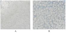
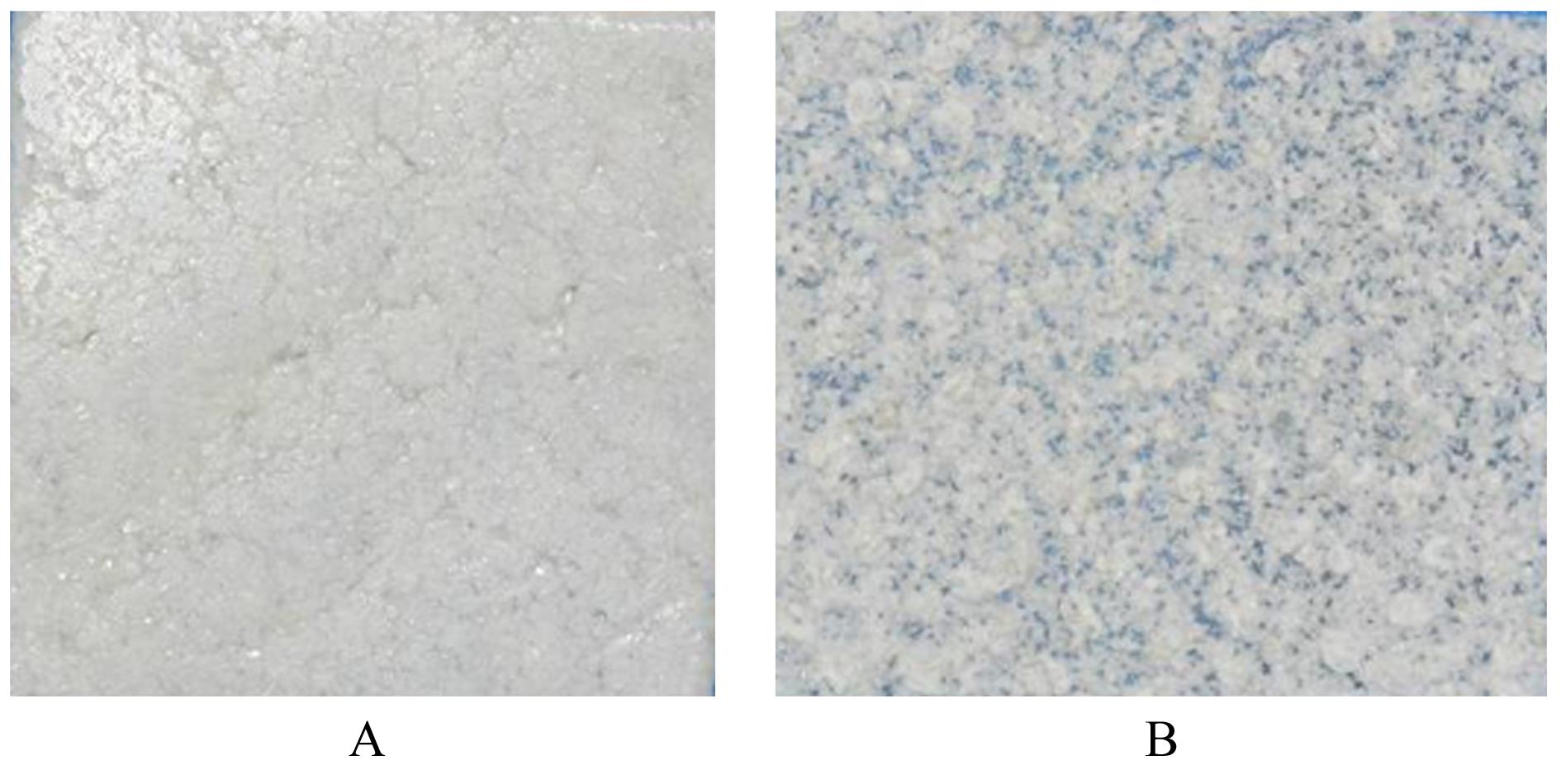
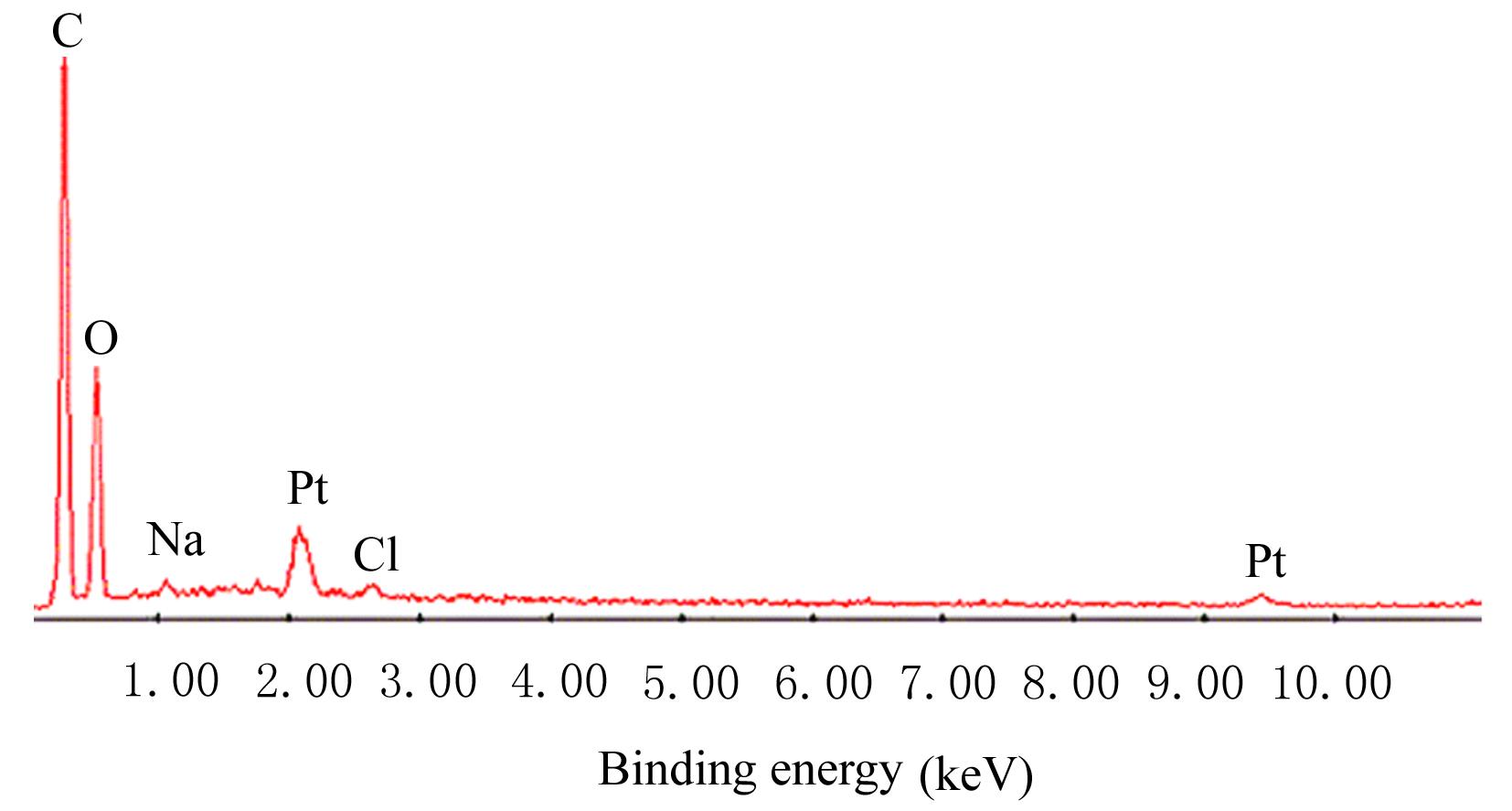
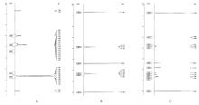
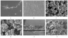
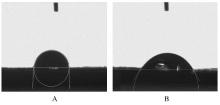
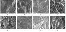
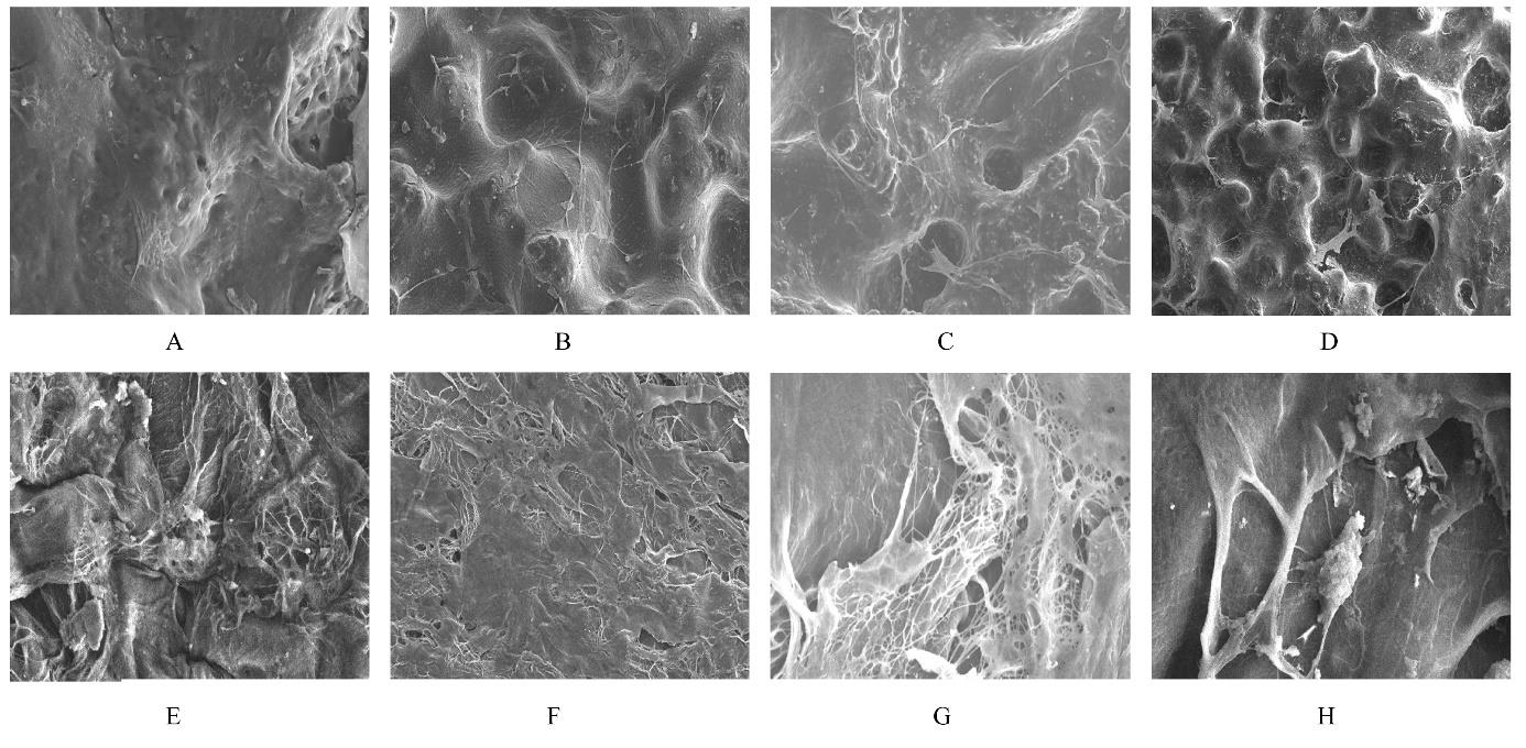
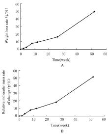
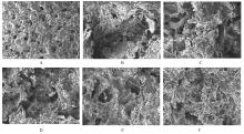
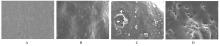
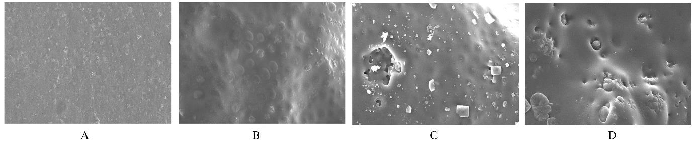
目的 制备与人体骨外膜结构相仿的聚碳酸1,2-丙二酯(PPC)/聚丁二酸丁二醇酯(PBS)生物膜,并评价其理化性能和生物学特征。 方法 采用盐析法制备PPC/PBS生物膜,核磁共振氢谱(1HRNM)观察PPC、PBS和 PPC/PBS的谱吸收峰,分析其共混前后化学结构变化。扫描电子显微镜(SEM)观察PPC/PBS生物膜超微形态表现,GPC凝膜渗透色谱仪检测其孔隙率、杨氏模量、断裂强度、断裂伸长率和静态接触角等理化性能。分离、培养和纯化原代SD大鼠成骨细胞,分为对照组(未加生物膜)、BME-10X胶原膜组和PPC/PBS生物膜组,SEM观察各组成骨细胞附着和生长情况,细胞计数法检测各组成骨细胞数量,碱性磷酸酶(ALP)试剂盒检测各组成骨细胞分化情况,兔背部肌肉降解实验检测PPC/PBS生物膜体内降解性能。 结果 PPC/PBS生物膜具有光滑面层和粗糙面层双层样结构,全层厚约为0.5 mm,平均孔径约为120 μm;孔隙率约为77.4%;杨氏模量约为38.1 MPa,断裂强度约为1.22 MPa,断裂伸长率约为7.4%;粗糙面接触角为85°,光滑面接触角为57°。SEM观察,培养1 d时,PPC/PBS生物膜表面附着的细胞较少;培养3、7和14 d时,可见大量成骨细胞附着于生物膜表面生长,细胞伸出伪足附着于生物膜上,胞体可呈悬空样分布于孔隙中。与对照组比较,培养1、3、7和14 d时,BME-10X胶原膜组和PPC/PBS复合膜组材料表面附着成骨细胞数量均减少(P<0.05)。与BME-10X胶原膜组比较,培养1、3、7和14 d时,PPC/PBS生物膜组材料表面附着成骨细胞数量均增加(P<0.05)。与对照组比较,培养1、3、7和14 d时,BME-10X胶原膜组和PPC/PBS生物膜组材料表面附着成骨细胞中ALP水平明显减少(P<0.01)。与BME-10X胶原膜组比较,培养1和3 d时,PPC/PBS生物膜组材料表面附着成骨细胞中ALP水平明显增加(P<0.01)。降解实验中,与降解0 周时比较,降解4 周时,PPC/PBS生物膜失重率和相对分子质量失重率均升高(P<0.05),断裂强度和断裂伸长率均明显减小(P<0.05或P<0.01)。自降解第2周开始,PPC/PBS生物膜粗糙面表面微孔样结构数逐渐增多,孔径为0~10 μm;至降解26周时,PPC/PBS生物膜粗糙面表面微孔样结构均匀分布;降解4周时,PPC/PBS生物膜光滑面表面出现脱屑样改变,但未见有微孔样结构;降解12周时,光滑面表面出现少量微孔样改变,微孔直径<5 μm;降解26周时,光滑面表面微孔样结构数增加,孔径<10 μm。 结论 PPC/PBS生物膜与人体骨外膜结构相似,呈双层样结构,亲水性好,光滑面层表面较为致密,粗糙面层孔隙率高,生物相容性好,且降解缓慢,是一种理想的引导再生生物膜。

关键词: 引导骨再生, 生物膜, 聚碳酸1,2-丙二酯, 聚丁二酸丁二醇酯, 骨缺损

Abstract:

Objective To prepare the polypropylene carbonate(PPC)/polybutylene succinate(PBS) biofilm that mimics the structure of human bone periosteum,and to evaluate its physicochemical properties and biological characteristics. Methods The PPC/PBS biofilm was prepared by salting-out method. 1H nuclear magnetic resonance spectra(1HNMR) was used to observe the spectral absorption peak of PPC, PBS, and PPC/PBS,and the chemical structure changes of PPC, PBS, and PPC/PBS were analyzed; the ultramorphology of the PPC/PBS biofilm was observed by scanning electron microscope (SEM); and the physicochemical properties, including porosity, Young’s modulus, fracture strength, fracture elongation, and static contact angle of the PPC/PBS biofilm were measured by GPC membrane permeation chromatography. The osteoblasts of the primary SD rat were isolated, cultured, and purified, and divided into control group (without biofilm), BME-10X collagen membrane group, and PPC/PBS biofilm group. SEM was used to observe the adhesion and growth of the osteoblasts in various groups; cell counting was performed to detect the number of osteoblasts;alkaline phosphatase (ALP) assay kit was used to the differentiation of the osteoblasts; rabbit muscle degradation experiment was used to evaluate the degradation performance of the PPC/PBS biofilm in vivo. Results The PPC/PBS biofilm had a dual-layer structure consisting of a smooth surface layer and a rough surface layer, with a total thickness of approximately 0.5 mm and an average pore size of about 120 μm;the porosity was approximately 77.4%, the Young’s modulus was approximately 38.1 MPa, the fracture strength was approximately 1.22 MPa, the fracture elongation was approximately 7.4%, and the contact angle on the rough surface was 85°, while the contact angle on the smooth surface was 57°. The SEM observation results showed that fewer cells adhered to the surface of the PPC/PBS biofilm 1 d after culture; 3, 7, and 14 d after culture, a large number of osteoblasts were observed to adhere and grow on the surface of the biofilm, with cell protrusions attached to the film and cell bodies distributed in the pores. Compared with control group, the numbers of osteoblasts attached to the surface of the materials in BME-10X collagen membrane group and PPC/PBS biofim group were decreased after cultured for 1, 3, 7, and 14 d (P<0.05). Compared with BME-10X collagen membrane group, the number of the osteoblasts attached to the surface of the materials in PPC/PBS biofilm group was increased after cultured for 1, 3, 7, and 14 d (P<0.05). Compared with control group, the ALP levels in the osteoblasts attached to the surface of the materials in BME-10X collagen membrane group and PPC/PBS biofilm group were significantly decreased after cultured for 1, 3, 7, and 14 d (P<0.01). Compared with BME-10X collagen membrane group, the ALP level in the osteoblasts attached to the surface of the materials in PPC/PBS biofilm group was significantly increased after cultured for 1 and 3 d (P<0.01). The degradation experiment results showed that compared with 0 week after degradation, the weight loss rate and weight loss rate of number-avarage molecular of the PPC/PBS biofilm were increased 4 weeks after degradation (P<0.05), while the fracture strength and fracture elongation were significantly decreased(P<0.05 or P<0.01). From the 2nd week after degradation,the number of microstructures on rough surface of the PPC/PBS biofilm was gradually increased, and the pore sizes was ranged from 0 to 10 μm; by 26 weeks after degradation,the number of micro-porous structures was evenly distributed on rough surface of the PPC/PBS biofilm; by 4 weeks after degradation, the smooth surface of the biofilm showed exfoliation changes, but no micro-porous structures were observed; by 12 weeks after degradation, a small number of micro-porous changes were observed on the smooth surface, and the pore diameters were less than 5 μm; by 26 weeks after degradation, the number of micro-porous structures on the smooth surface was increased,and the pore diameters were less than 10 μm. Conclusion The structure of PPC/PBS biofilm is similar to that of human bone outer membrane,with a double-layer structure,good hydrophilicity,dense smooth surface,high porosity of rough surface,good biocompatibility,and slow degradation;it is an ideal guided regeneration biofilm.

Key words: Guided bone regeneration, Biofilm, Poly propylene carbonate, Poly butylene succinate, Bone defect

中图分类号: 

  • R783.1