吉林大学学报(医学版) ›› 2023, Vol. 49 ›› Issue (6): 1604-1609.doi: 10.13481/j.1671-587X.20230626

• 临床医学 • 上一篇    下一篇

抗GAD65和抗GABABR双抗体阳性自身免疫性脑炎1例报告及文献复习

苏杭,李佳,曾慧宾,陈加俊()   

  1. 吉林大学中日联谊医院神经内科,吉林 长春 130033
  • 收稿日期:2023-02-10 出版日期:2023-11-28 发布日期:2023-12-22
  • 通讯作者: 陈加俊 E-mail:cjj@jlu.edu.cn
  • 作者简介:苏 杭(1997-),女,吉林省辽源市人,在读硕士研究生,主要从事神经系统疾病诊断和治疗方面的研究。
  • 基金资助:
    吉林省科技厅科研项目(20200201451JC);吉林省神经系统疾病精准医学诊疗中心项目(20200602045ZP)

Autoimmune encephalitis with positive anti-GAD65 and anti-GABABR double antibodies:One case report and literature review

Hang SU,Jia LI,Huibin ZENG,Jiajun CHEN()   

  1. Department of Neurology,China-Japan Union Hospital,Jilin University,Changchun 130033,China
  • Received:2023-02-10 Online:2023-11-28 Published:2023-12-22
  • Contact: Jiajun CHEN E-mail:cjj@jlu.edu.cn

摘要:

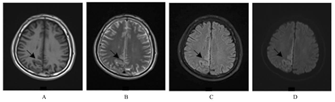
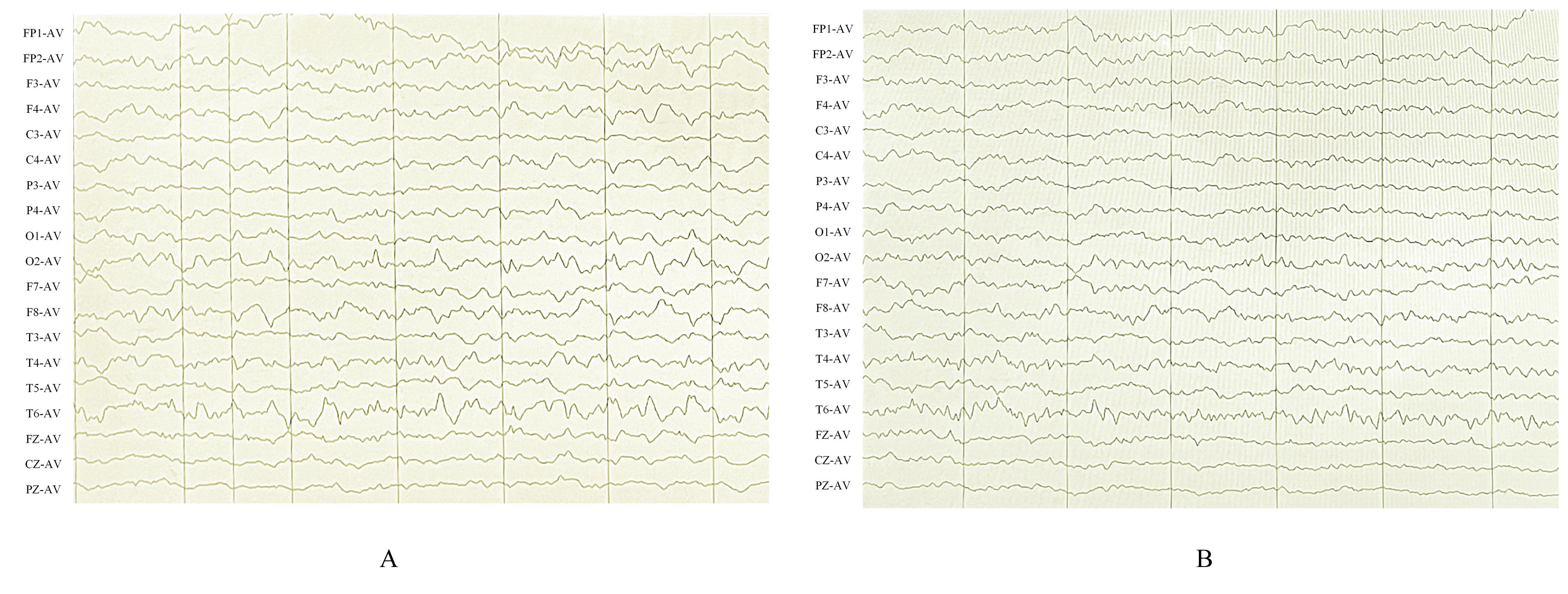
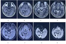
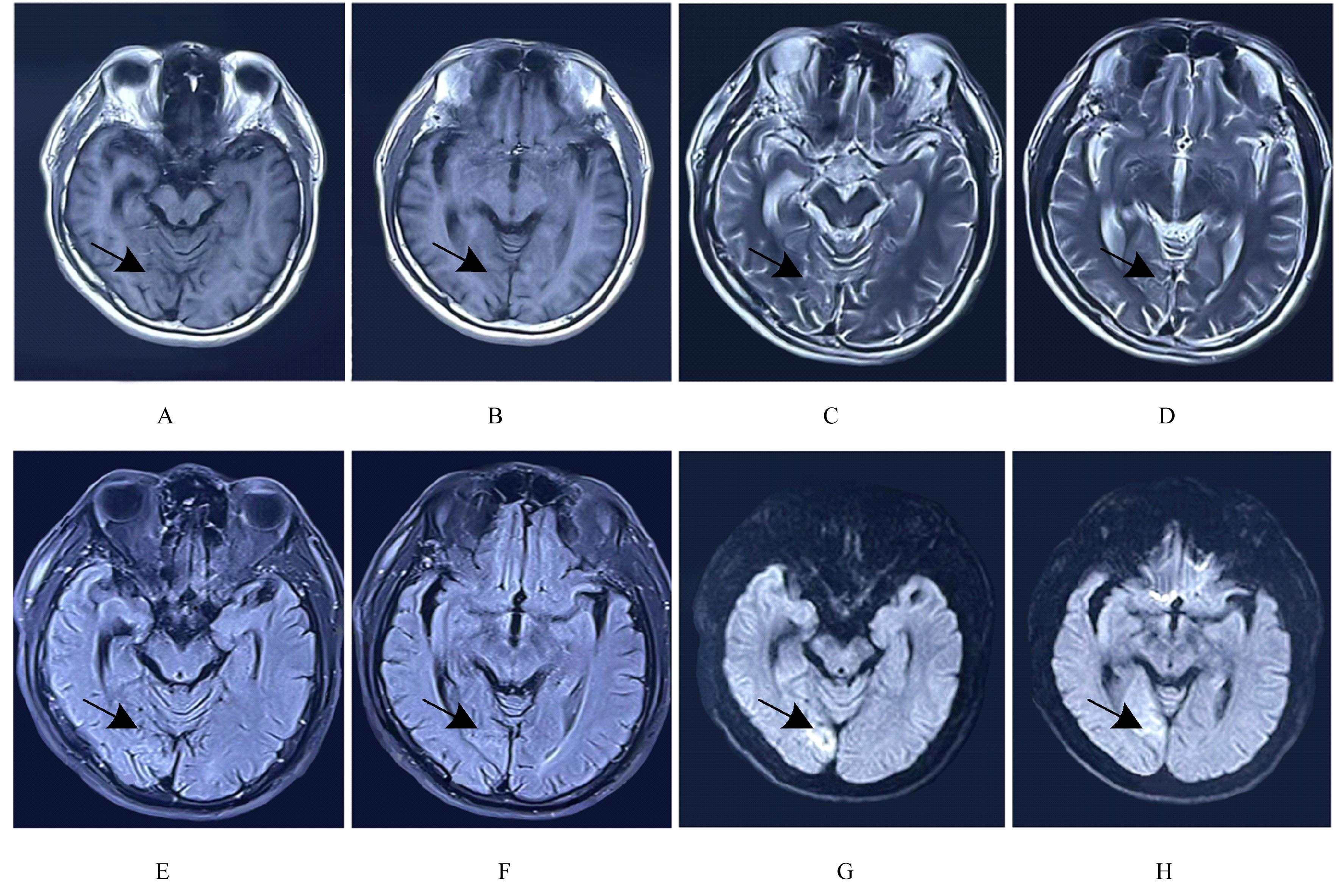
目的 分析脑脊液(CSF)双抗体阳性自身免疫性脑炎(AE)患者的临床表现和诊疗过程,为该类患者的诊断和治疗提供参考。 方法 回顾性分析1例CSF中抗谷氨酸脱羧酶(GAD)65和抗γ-氨基丁酸B型受体(GABABR)双抗体阳性AE患者的临床表现、头部核磁共振成像(MRI)、脑电图(EEG)、CSF特征及预后,并结合文献进行复习。 结果 患者,男性,47岁,亚急性起病,病情逐渐加重,主要表现为头痛和发作性抽搐,意识模糊,头部MRI提示病灶位于大脑镰右侧额顶枕叶,CSF检测抗GAD65和抗GABABR双抗体阳性,EEG有异常的尖波及慢波,诊断为AE,患者经抗炎等对症治疗逐渐好转并出院,继续口服激素治疗,5个月后再次复发,急性起病,表现为抽搐伴口角流涎,头部MRI提示右侧颞叶异常高信号影,行糖皮质激素治疗后患者好转。 结论 CSF双抗体阳性AE患者复发可能性较大,激素抗炎治疗有效,在颅内病变定位于额顶枕叶时需考虑出现抽搐等症状,并及早完善EEG等检查。

关键词: 谷氨酸脱羧酶65, γ-氨基丁酸B型受体, 自身免疫性脑炎, 脑脊液, 脑电图

Abstract:

Objective: To analyze the clinical performance and diagnosis process of the autoimmune encephalitis (AE) patients with positive double antibodies in the cerebrospinal fluid(CSF), and to provide the references for the diagnosis and treatment of such patients. Methods The clinical manifestations, magnetic resonance imaging (MRI) of the head, electroencephalogram (EEG), CSF characteristics, and prognosis of one patient with AE positive for anti-glutamic acid decarboxylase (GAD) 65 and anti-γ-aminobutyric acid B receptor (GABABR) double antibodies in the CSF were retrospectively analyzed,and the literatures were reviewed. Results The patient was a 47-year-old male with subacute onset and progressively aggravated symptoms, mainly presenting with headaches and episodic convulsions, and blurred consciousness.The MRI results of the head suggested that the lesions were located on the right side of the cerebral falx in the frontal and parieto-occipital lobes;the CSF was positive for the anti-GAD65 and anti-GABABR double antibodies, and the EEG results showed the abnormal spike and slow waves, so the patient was diagnosed as AE. After anti-inflammatory and other symptomatic treatments, the patient was gradually improved and was discharged;the patient was given continuous oral corticosteroid treatment, but after 5 months, the patient was relapsed with acute onset, presenting with convulsion accompanied by drooling from the corner of the mouth. The head MRI results showed there was an abnormal high signal in the right temporal lobe. After corticosteroid treatment, the patient was improved. Conclusion The AE patients with positive double antibodies in CSF are more likely to relapse. Steroid anti-inflammatory treatment is effective. When intracranial lesions are located in the frontal and parieto-occipital lobes, it should be considered the possibility of symptoms such as convulsions. It is necessary to complete the EEG and the other inspections as soon as possible.

Key words: Glutamic acid decarboxylase 65, γ-aminobutyric acid B receptor, Autoimmune encephalitis, Cerebrospinal fluid, Electroencephalogram

中图分类号: 

  • R741