吉林大学学报(医学版) ›› 2024, Vol. 50 ›› Issue (4): 1150-1155.doi: 10.13481/j.1671-587X.20240431

• 影像学 • 上一篇    下一篇

间皮瘤患者18F-FDG PET/CT影像学表现及其在诊断中的应用

赵爽言,赵红光,林秋玉,焦本蒸,林承赫()   

  1. 吉林大学第一医院核医学科,吉林 长春 130021
  • 收稿日期:2023-11-04 出版日期:2024-07-28 发布日期:2024-08-01
  • 通讯作者: 林承赫 E-mail:linchh1967@163.com
  • 作者简介:赵爽言 (1997-),女,吉林省长春市人,在读硕士研究生,主要从事PET/CT诊断方面的研究。
  • 基金资助:
    科技部重大项目(2008ZX-00);吉林省科技厅科研基金资助项目(20075150)

Imaging findings of 18F-FDG PET/CT in mesothelioma patients and its application in diagnosis

Shuangyan ZHAO,Hongguang ZHAO,Qiuyu LIN,Benzheng JIAO,Chenghe LIN()   

  1. Department of Nuclear Medicine,First Hospital,Jilin University,Changchun 130021,China
  • Received:2023-11-04 Online:2024-07-28 Published:2024-08-01
  • Contact: Chenghe LIN E-mail:linchh1967@163.com

摘要:

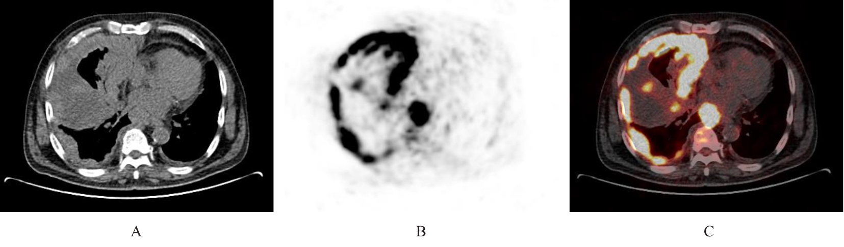
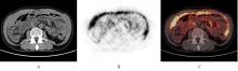
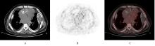
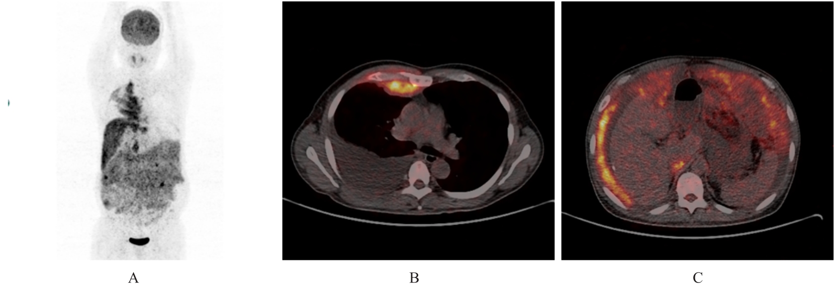
目的 分析胸膜和腹膜间皮瘤患者18F-氟代脱氧葡萄糖正电子发射扫描/计算机断层扫描(18F-FDG PET/CT)影像学表现,以期提高对该病的诊断水平。 方法 回顾性分析经病理证实的22例胸膜和腹膜间皮瘤患者(其中恶性间皮瘤21例,良性间皮瘤1例)18F-FDG PET/CT影像资料和免疫组织化学结果,对其影像表现和葡萄糖代谢特征进行归纳总结。 结果 恶性胸膜间皮瘤患者多表现为单侧胸膜弥漫性增厚伴放射性摄取增高,厚度为1.0~10.6 cm,平均半定量最大标准摄取值(SUVmax)为10.1,其中半数以上并发少量胸腔积液;恶性腹膜间皮瘤患者多数表现为腹膜、网膜和肠系膜弥漫增厚伴放射性摄取增高,厚度为1.2~6.6 cm,平均SUVmax为8.4,其中半数以上并发大量腹腔积液。恶性间皮瘤患者除原发部位外其他转移部位出现结节状、条片状和团块状不同程度异常放射性摄取17例,考虑为转移灶,平均SUVmax为7.4,大部分以病灶周围淋巴结转移为主,恶性胸膜间皮瘤患者可见骨骼和肌肉转移,恶性腹膜间皮瘤患者未见骨骼和肌肉转移。1例良性胸膜间皮瘤患者表现为双侧胸膜弥漫性增厚, 厚度约 3.5 cm, 未见明确异常放射性摄取, 并发少量胸腔积液。 结论 胸膜和腹膜间皮瘤患者18F-FDG PET/CT影像学表现具有一定的特征性,通过其胸膜和腹膜增厚方式及厚度、有无氟代脱氧葡萄糖(FDG)摄取及摄取程度可初步判断间皮瘤的良恶性,对间皮瘤临床早期诊断有一定的参考价值。PET/CT全身显像可确定间皮癌患者有无其他部位转移,对临床分期有一定的帮助。

关键词: 间皮瘤, 体层摄影术, 18F脱氧葡萄糖, 正电子发射扫描/计算机断层扫描

Abstract:

Objective To analyze the imaging manifestations of 18F-fluorodeoxyglucose positron emission tomography/computed tomography (18F-FDG PET/CT) in the patients with pleural and peritoneal mesothelioma, and to enhance the diagnostic accuracy for this disease. Methods A retrospective analysis was conducted on the 18F-FDG PET/CT imaging and immunohistochemical results of 22 patients confirmed pleural and peritoneal mesothelioma (21 malignant and 1 benign) by pathology. The imaging features and glucose metabolism characteristics were summarized. Results The majority of the patients with malignant pleural mesothelioma presented with unilateral pleural diffuse thickening accompanied by increased radiotracer uptake, and the thicknesses were ranged from 1.0 to 10.6 cm and the average semi-quantitative maximum standard uptake value (SUVmax) was 10.1. Over half of these patients also had a small amount of pleural effusion. The patients with malignant peritoneal mesothelioma mostly showed diffuse thickening of the peritoneum, omentum, and mesentery with increased radiotracer uptake, and the thicknesses were from 1.2 to 6.6 cm and the average SUVmax was 8.4, and over half of these patients had a significant amount of abdominal ascites. Besides the primary sites, nodular, striated, and mass-like abnormal radiotracer uptakes were observed in other metastatic sites in 17 cases of malignant mesothelioma, suggesting metastasis, and the average SUVmax was 7.4, predominantly surrounding lymph node metastasis. Bone and muscle metastases were visible in the patients with malignant pleural mesothelioma, while no such metastasis were seen in those with malignant peritoneal mesothelioma. One patient with benign pleural mesothelioma presented with bilateral pleural diffuse thickening approximately 3.5 cm thick, without significant abnormal radiotracer uptake and with a minor pleural effusion. Conclusion The 18F-FDG PET/CT imaging manifestations of pleural and peritoneal mesothelioma exhibit the distinctive characteristics. The mode and thickness of pleural and peritoneal thickening, the presence and degree of 18F-fluorodeoxyglucose uptake, can preliminarily differentiate between benign and malignant mesothelioma, thus providing valuable references for the early clinical diagnosis of mesothelioma. PET/CT based on whole-body imaging can determine whether there are other sites of metastasis, which is helpful for clinical staging.

Key words: Mesothelioma, Tomography, 18F deoxyglucose, Positron emission tomography/computed tomography

中图分类号: 

  • R734.3