吉林大学学报(医学版) ›› 2024, Vol. 50 ›› Issue (5): 1275-1285.doi: 10.13481/j.1671-587X.20240511

• 基础研究 • 上一篇    

水泡性口炎病毒对体外血管内皮屏障功能的损伤作用及其机制

曹宇璇1,陈为2,3,孙成彪2,赵娜2,王燕2,董明鑫2,许娜4,刘文森2(),李咏梅1()   

  1. 1.北华大学基础医学院病原生物学教研室, 吉林 吉林 132013
    2.中国农业科学院长春兽医研究所动物性食品安全与生物交叉研究室, 吉林 长春 130122
    3.河南科技大学应用工程学院食品医药 教研组, 河南 三门峡 472099
    4.吉林医药学院教务处, 吉林 吉林 132013
  • 收稿日期:2023-10-24 出版日期:2024-09-28 发布日期:2024-10-28
  • 通讯作者: 刘文森,李咏梅 E-mail:liuws85952@163.com;lymjyx@126.com
  • 作者简介:曹宇璇(1999-),女,安徽省合肥市人,在读硕士研究生,主要从事病原生物致病机制方面的研究。
  • 基金资助:
    吉林省卫健委卫生健康科技能力提升项目(2022JC021)

Damage effect of VSV on vascular endothelial barrier function in vitro and its mechanism

Yuxuan CAO1,Wei CHEN2,3,Chengbiao SUN2,Na ZHAO2,Yan WANG2,Mingxin DONG2,Na XU4,Wensen LIU2(),Yongmei LI1()   

  1. 1.Department of Pathogen Biology,School of Basic Medical Sciences,Beihua University,Jilin 132013,China
    2.Animal Food Safety and Biological Intersection Research Laboratory,Changchun Institute of Veterinary Medicine,Chinese Agricultural Sciences,Changchun 130122,China
    3.Food and Medicine Teaching and Research Group,School of Applied Engineering,Henan University of Science and Technology,Sanmenxia 472099,China
    4.Academic Affairs Office,Jilin Medical University,Jilin 132013,China
  • Received:2023-10-24 Online:2024-09-28 Published:2024-10-28
  • Contact: Wensen LIU,Yongmei LI E-mail:liuws85952@163.com;lymjyx@126.com

摘要:

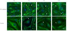
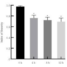
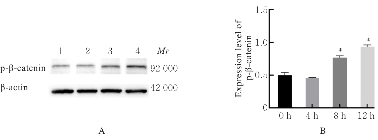
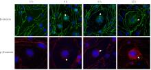
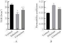
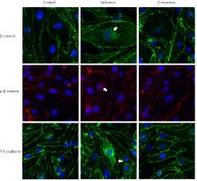
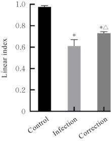
目的 探讨水泡性口炎病毒(VSV)对血管内皮(VE)屏障的损伤作用,并阐明其作用机制。 方法 采用犬肾细胞对VSV进行扩增,采用小鼠脑血管内皮瘤bEnd.3细胞检测VSV的半数组织培养感染剂量(TCID50),采用300倍TCID50进行后续实验。将bEnd.3细胞分为感染0 h组、感染4 h组、感染8 h组和感染12 h组,进行VSV感染损伤VE屏障实验;将bEnd.3细胞分为对照组、感染组和纠正组,进行抑制VSV复制与VE屏障恢复实验。将bEnd.3细胞接种于Transwell小室,构建体外VE屏障模型,采用细胞电压电阻仪检测感染VSV不同时间后各组bEnd.3细胞中跨上皮电阻(TER),采用异硫氰酸荧光素-葡聚糖渗漏实验检测各组渗透系数,采用免疫荧光染色法观察VSV感染后各组bEnd.3细胞骨架及黏附连接(AJs)中VE-钙黏蛋白、β-连环蛋白(β-catenin)和磷酸化β-连环蛋白(p-β-catenin)定位变化,采用实时荧光定量PCR(RT-qPCR)法检测各组细胞中Wnt和β-catenin mRNA表达水平,采用Western blotting法检测各组细胞中Wnt、β-catenin和p-β-catenin表达水平。 结果 VSV的TCID50为10-4.5·100 μL-1。Transwell小室实验检测,与感染0 h比较,其他各组细胞中TER明显降低(P<0.05),渗透系数明显升高(P<0.05)。免疫荧光染色,与对照组比较,感染组bEnd.3细胞骨架紊乱,细胞间隙增大,AJs线性指数明显降低(P<0.05),β-catenin和p-β-catenin从细胞膜转移至细胞核周围。RT-qPCR法检测,与感染0 h比较,其他各组细胞中Wnt mRNA表达水平明显降低(P<0.05),β-catenin mRNA表达水平差异无统计学意义(P>0.05)。Western blotting法检测,与感染0 h比较,其他各组细胞中Wnt蛋白表达水平明显降低(P<0.05),β-catenin表达水平差异无统计学意义(P>0.05),p-β-catenin表达水平明显升高(P<0.05)。在抑制VSV复制并纠正低密度脂蛋白受体(LDLR)异常后,Transwell小室实验检测,与感染组比较,纠正组 bEnd.3细胞中TER明显升高(P<0.05),渗透系数明显降低(P<0.05)。免疫荧光染色,与感染组比较,纠正组细胞间隙减小,细胞中β-catenin和p-β-catenin核周聚集现象有所改善。RT-qPCR法检测,与感染组比较,纠正组细胞中Wnt mRNA表达水平明显升高(P<0.05)。Western blotting法检测,与感染组比较,纠正组细胞中Wnt蛋白表达水平明显升高(P<0.05),β-catenin表达水平差异无统计学意义(P>0.05),p-β-catenin表达水平明显降低(P<0.05)。 结论 VSV感染后可引起LDLR失活,降低Wnt蛋白表达水平,造成β-catenin磷酸化水平升高并发生内化,破坏AJs稳定性,最终导致VE屏障损伤。

关键词: 水泡性口炎病毒, 血管内皮屏障, 黏附连接, 低密度脂蛋白受体, Wnt/β-连环蛋白信号通路

Abstract:

Objective To discuss the damage effect of vesicular stomatitis virus (VSV) on the vascular endothelial (VE) barrier, and to clarify its mechanism. Methods The canine kidney cells were used to amplify VSV. The half tissue culture infective dose (TCID50) of VSV was determined using mouse brain endothelial tumor bEnd.3 cells, and subsequent experiment was conducted using 300 times the TCID50. The bEnd.3 cells were divided into infection 0 h group, infection 4 h group, infection 8 h group, and infection 12 h group for VE barrier damage experiments due to VSV infection. The bEnd.3 cells were also divided into control group, infection group, and correction group for experiments to inhibit the VSV replication and restore the VE barrier. The bEnd.3 cells were inoculated into Transwell chambers to construct an in vitro VE barrier model. Cell voltage resistance meter was used to detect the transepithelial resistance (TER) in various groups after the bEnd.3 cells were infected with VSV at different time points;fluorescein isothiocyanate-dextran leakage assay was used to detect the permeability coefficients of the cells in various groups; immunofluorescence staining was used to observe the localization changes of VE-cadherin, β-catenin, and phosphorylated β-catenin (p-β-catenin) in cytoskeleton and adherens junctions (AJs) of the bEnd.3 cells after VSV infection; real-time fluorescence quantitative PCR (RT-qPCR) method was used to detect the expression levels of Wnt and β-catenin mRNA in the cells in various groups; Western blotting method was used to detect the expression levels of Wnt, β-catenin, and p-β-catenin proteins in the cells in various groups. Results The TCID50 of VSV was 10-4.5·100 μL-1. The Transwell chamber experiment results showed that compared with infection 0 h group, the TERs in the cells in the other groups were significantly decreased (P<0.05), and the permeability coefficients were significantly increased (P<0.05). The immunofluorescence staining results showed that compared with control group, the cytoskeleton of the bEnd.3 cells in infection group was disordered, the cell gaps was increased, the linear index of AJs was significantly decreased (P<0.05), and β-catenin and p-β-catenin translocated from the cell membrane to the perinuclear area. The RT-qPCR results showed that compared with infection 0 h group, the expression levels of Wnt mRNA in the cells in the other groups were significantly decreased (P<0.05), while the expression levels of β-catenin mRNA showed no statistically significant difference (P>0.05). The Western blotting results showed that compared with infection 0 h group, the expression levels of Wnt protein in the cells in the other groups were significantly decreased (P<0.05), the expression levels of β-catenin showed no statistically significant differences (P>0.05), and the expression levels of p-β-catenin were significantly increased (P<0.05).After inhibiting the VSV replication and correcting the low density lipoprotein receptor (LDLR) abnormalities, the Transwell chamber experiment results showed that compared with infection group, the TER in the cells in correction group was significantly increased (P<0.05), and the permeability coefficient was significantly decreased (P<0.05).The immunofluorescence staining results showed that compared with infection group, the gaps in the cells in correction group were reduced, and the perinuclear aggregation of β-catenin and p-β-catenin in the cells was restrained. The RT-qPCR results showed that compared with infection group, the expression level of Wnt mRNA in the cells in correction group was significantly increased (P<0.05). The Western blotting results showed that compared with infection group, the expression level of Wnt protein in the cells in correction group was significantly increased (P<0.05), the expression level of β-catenin showed no statistically significant difference (P>0.05), and the expression level of p-β-catenin was significantly decreased (P<0.05). Conclusion VSV infection can cause the LDLR inactivation, reduce the expression level of Wnt protein, increase the phosphorylation level of β-catenin and cause its internalization, disrupt the stability of AJs, and ultimately lead to VE barrier damage.

Key words: Vesicular stomatitis virus, Vascular endothelial barrier, Adherent junction, Low density lipoprotein receptor, Wnt/β-catenin signaling pathway

中图分类号: 

  • S852.65