| [1] |
FILHO A M, LAVERSANNE M, FERLAY J, et al. The GLOBOCAN 2022 cancer estimates: Data sources, methods, and a snapshot of the cancer burden worldwide[J]. Int J Cancer, 2025, 156(7): 1336-1346.
|
| [2] |
BUKOWSKI K, KCIUK M, KONTEK R. Mechanisms of multidrug resistance in cancer chemotherapy[J]. Int J Mol Sci, 2020, 21(9): 3233.
|
| [3] |
CHAN L S, LIU J, LI M S C, et al. Selenite as a dual apoptotic and ferroptotic agent synergizes with EGFR and KRAS inhibitors with epigenetic interference[J]. Clin Epigenetics, 2023, 15(1): 36.
|
| [4] |
CHEN W W, AN J J, GUO J W, et al. Sodium selenite attenuates lung adenocarcinoma progression by repressing SOX2-mediated stemness[J]. Cancer Chemother Pharmacol, 2018, 81(5): 885-895.
|
| [5] |
MORO C F, SELVAM A K, GHADERI M, et al. Drug-induced tumor-specific cytotoxicity in a whole tissue ex vivo model of human pancreatic ductal adenocarcinoma[J]. Front Oncol, 2022, 12: 965182.
|
| [6] |
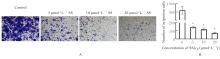
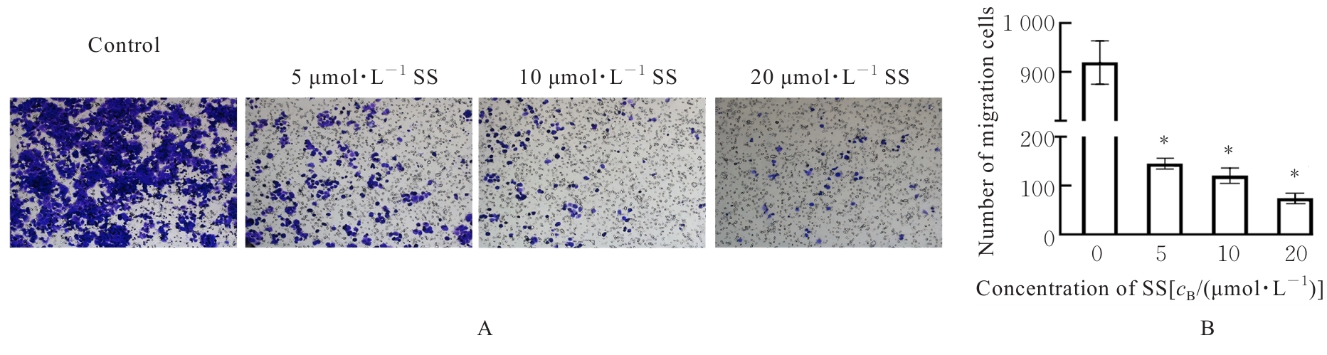
韩宇晨, 陈微微, 白 玉, 等. 亚硒酸钠对肺癌细胞迁移和血管生成的影响及其机制探究[J]. 中国病理生理杂志, 2024, 40(9): 1598-1605.
|
| [7] |
刘 啸. 亚硒酸钠对肾细胞癌生物学行为影响及其作用机制研究[D]. 济南: 山东大学, 2023.
|
| [8] |
徐梦辰. 亚硒酸钠诱导人源三阴性乳腺癌细胞铁死亡的机制研究[D]. 兰州: 兰州大学, 2023.
|
| [9] |
CAFFREY P B, FRENKEL G D. Selenite enhances and prolongs the efficacy of cisplatin treatment of human ovarian tumor xenografts[J]. In Vivo, 2012, 26(4): 549-552.
|
| [10] |
WANG J, KOBAYASHI M, SAKURADA K, et al. Possible roles of an adult T-cell leukemia (ATL)- derived factor/thioredoxin in the drug resistance of ATL to adriamycin[J]. Blood, 1997, 89(7): 2480-2487.
|
| [11] |
XU X, HOU Y Q, LIN S M, et al. Sodium selenite inhibits proliferation of lung cancer cells by inhibiting NF-κB nuclear translocation and down-regulating PDK1 expression which is a key enzyme in energy metabolism expression[J]. J Trace Elem Med Biol, 2023, 78: 127147.
|
| [12] |
LV C Q, ZENG Q Y, QI L, et al. Sodium selenite induces autophagy and apoptosis in cervical cancer cells via mitochondrial ROS-activated AMPK/mTOR/FOXO3a pathway[J]. Antioxidants, 2024, 13(8): 1004.
|
| [13] |
李艳梅, 马琳, 刘单, 等. 亚硒酸钠通过增加ROS抑制PI3K/AKT通路改善肺癌PC-9/GR细胞对吉非替尼的耐药性[J]. 现代肿瘤医学, 2024, 32(4): 636-641.
|
| [14] |
SKINNER K T, PALKAR A M, HONG A L. Genetics of ABCB1 in cancer[J]. Cancers, 2023, 15(17): 4236.
|
| [15] |
CHENG G R, PI Z F, ZHUANG X Y, et al. The effects and mechanisms of aloe-emodin on reversing adriamycin-induced resistance of MCF-7/ADR cells[J]. Phytother Res, 2021, 35(7): 3886-3897.
|
| [16] |
CHEN H K, CHEN Y L, WANG C Y, et al. ABCB1 regulates immune genes in breast cancer[J]. Breast Cancer, 2023, 15: 801-811.
|
| [17] |
IBRAHIM S M, KARIM S, ABUSAMRA H, et al. Genomic amplification of chromosome 7 in the Doxorubicin resistant K562 cell line[J]. Bioinformation, 2018, 14(9): 587-593.
|
| [18] |
REED K, HEMBRUFF S L, LABERGE M L, et al. Hypermethylation of the ABCB1 downstream gene promoter accompanies ABCB1 gene amplification and increased expression in docetaxel-resistant MCF-7 breast tumor cells[J]. Epigenetics, 2008, 3(5): 270-280.
|
| [19] |
WANG Y C, JURIC D, FRANCISCO B, et al. Regional activation of chromosomal arm 7q with and without gene amplification in taxane-selected human ovarian cancer cell lines[J]. Genes Chromosom Cancer, 2006, 45(4): 365-374.
|
| [20] |
HU B Y, ZOU T T, QIN W, et al. Inhibition of EGFR overcomes acquired lenvatinib resistance driven by STAT3-ABCB1 signaling in hepatocellular carcinoma[J]. Cancer Res, 2022, 82(20): 3845-3857.
|
| [21] |
HAN X W, MO J G, YANG Y M, et al. Crucial roles of LncRNAs-mediated autophagy in breast cancer[J]. Int J Med Sci, 2022, 19(6): 1082-1092.
|
| [22] |
IBRAGIMOVA M, TSYGANOV M, LITVIAKOV N. Tumour stem cells in breast cancer[J]. Int J Mol Sci, 2022, 23(9): 5058.
|
| [23] |
LIAO M M, WANG C W, YANG B W, et al. Corrigendum: autophagy blockade by Ai Du Qing formula promotes chemosensitivity of breast cancer stem cells via GRP78/β-catenin/ABCG2 axis[J]. Front Pharmacol, 2022, 13: 809565.
|
| [24] |
QIAN M Q, MA X D, PAN G W. Curcumin improving drug resistance of MDA-MB-231/DDP tumor treatment by enhancing autophagy[J]. International J Pharmacology, 2022, 18(4): 806-816.
|
| [25] |
SHAHVERDI M, HAJIASGHARZADEH K, SORKHABI A D, et al. The regulatory role of autophagy-related miRNAs in lung cancer drug resistance[J]. Biomed Pharmacother, 2022, 148: 112735.
|
| [26] |
YUN C W, JEON J, GO G, et al. The dual role of autophagy in cancer development and a therapeutic strategy for cancer by targeting autophagy[J]. Int J Mol Sci, 2020, 22(1): 179.
|
 ),安佳佳1(