吉林大学学报(医学版) ›› 2026, Vol. 52 ›› Issue (2): 469-482.doi: 10.13481/j.1671-587X.20260219

• 临床研究 • 上一篇    下一篇

GSTO1在卵巢癌组织中的表达及其N-糖基化位点突变对上皮性卵巢癌细胞生物学行为的影响

李红1,2,于盼盼2,赵邹宇1,2,孙崇凤1,2,乔慧1,2,杨萍1,2,3()   

  1. 1.石河子大学第一附属医院妇科,新疆 石河子 832008
    2.石河子大学医学院,新疆 石河子 832008
    3.新疆生产建设兵团医院妇科,新疆 乌鲁木齐 830001
  • 收稿日期:2025-04-25 接受日期:2025-07-06 出版日期:2026-03-28 发布日期:2026-04-15
  • 通讯作者: 杨萍 E-mail:pingy2018@163.com
  • 作者简介:李 红(1992-),女,安徽省淮南市人,在读硕士研究生,主要从事妇科肿瘤方面的研究。
  • 基金资助:
    国家自然科学基金地区科学基金项目(82360541);人才发展基金-兵团临床医学研究中心-兵团妇产疾病临床医学研究中心项目(CZ001230);天山英才科技创新领军人才-高层次领军人才项目(2022TSYCLJ0055)

Expression of GSTO1 in ovarian cancer tissue and effect of N-glycosylation site mutations on biological behaviors of epithelial ovarian cancer cells

Hong LI1,2,Panpan YU2,Zouyu ZHAO1,2,Chongfeng SUN1,2,Hui QIAO1,2,Ping YANG1,2,3()   

  1. 1.Department of Gynecology,First Affiliated Hospital,Shihezi University,Shihezi 832008,China
    2.School of Medicine,Shihezi University,Shihezi 832008,China
    3.Department of Gynecology,Xinjiang Production and Construction Corps Hospital,Urumqi 830001,China
  • Received:2025-04-25 Accepted:2025-07-06 Online:2026-03-28 Published:2026-04-15
  • Contact: Ping YANG E-mail:pingy2018@163.com

摘要:

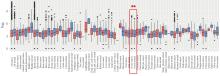
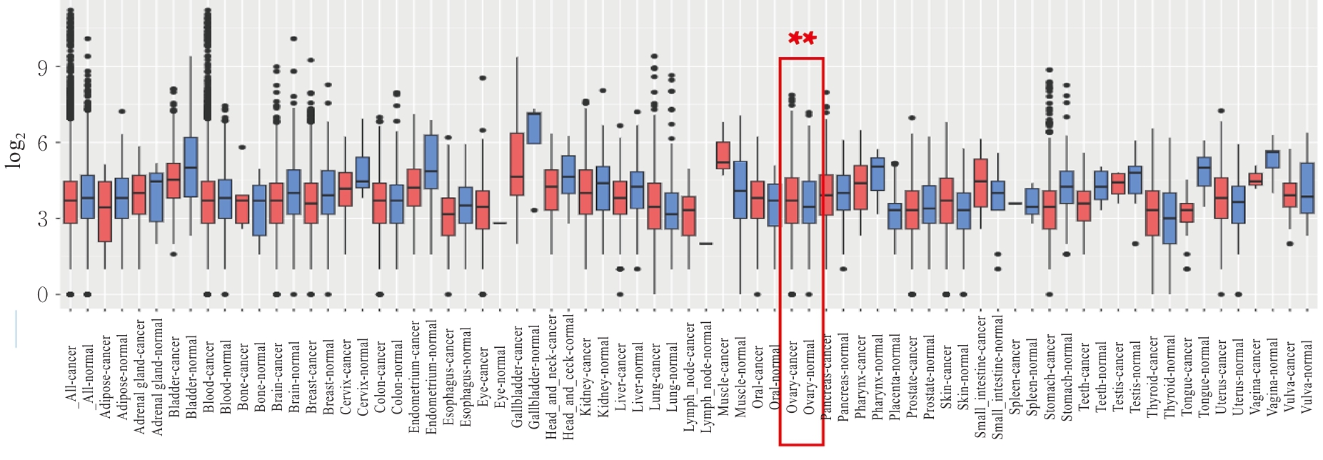
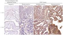
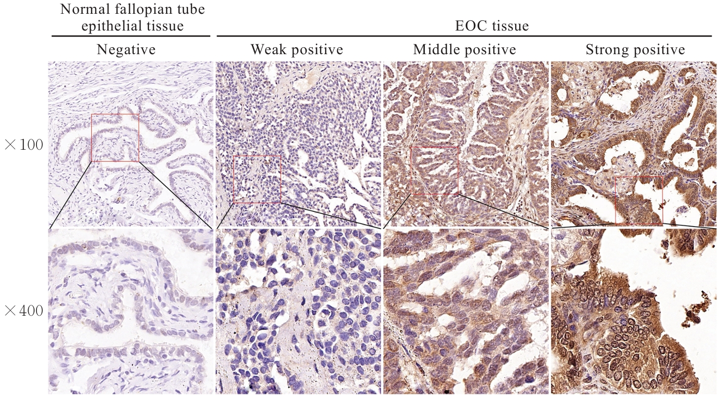
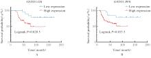
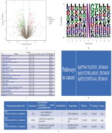
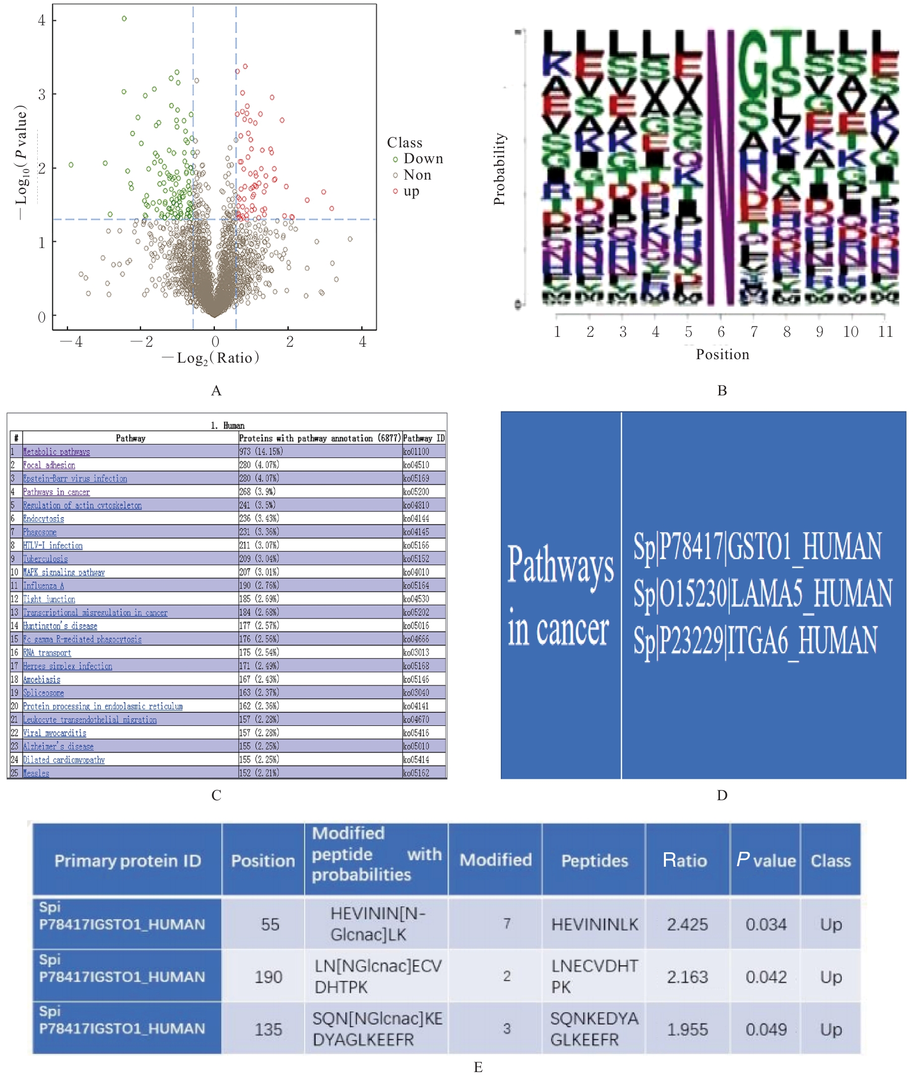
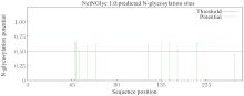
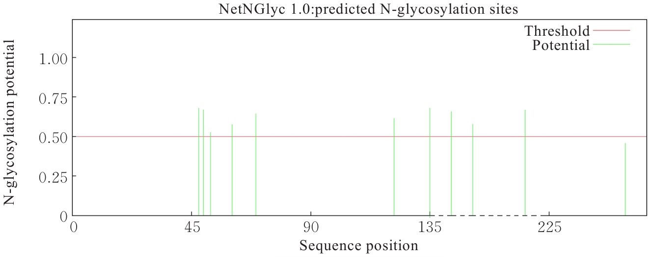
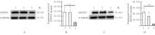
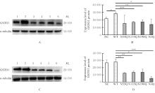
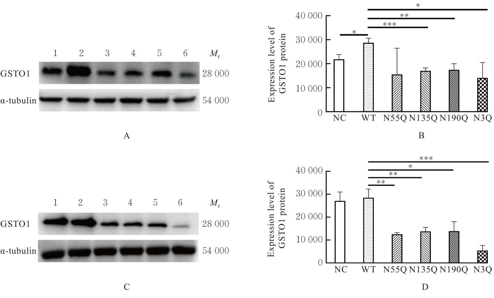
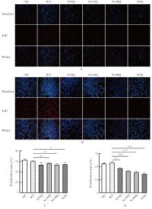
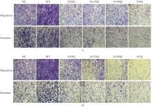
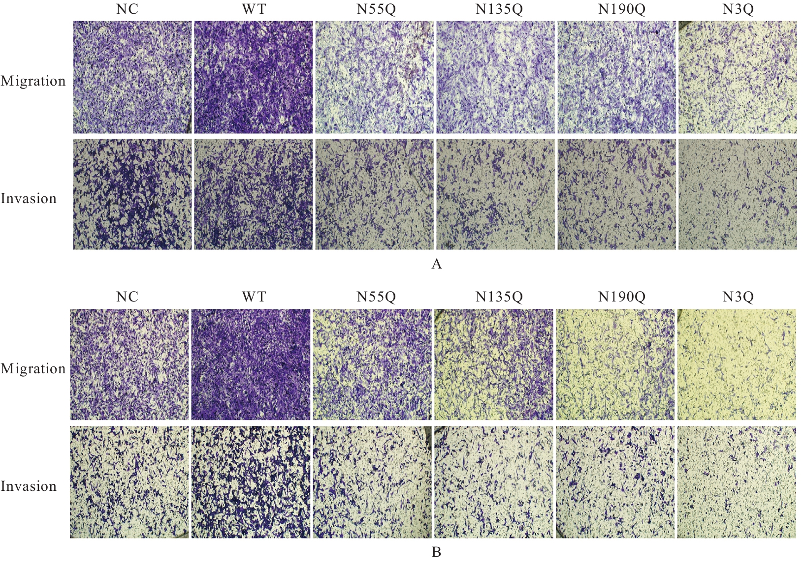
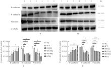
目的 探讨谷胱甘肽S-转移酶Omega-1蛋白(GSTO1)在上皮性卵巢癌(EOC)组织中的表达及其与患者临床病理特征和预后的关系,并探讨其N-糖基化修饰对卵巢癌A2780和SKOV3细胞生物学行为的影响,阐明其可能的机制。 方法 利用GENT2数据库分析卵巢癌组织和正常卵巢上皮组织中GSTO1 mRNA表达水平。收集于石河子大学第一附属医院妇科就诊并手术的88例EOC患者临床信息,采集组织样本,定期随访患者预后情况,记录患者总生存期 (OS) 和无进展生存期 (PFS)。采用免疫组织化学法检测EOC患者组织中GSTO1蛋白表达情况,分析其与患者临床病理特征及预后的关系。采用单因素和多因素Cox回归分析评估影响EOC患者预后的危险因素。采用质谱分析比较卵巢癌高转移ES-2细胞与亲本SKOV3细胞中GSTO1蛋白的N-糖基化修饰差异,NetNGlyc 1.0 server数据库鉴定其修饰位点。使用衣霉素抑制细胞整体N-糖基化,将卵巢癌A2780和SKOV3细胞分为对照组、二甲亚砜(DMSO)组及衣霉素组。采用慢病毒转染方式稳定构建不同N-糖基化修饰水平的卵巢癌A2780和SKOV3细胞,分为NC组、WT组、N55Q组、N135Q组、N190Q组和N3Q组。采用5-乙炔基-2'-脱氧尿苷(EdU)实验检测各组细胞增殖率,Transwell小室实验检测各组迁移细胞数和侵袭细胞数;Western blotting法检测各组细胞中GSTO1蛋白和上皮-间充质转化(EMT)相关蛋白E-钙黏蛋白(E-cadherin)、N-钙黏蛋白(N-cadherin)及波形蛋白(Vimentin)表达水平。 结果 GENT2数据库,与正常卵巢上皮组织比较,卵巢癌组织中GSTO1 mRNA表达水平明显升高(P<0.01)。免疫组织化学法,EOC患者肿瘤组织中GSTO1蛋白高表达者66例,低表达者22例。EOC患者肿瘤组织中GSTO1蛋白高表达与FIGO分期、淋巴脉管间隙浸润和肿瘤直径有关联(P<0.05)。单因素和多因素Cox回归分析,FIGO高分期、有淋巴结转移和GSTO1高表达均是影响EOC患者OS)及PFS的独立危险因素。质谱分析,与亲本SKOV3细胞比较,卵巢癌高转移ES-2细胞中GSTO1 N-糖基化修饰水平明显升高(P<0.05),且其N-糖基化修饰位点分别为Asn55、Asn135和Asn190。与对照组比较,衣霉素组A2780和SKOV3细胞中GSTO1蛋白表达水平明显降低(P<0.05);与WT组比较,N135Q、N190Q和N3Q组 A2780及SKOV3细胞中GSTO1蛋白表达水平明显降低(P<0.05)。与WT组比较,N135Q、N190Q和N3Q组A2780及SKOV3细胞增殖率明显降低(P<0.05)。与WT组比较,N55Q、N135Q、N190Q和N3Q组A2780及SKOV3细胞的迁移细胞数和侵袭细胞数均明显降低(P<0.05)。与WT组比较,N3Q组A2780和SKOV3细胞中E-cadherin蛋白表达水平明显升高(P<0.05),N190Q和N3Q组A2780及SKOV3细胞中N-cadherin蛋白和Vimentin蛋白表达水平明显降低(P<0.05)。 结论 GSTO1在EOC组织和细胞中呈高表达且与患者不良预后相关,其N-糖基化位点突变可抑制卵巢癌细胞增殖、迁移、侵袭和EMT进程。

关键词: 上皮性卵巢癌, 谷胱甘肽S-转移酶Omega-1蛋白, N-糖基化, 细胞迁移, 细胞侵袭, 细胞增殖, 上皮-间充质转化

Abstract:

Objective To discuss the expression of glutathione S-transferase Omega-1 protein (GSTO1) in epithelial ovarian cancer (EOC) tissue and its relationship with the clinicopathological characteristics and prognosis of the patients, and to investigate the effect of its N-glycosylation modification on the biological behaviors of ovarian cancer A2780 and SKOV3 cells, and to clarify its possible mechanism. Methods GENT2 database was used to analyze the expression levels of GSTO1 mRNA in ovarian cancer tissue and normal ovarian epithelial tissue. The clinical information of 88 EOC patients who visited and underwent surgery in the Department of Gynecology of the First Affiliated Hospital of Shihezi University was collected, the tissue samples were collected, and the prognosis of the patients was followed up regularly. Immunohistochemical method was used to detect the expression of GSTO1 protein in ovarian tissue of EOC patients, and its relationship with the clinicopathological characteristics and prognosis of the patients was analyzed. Univariate and multivariate Cox regression analyses were used to evaluate the risk factors affecting the prognosis of EOC patients. Mass spectrometry was used to compare the difference in N-glycosylation modification of GSTO1 protein between highly metastatic ovarian cancer ES-2 cells and parental SKOV3 cells, and NetNGlyc 1.0 server database was used to identify its modification sites. Tunicamycin was used to inhibit the overall N-glycosylation of cells, and ovarian cancer A2780 and SKOV3 cells were divided into control group, dimethyl sulfoxide (DMSO) group, and tunicamycin group. The ovarian cancer A2780 and SKOV3 cells with different levels of N-glycosylation modification were stably constructed by lentiviral transfection and divided into NC group, WT group, N55Q group, N135Q group, N190Q group, and N3Q group. 5-Ethynyl-2'-deoxyuridine (EdU) assay was used to detect the proliferation rates of the cells in various groups; Transwell chamber assay was used to detect the numbers of migration cells and invasion cells in various groups; Western blotting method was used to detect the expression levels of GSTO1 protein and epithelial-mesenchymal transition (EMT)-related proteins E-cadherin, N-cadherin, and Vimentin in the cells in various groups. Results According to GENT2 database, compared with normal ovarian epithelial tissue, the expression level of GSTO1 mRNA in ovarian cancer tissue was significantly increased (P<0.01). Immunohistochemical staining results showed that there were 66 cases with high expression of GSTO1 and 22 cases with low expression of GSTO1 in tumor tissues of EOC patients.The high expression of GSTO1 protein in tumor tissue of EOC patients was associated with FIGO stage, lymphovascular space invasion, and tumor diameter (P<0.05). The univariate and multivariate Cox regression analyses results showed that high FIGO stage, lymph node metastasis, and high GSTO1 expression were independent risk factors affecting the overall survival (OS) and progression-free survival (PFS) of EOC patients. The mass spectrometry results showed that compared with parental SKOV3 cells, the level of N-glycosylation modification of GSTO1 in highly metastatic ovarian cancer ES-2 cells was significantly increased (P<0.05), and its N-glycosylation modification sites were Asn55, Asn135, and Asn190, respectively. Compared with control group, the expression levels of GSTO1 protein in A2780 and SKOV3 cells in tunicamycin group were significantly decreased (P<0.05). Compared with WT group, the expression levels of GSTO1 protein in A2780 and SKOV3 cells in N135Q, N190Q, and N3Q groups were significantly decreased (P<0.05). Compared with WT group, the proliferation rates of A2780 and SKOV3 cells in N135Q, N190Q, and N3Q groups were significantly decreased (P<0.05). Compared with WT group, the numbers of migration cells and invasion cells of A2780 and SKOV3 cells in N55Q, N135Q, N190Q, and N3Q groups were significantly decreased (P<0.05). Compared with WT group, the expression levels of E-cadherin protein in A2780 and SKOV3 cells in N3Q group were significantly increased (P<0.05), and the expression levels of N-cadherin protein and Vimentin protein in A2780 and SKOV3 cells in N190Q and N3Q groups were significantly decreased (P<0.05). Conclusion GSTO1is highly expressed in EOC tissue and cells and is associated with poor prognosis of patients. Mutation of its N-glycosylation sites can inhibit the proliferation, migration, invasion, and EMT process of ovarian cancer cells.

Key words: Epithelial ovarian cancer, Glutathione S-transferase Omega-1 protein, N-glycosylation, Cell migration, Cell invasion, Cell proliferation, Epithelial-mesenchymal transition

中图分类号: 

  • R737.33