吉林大学学报(医学版) ›› 2021, Vol. 47 ›› Issue (2): 299-306.doi: 10.13481/j.1671-587X.20210207

• 基础研究 • 上一篇    下一篇

疏肝化癥方对小鼠皮下移植性三阴性乳腺癌生长的抑制作用及其机制

王博1,杨焱2,费瑞3(),景年财2,李兆东3,卢义2,肖宏宇2,张越1,2()   

  1. 1.长春中医药大学中医学院中医内科,吉林 长春 130117
    2.吉林省肿瘤医院中西医结合科,吉林 长春 130012
    3.吉林大学基础医学院细胞生物学系,吉林 长春 130021
  • 收稿日期:2020-08-15 出版日期:2021-03-28 发布日期:2021-03-25
  • 通讯作者: 费瑞,张越 E-mail:feirui@jlu.edu.cn;zhangyuejlcc@163.com
  • 作者简介:王 博(1990-),男,吉林省长春市人,主治医师,在读博士研究生,主要从事中西医结合治疗肿瘤等方面的研究。
  • 基金资助:
    吉林省科技厅科技发展计划项目(201603046YY)

Inhibitory effect of Shuganhuazheng Formula on growth of triple negative breast cancer of subcutaneous transplantation in mice

Bo WANG1,Yan YANG2,Rui FEI3(),Niancai JING2,Zhaodong LI3,Yi LU2,Hongyu XIAO2,Yue ZHANG1,2()   

  1. 1.Department of Internal Medicine of Chinese Medicine,College of Chinese Medicine,Changchun University of Chinese Medicine,Changchun 130117,China
    2.Department of Integrated Traditional Chinese and Western Medicine,Jilin Tumor Hospital,Changchun 130021,China
    3.Department of Cell Biology,School of Basic Medical Sciences,Jilin University,Changchun 130021,China
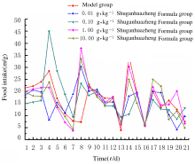
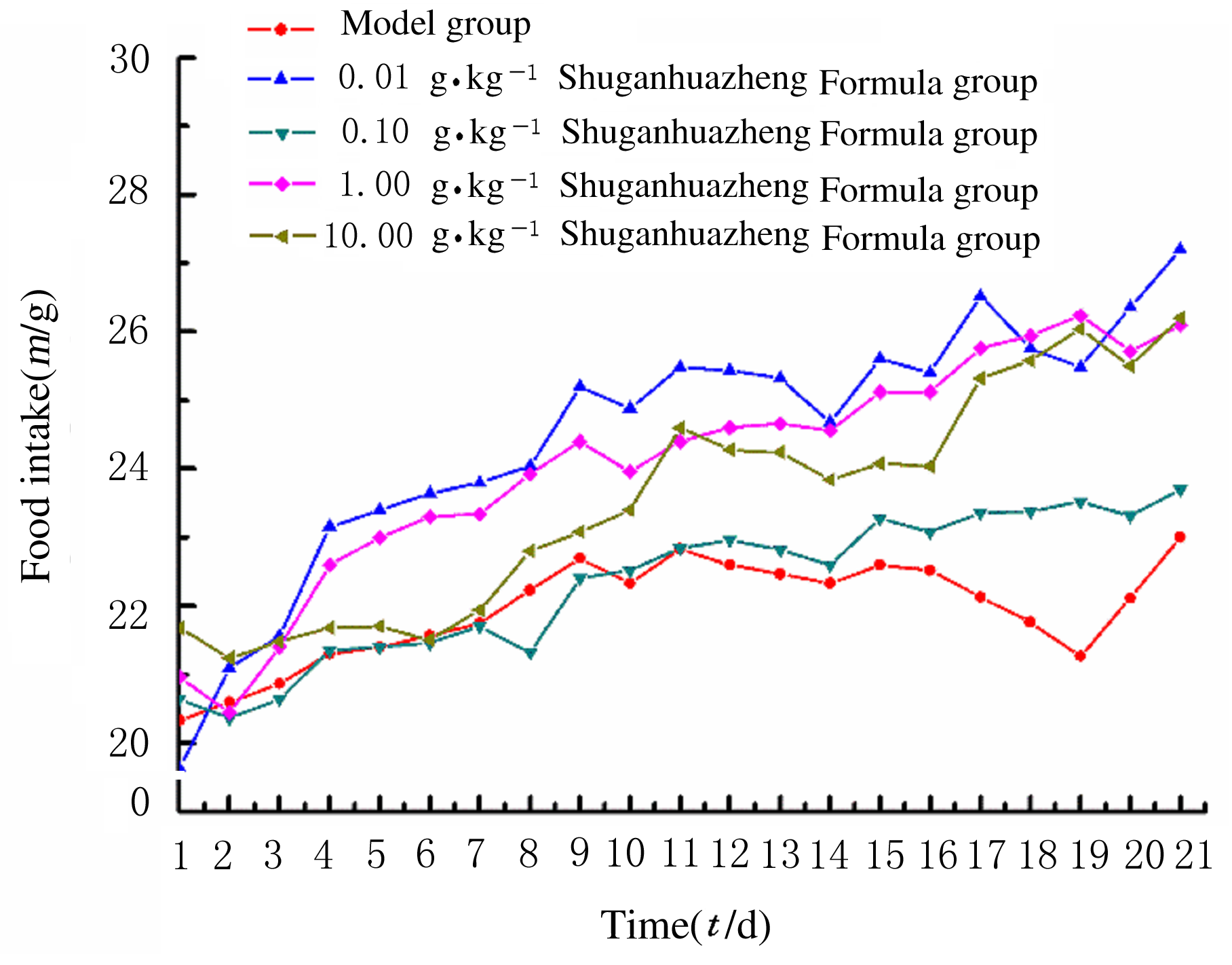
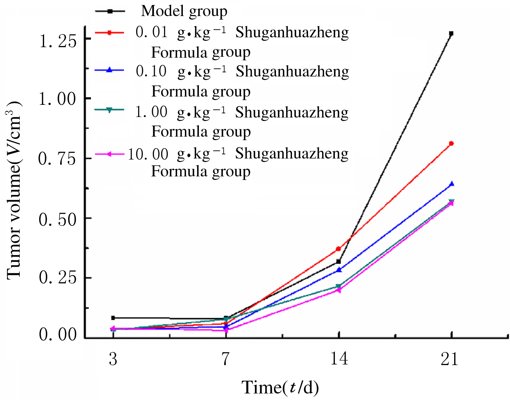
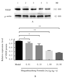
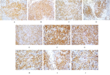
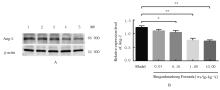
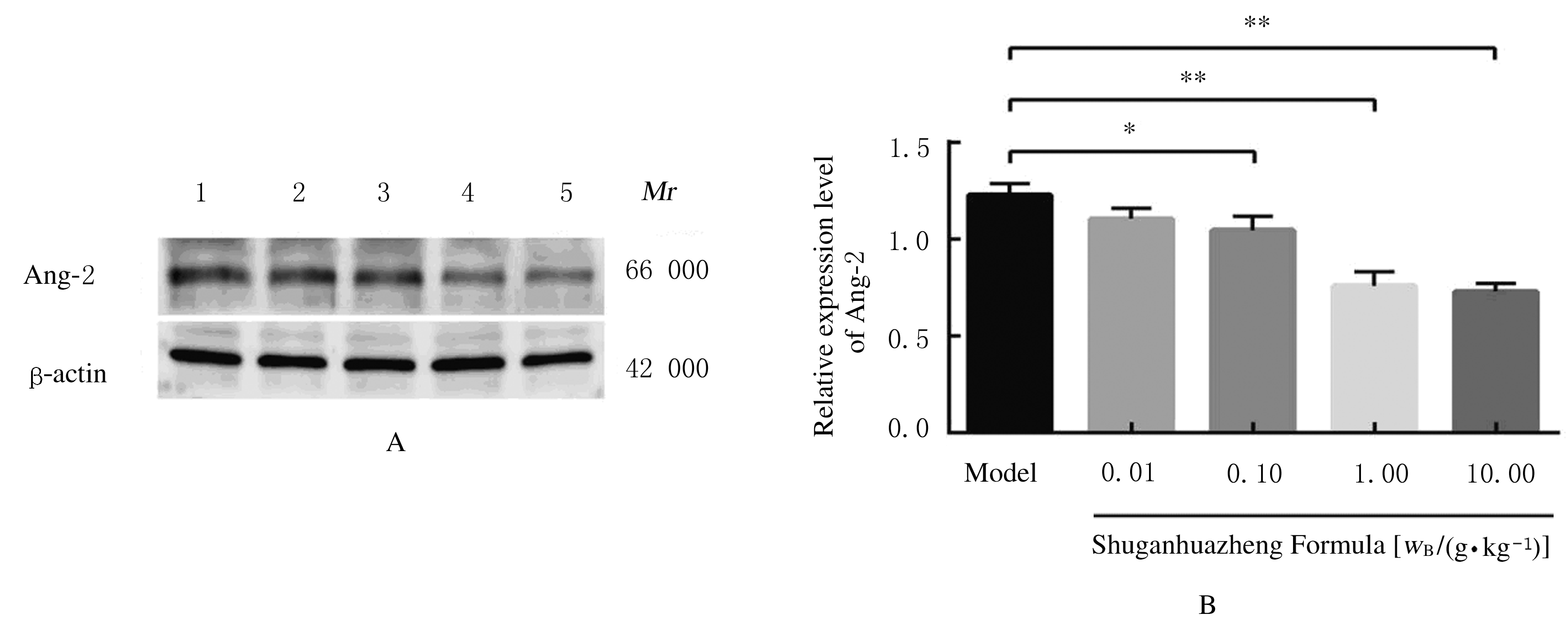
  • Received:2020-08-15 Online:2021-03-28 Published:2021-03-25
  • Contact: Rui FEI,Yue ZHANG E-mail:feirui@jlu.edu.cn;zhangyuejlcc@163.com

摘要: 目的

探讨疏肝化癥方对乳腺癌小鼠的抑瘤作用及其对肿瘤血管生成的影响,并阐明其可能的作用机制。

方法

将40只雌性BALB/c小鼠随机分为模型组和0.01、0.10、1.00及10.00 g·kg-1疏肝化癥方组,每组8只。采用小鼠腋下胸骨前缘乳腺脂肪垫内接种4T1细胞法构建三阴性乳腺癌(TNBC)小鼠模型。模型构建成功后,疏肝化癥方组小鼠灌胃给予相应剂量的疏肝化癥方液,模型组小鼠给予等量蒸馏水,持续21 d。每天检测各组小鼠食量和体质量;并在模型构建成功后第3、7、14和21天时,采用游标卡尺测量瘤体长径和短径,计算各组小鼠肿瘤体积和抑瘤率。21 d后处死小鼠,取瘤体组织,采用免疫组织化学法和Western blotting 法检测各组小鼠肿瘤组织中血管内皮生长因子(VEGF)和血管生成素样蛋白2(Ang-2)蛋白表达水平。

结果

与模型组比较,不同剂量疏肝化癥方组小鼠每日食量随天数增加均降低,但差异无统计学意义(P>0.05)。在第14和21天时,模型组小鼠体质量增长缓慢;但10.00 g·kg-1疏肝化癥方组小鼠体质量明显增加,与模型组比较差异有统计学意义(P<0.01)。与模型组比较,第21天时0.10、1.00和10.00 g·kg-1疏肝化癥方组小鼠抑瘤率明显升高(P<0.05或P<0.01)。免疫组织化学检测,模型组小鼠肿瘤组织细胞质棕黄色较深;各剂量疏肝化癥方组随剂量的升高,肿瘤组织细胞着色逐渐减轻,各剂量疏肝化癥方组小鼠肿瘤组织细胞质棕黄色逐渐减少,而细胞核淡蓝色逐渐加大。阳性信号吸光度(A)值检测,与模型组比较,各剂量疏肝化癥方组VEGF和Ang-2蛋白表达水平明显降低,差异有统计学意义(P<0.05或P<0.01),且以10.00 g·kg-1疏肝化癥方组降低最明显。Western blotting法检测,与模型组比较,各剂量疏肝化癥方组小鼠肿瘤组织中VEGF和Ang-2蛋白表达水平明显降低(P<0.05或P<0.01),且呈剂量依赖性。

结论

疏肝化癥方对乳腺癌荷瘤小鼠的瘤体生长有抑制作用,其作用机制可能与抑制肿瘤组织中VEGF和Ang-2蛋白表达有关。

关键词: 疏肝化癥方, 三阴性乳腺癌, 血管生成, 血管内皮生长因子, 血管生成素样蛋白2

Abstract: Objective

To investigate the anti-tumor effect of Shuganhuazheng Formula and the influence in the tumor angiogenesis in the mice with breast cancer, and to clarify its posible mechanism.

Methods

Forty female BALB/c mice were randomly divided into model group and 0.01, 0.10, 1.00 and 10.00 g·kg-1 Shuganhuazheng Formula groups, with 8 mice in each group. The mouse models of three-negative breast cancer (TNBC) were established by inoculation of 4T1 cells at the breast fat pad of the anterior axillary sternum in the mice. After the models were built successfully, the mice in Shuganhuazheng Formula groups were given the corresponding doses of Shuganhuazheng Formula by gavage, and the mice in model group were given the same amount of distilled water, lasted for 21 d. The food intakes and body weights of the mice were measured daily. On the 3rd, 7th, 14th and 21st days after the modeling, vernier calipers were used to measure the long and short diameters of the tumor, and the tumor volumes and the inhibitory rate of tumor of the mice in various groups were calculated. After 21 d, the mice were sacrificed and tumor tissue was taken. Immunohistochemistry and Western blotting methods were used to detect the expression levels of vascular endothelial growth factor (VEGF) and angiogenin-like protein-2 (Ang-2) proteins in tumor tissue of the mice in various groups.

Results

Compared with model group, the daily food intakes of the mice in different doses of Shuganhuazheng Formula groups were decreased with the increase of time, but the difference was not statistically significant (P>0.05). On the 14th and 21st days, the body weight of the mice in model group was increased slowly, but the weights of the mice in 10.00 g·kg-1 Shuganhuazheng Formula group were significantly increased,and the difference was statistically significant compared with model group (P<0.01). Compared with model group, the inhibitory rates of tumor of the mice in 0.10, 1.00 and 10.00 g·kg-1 Shuganhuazheng Formula groups were significantly increased on the 21st day (P<0.05 or P<0.01).The immunohistochemical results showed that the cytoplasm of the mice in model group had darker brownish yellow color, while the staining was gradually decreased with the increase of dose in each Shuganhuazheng Formula group. The brownish yellow color in cytoplasm in each Shuganhuazheng Formula group was decreased gradually, while the pale blue color in nucleus was increased gradually.The positive signal absorbance(A) value detection results showed that the expression levels of VEGF and Ang-2 proteins in tumor tissue of the mice in different doses of Shuganhuazheng Formula groups were significantly decreased compared with model group (P<0.05 or P<0.01),especially in 10.00 g·kg-1 Shuganhuazheng Formula group. The Western blotting results showed that compared with model group, the expression levels of VEGF and Ang-2 proteins in tumor tissue of the mice in different doses of Shuganhuazheng Formula groups were decreased (P<0.05 or P<0.01) in a does-dependent manner.

Conclusion

Shuganhuazheng Formula has an inhibitory effect on the tumor growth of the breast cancer-bearing mice, and its mechanism may be related to inhibition of VEGF and Ang-2 protein expressions in tumor tissue.

Key words: Shuganhuazheng Formula, triple negative breast cancer, angiogenesisa, vascular endothelial growth factor, angiogenic protein-2

中图分类号: 

  • R737.9