吉林大学学报(医学版) ›› 2024, Vol. 50 ›› Issue (5): 1259-1265.doi: 10.13481/j.1671-587X.20240509

• 基础研究 • 上一篇    

基于下颌第一前磨牙根管预备前后牙本质应力分布差异的CBCT和三维有限元分析

姜鑫淼,徐智博,甄雨琦,白玛曲珍,孟秀萍()   

  1. 吉林大学口腔医院牙体牙髓科,吉林 长春 130021
  • 收稿日期:2023-09-25 出版日期:2024-09-28 发布日期:2024-10-28
  • 通讯作者: 孟秀萍 E-mail:mengxp@jlu.edu.cn
  • 作者简介:姜鑫淼(1998-),女,吉林省长春市人,在读硕士研究生,主要从事牙体牙髓病学方面的研究。
  • 基金资助:
    吉林省财政厅科技项目(JCSZ2021893-23)

CBCT and three-dimensional finite element analysis based on differences in dentin stress distribution before and after root canal preparation of mandibular first premolar teeth

Xinmiao JIANG,Zhibo XU,Yuqi ZHEN,Quzhen BAIMA,Xiuping MENG()   

  1. Department of Endodontics,Stomatology Hospital,Jilin University,Changchun 130021,China
  • Received:2023-09-25 Online:2024-09-28 Published:2024-10-28
  • Contact: Xiuping MENG E-mail:mengxp@jlu.edu.cn

摘要:

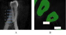
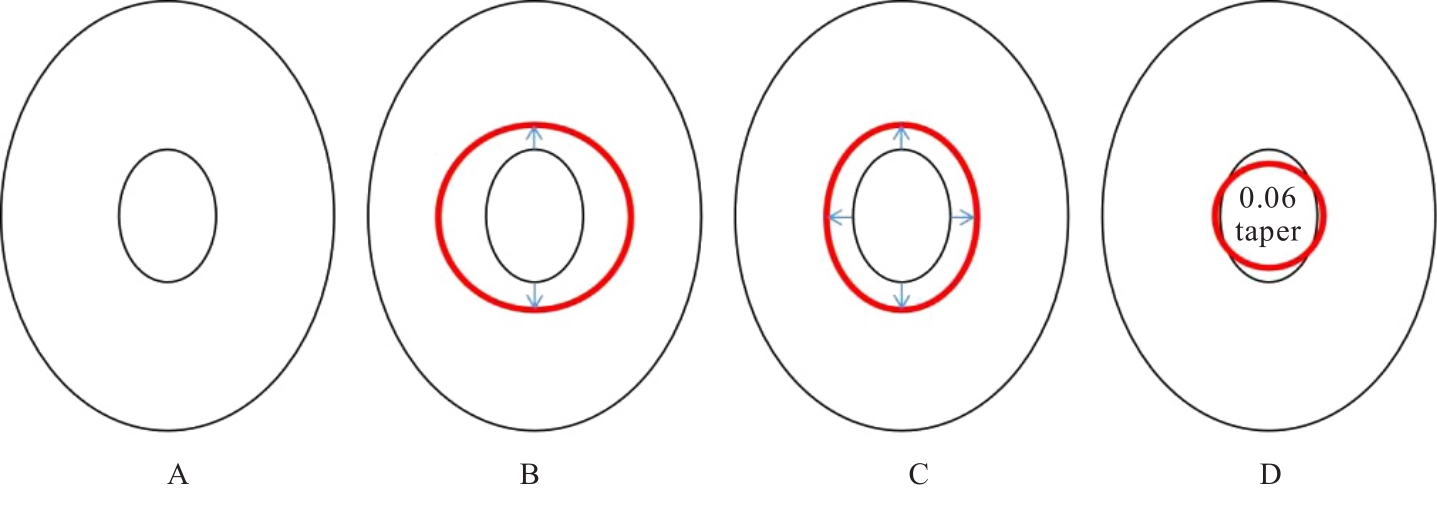
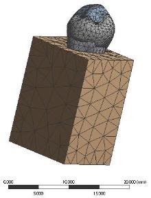
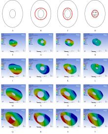
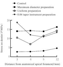
目的 分析下颌第一前磨牙根管直径并利用有限元分析法模拟3种不同预备方式下牙本质的应力分布,为临床下颌第一前磨牙根管预备策略提供依据。 方法 选择21例锥形束计算机断层扫描(CBCT)图像资料完整的患者,将CBCT原始数据DICOM格式文件导入Mimics 21.0软件,测量距下颌第一磨牙根尖孔3、6、9和12 mm处根管直径并分段计算根管锥度,在此基础上构建牙体组织和牙周组织三维有限元模型,分为对照组、最大直径预备组、均匀预备组和0.06锥度器械预备组。在ANSYS Workbench 17.0有限元分析软件中,对各组颊尖舌斜面分别施加200 N载荷,分析各组下颌第一前磨牙牙本质所受应力。 结果 距离下颌第一前磨牙根尖孔3~6 mm段、6~9 mm段和9~12 mm段的根管锥度分析,近远中向距根尖孔3~6 mm根管锥度基本相同;颊舌向距根尖孔6~9 mm段根管锥度平均为0.29,大于根尖1/3和根上1/3。在相同的载荷下,各组下颌第一前磨牙牙本质的应力峰值依次增大,分别为4.693 6、16.304 0、14.278 0和18.682 0 MPa。最大直径预备组受力集中于根管壁且应力最大,均匀预备组受力集中于牙根表面且各截面应力均小于最大直径预备组,0.06锥度器械预备组应力集中于根尖1/3牙根表面。 结论 下颌第一前磨牙根管具有椭圆形变锥度的特殊形态,近远中向和颊舌向根管直径与锥度相差较大,不同预备方式对根管壁的应力不同,均匀预备扩大根管的预备方式最佳。

关键词: 牙根纵裂, 根管直径, 根管预备, 有限元分析, 应力分析

Abstract:

Objective To analyze the root canal diameter of the mandibular first premolar by using finite element analysis to simulate the stress distribution of dentin under three different preparation methods,and to provide the basis for clinical root canal preparation strategies of the mandibular first premolars. Methods Twenty-one patients with complete cone beam computed tomography (CBCT) images were selected. The original DICOM format data from CBCT were imported into Mimics 21.0 software to measure the root canal diameter at 3, 6, 9, and 12 mm from the apex and the root canal taper was segmentally calculated. Based on this, three-dimensional finite element models of the dental and periodontal tissues were constructed. Control group, maximum diameter preparation group, uniform preparation group, and 0.06 taper instrument preparation group were designed. In ANSYS Workbench 17.0 finite element analysis software, a 200 N load was applied to the buccal, lingual, and occlusal surfaces in various groups, and the stresses on dentin in various groups were analyzed. Results The analysis of root canal taper at 3-6 mm, 6-9 mm, and 9-12 mm from the apex of mandibular first premolar teeth showed that the taper was similar in the mesial-distal direction at 3-6 mm from the apex. The average taper in the buccal-lingual direction at 6-9 mm from the apex was 0.29, which was greater than the taper in the apical 1/3 and coronal 1/3. Under the same load, the peak stress values in dentin of mandibular first premolar teech in various groups were increased sequentially: 4.693 6, 16.304 0, 14.278 0, and 18.682 0 MPa. The stress in maximum diameter preparation group concentrated on the canal wall with the highest stress value. The stress in uniform preparation group concentrated on the root surface,and the stress values on each section were lower than those in maximum diameter preparation group. The stress in 0.06 taper instrument preparation group concentrated on the apical 1/3 of the root surface. Conclusion The root canal of the mandibular first premolar has a unique elliptical taper shape, and there are significant differences in diameter and taper between the mesial-distal and buccal-lingual directions. Different preparation methods result in different stress distributions on the canal wall, and the uniform preparation is the best method for enlarging the canal.

Key words: Vertical root fracture, Root canal diameter, Root canal preparation, Finite element analysis, Stress analysis

中图分类号: 

  • R78