吉林大学学报(医学版) ›› 2025, Vol. 51 ›› Issue (2): 325-332.doi: 10.13481/j.1671-587X.20250206

• 基础研究 • 上一篇    

小鼠骨髓间充质干细胞通过JAK2/STAT3信号通路对成纤维细胞增殖和胶原表达水平的影响

黎涵玥,阳莲,刘剑锋,张舒飞,洪莉()   

  1. 武汉大学人民医院妇产科,湖北 武汉 430060
  • 收稿日期:2024-06-14 接受日期:2024-07-12 出版日期:2025-03-28 发布日期:2025-04-22
  • 通讯作者: 洪莉 E-mail:drhongli@gmail. com
  • 作者简介:黎涵玥(1999-),女,福建省南平市人,在读硕士研究生,主要从事女性盆底功能障碍性疾病方面的研究。
  • 基金资助:
    国家自然科学基金项目(82371639);国家重点研发计划项目(2021YFC2701300);湖北省 科技厅自然科学基金项目(2022CFB124);湖北省科技厅重点研发项目(2022BCA045);湖北省领军人才项目(〔2019〕47)

Effect of bone marrow mesenchymal stem cells of mice on proliferation and collagen expression levels of fibroblasts through JAK2/STAT3 signaling pathway

Hanyue LI,Lian YANG,Jianfeng LIU,Shufei ZHANG,Li HONG()   

  1. Department of Obstetrics and Gynecology,Renmin Hospital,Wuhan University,Wuhan 430060,China
  • Received:2024-06-14 Accepted:2024-07-12 Online:2025-03-28 Published:2025-04-22
  • Contact: Li HONG E-mail:drhongli@gmail. com

摘要:

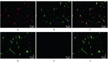
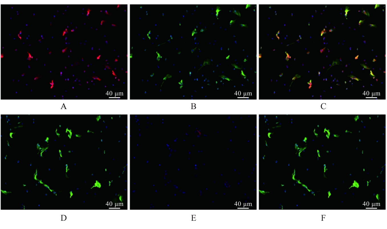
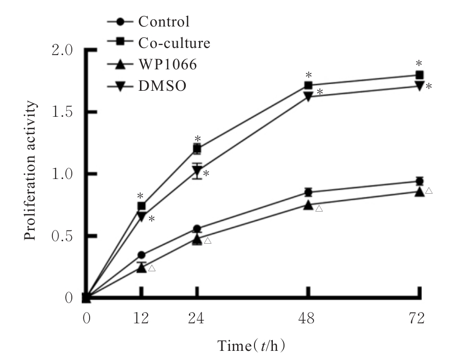
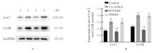
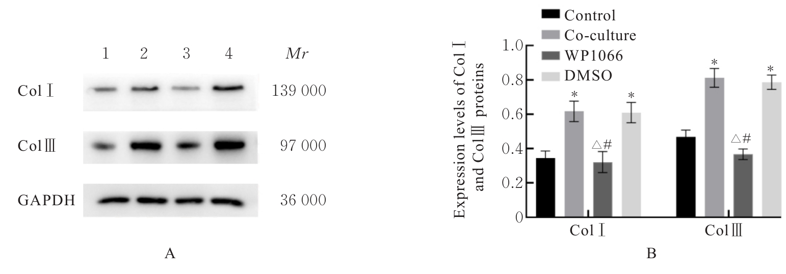
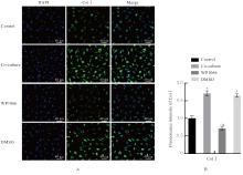
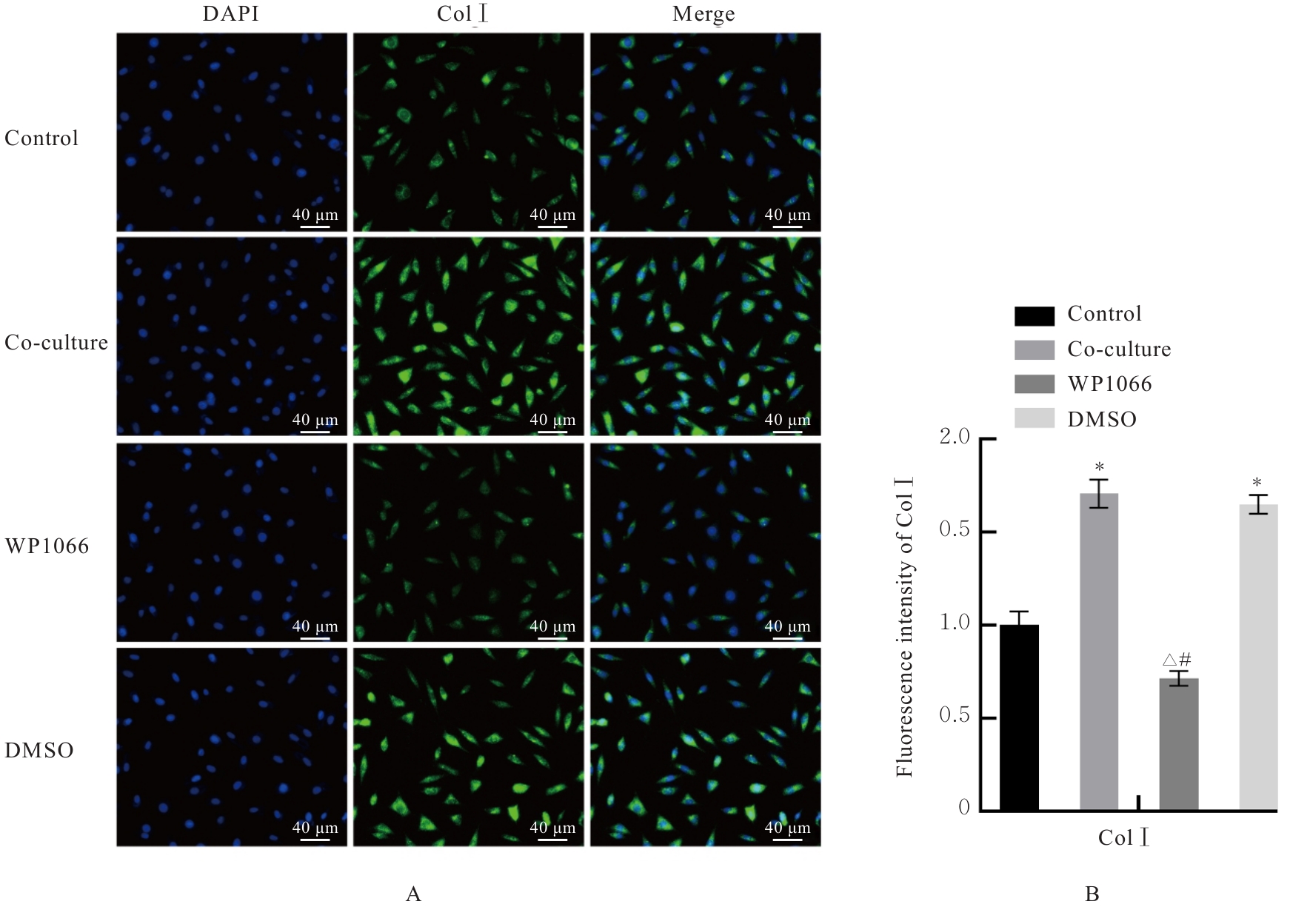
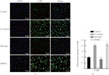
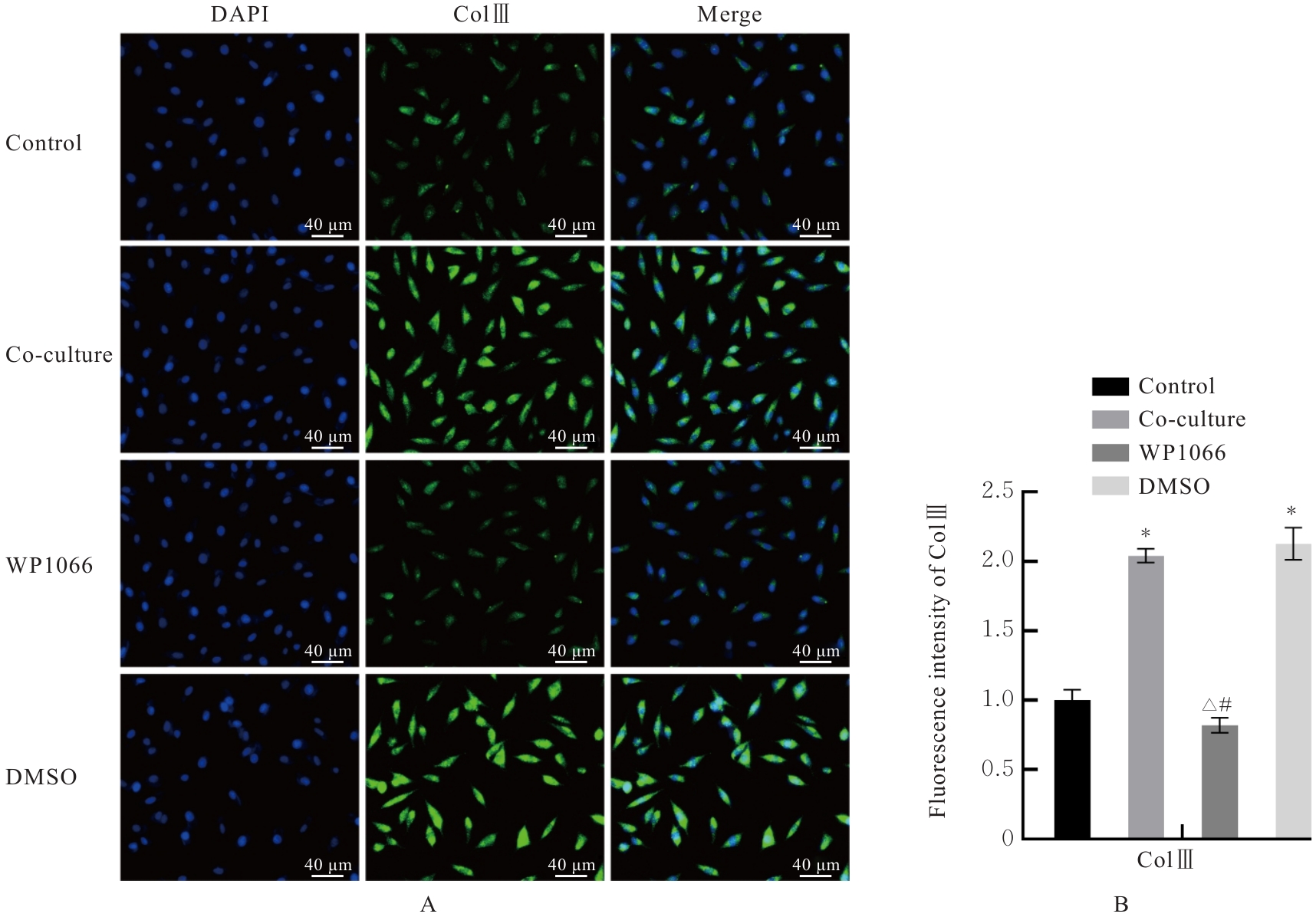
目的 探讨骨髓间充质干细胞(BMSCs)对L929细胞增殖和其胶原表达水平的影响,阐明其相关作用机制。 方法 提取4周龄C57BL/6小鼠的BMSCs。采用免疫荧光染色鉴定BMSCs的表型。将L929细胞分为对照组(L929细胞)、共培养组(L929细胞与BMSCs)、Janus激酶(JAK)抑制剂WP1066组(WP1066处理L929细胞与BMSCs)和二甲基亚砜(DMSO)组(DMSO处理L929细胞与BMSCs)。细胞计数试剂盒8(CCK-8)法检测不同时间点各组L929细胞增殖活性,Western blotting法检测各组L929细胞中Ⅰ型胶原蛋白(ColⅠ)和Ⅲ型胶原蛋白(ColⅢ)表达水平,免疫荧光染色法检测各组L929细胞中ColⅠ和ColⅢ蛋白表达。 结果 BMSCs表面抗原(SA)荧光检测,BMSCs表达表面标志物CD29+、CD45-、CD90+和CD105+。CCK-8法检测,与对照组比较,共培养组L929细胞增殖活性明显升高(P<0.01),DMSO组L929细胞增殖活性明显升高(P<0.01);与共培养组比较,WP1066组L929细胞增殖活性明显降低(P<0.01)。Western blotting法检测,与对照组比较,共培养组和DMSO组L929细胞中ColⅠ及ColⅢ蛋白表达水平明显升高(P<0.01);与共培养组比较,WP1066组L929细胞中ColⅠ和ColⅢ蛋白表达水平明显降低(P<0.01);与DMSO组比较,WP1066组L929细胞中ColⅠ和ColⅢ蛋白表达水平明显降低(P<0.01)。免疫荧光染色法检测,与对照组比较,共培养组和DMSO组L929细胞中ColⅠ及ColⅢ荧光强度明显升高(P<0.01);与共培养组比较,WP1066组L929细胞中ColⅠ和ColⅢ荧光强度明显降低(P<0.01);与DMSO组比较,WP1066组L929细胞中ColⅠ和ColⅢ荧光强度明显降低(P<0.01)。 结论 间充质干细胞可通过JAK2/信号转导和转录激活子3(STAT3)信号通路促进小鼠L929细胞增殖及胶原生成。

关键词: 盆底功能障碍性疾病, 骨髓间充质干细胞, 成纤维细胞, Ⅰ型胶原蛋白, Ⅲ型胶原蛋白, Janus激酶2, 信号转导和转录激活子3

Abstract:

Objective To investigate the effect of bone marrow mesenchymal stem cells (BMSCs) on the proliferation and collagen expression levels of L929 cells, and to clarify its related mechanism. Methods The BMSCs were extracted from the 4-week-old C57BL/6 mice. The phenotypes of BMSCs were identified by immunofluorescence staining. The L929 cells were divided into control group (L929 cells), co-culture group (L929 cells and BMSCs), inhibitor of Janus kinase(JAK) WP1066 group (WP1066-treated L929 cells and BMSCs), and dimethyl sulfoxide (DMSO) group (DMSO-treated L929 cells and BMSCs). Cell counting kit-8 (CCK-8) assay was used to detect the proliferation activities of the L929 cells in various groups at different time points; Western blotting method was used to detect the expression levels of type Ⅰ collagen (ColⅠ) and type Ⅲ collagen (ColⅢ) in the L929 cells in various groups; immunofluorescence staining was used to detect the expressions of ColⅠ and ColⅢ proteins in the L929 cells in various groups. Results The fluorescence assay results of surface antigen(SA) showed that the surface markers CD29+, CD45-, CD90+ and CD105+ were found in the BMSCs. The CCK-8 assay results showed that compared with control group, the proliferation activities of the L929 cells in co-culture group and DMSO group were significantly increased(P<0.01); compared with co-culture group, the proliferation activity of the L929 cells in WP1066 group was significantly decreased (P<0.01). The Western blotting method results showed that compared with control group, the expression levels of ColⅠ and ColⅢ proteins in the L929 cells in co-culture group and DMSO group were significantly increased (P<0.01); compared with co-culture group, the expression levels of ColⅠ and ColⅢ proteins in the L929 cells in WP1066 group were significantly decreased (P<0.01); compared with DMSO group, the expression levels of ColⅠ and ColⅢ proteins in the L929 cells in WP1066 group were significantly decreased (P<0.01). The immunofluorescence staining results showed that compared with control group, the fluorescence intensities of ColⅠ and ColⅢ proteins in the L929 cells in co-culture group and DMSO group were significantly increased(P<0.01); compared with co-culture group, the fluorescence intensities of ColⅠ and ColⅢ in the L929 cells in WP1066 group were significantly decreased(P<0.01); compared with DMSO group, the fluorescence intensities of ColⅠ and ColⅢ in the L929 cells in WP1066 group were significantly decreased (P<0.01). Conclusion MSCs can promote the proliferation and collagen production of the L929 cells of the mice through the JAK2/signal transducer and activator of transcription 3(STAT3) signaling pathway.

Key words: Pelvic floor dysfunction, Bone marrow mesenchymal stem cell, Fibroblast, Type Ⅰ collagen, Type Ⅲ collagen, Janus kinase 2, Signal transducer and activator of transcription 3

中图分类号: 

  • R711