吉林大学学报(医学版) ›› 2022, Vol. 48 ›› Issue (3): 606-614.doi: 10.13481/j.1671-587X.20220308

• 基础研究 • 上一篇    

骨髓间充质干细胞来源外泌体诱导自噬对MPP+抑制SH-SY5Y细胞存活的影响及其机制

汪文涛1,米旭光2,周阳1,蒲文星1,高佳旭3,景猛4,孟繁凯4()   

  1. 1.长春中医药大学临床医学院外科学,吉林 长春 130117
    2.吉林省人民医院中心实验室,吉林 长春 130021
    3.吉林释然司法鉴定中心法医组,吉林 长春 130022
    4.吉林省人民医院神经外科,吉林 长春 130021
  • 收稿日期:2021-12-28 出版日期:2022-05-28 发布日期:2022-06-21
  • 通讯作者: 孟繁凯 E-mail:mfk200372019@126.com
  • 作者简介:汪文涛(1992-),男,河南省南阳市人,在读硕士研究生,主要从事脑损伤保护机制方面的研究。
  • 基金资助:
    吉林省科技厅科技发展计划项目(20200404125YY)

Effect of autophagy induced by exosomes derived from bone marrow mesenchymal stem cells on survival of SH-SY5Y cells inhibited by MPP+ and its mechanism

Wentao WANG1,Xuguang MI2,Yang ZHOU1,Wenxing PU1,Jiaxu GAO3,Meng JING4,Fankai MENG4()   

  1. 1.Department of Surgery, Changchun University of Chinese Medicine, Changchun 130117, China
    2.Central Laboratory, People’s Hospital, Jilin Province, Changchun 130021, China
    3.Department of Forensic Medicine, Jilin Shiran Forensic Expertise Center, Changchun 130022, China
    4.Department of Neurosurgery, People’s Hospital, Jilin Province, Changchun 130021, China
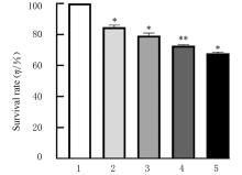
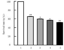
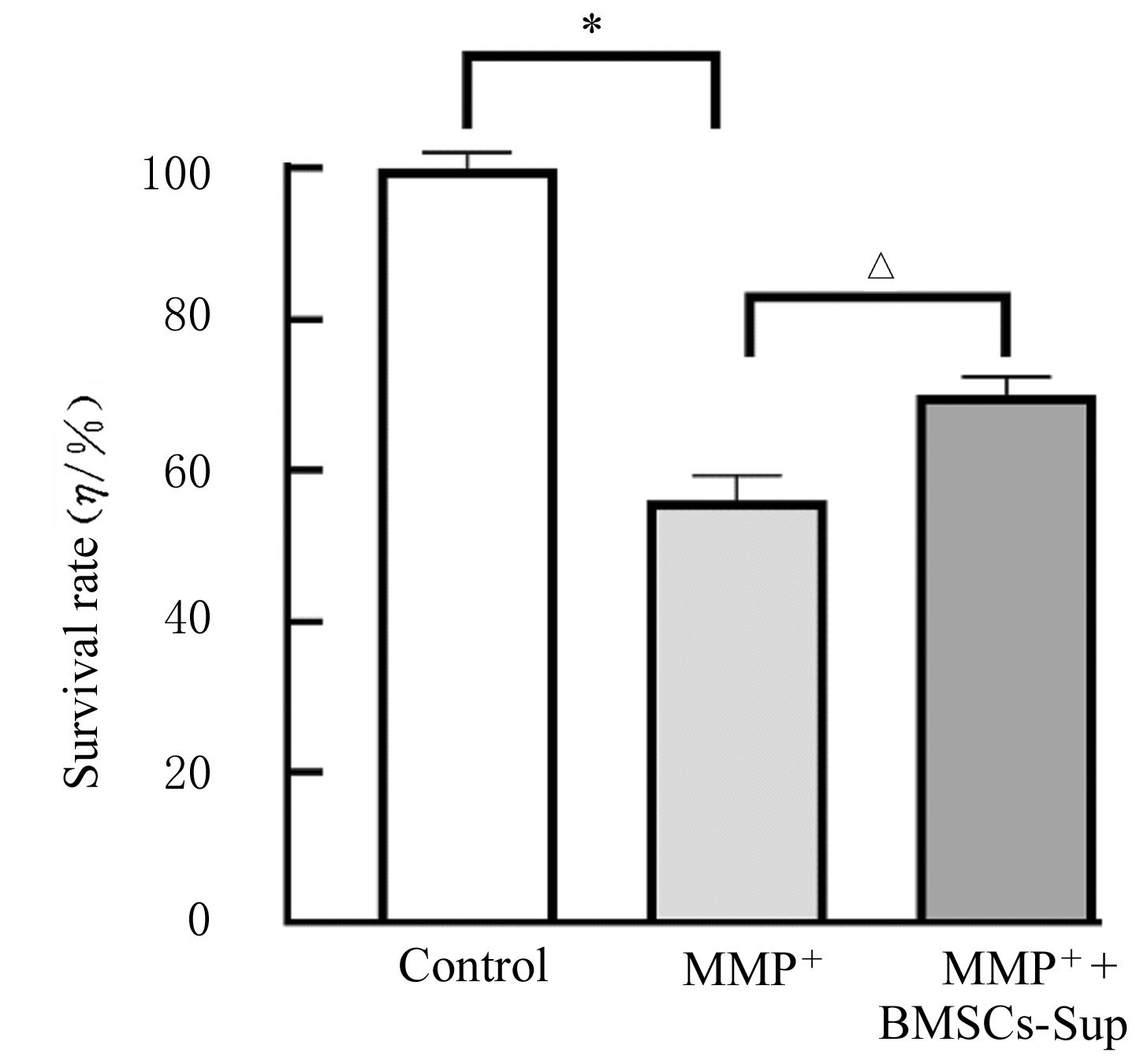
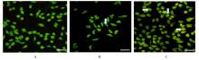
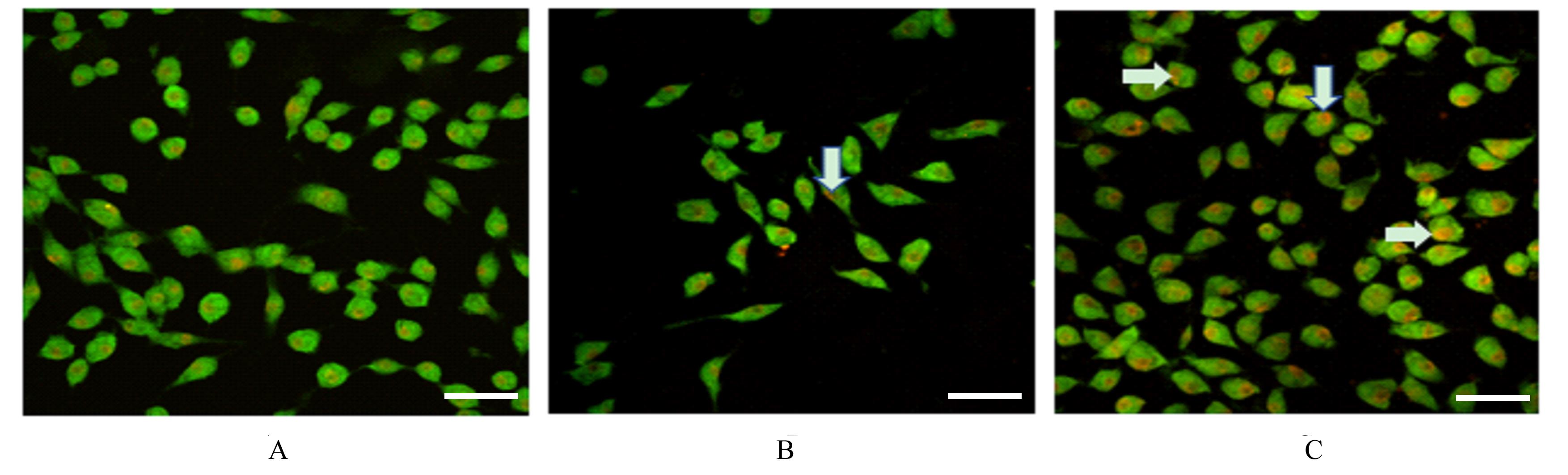
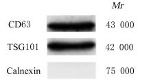
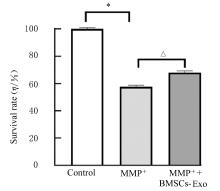
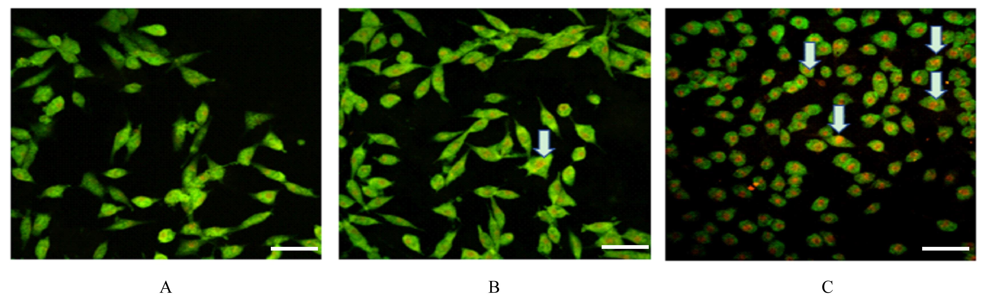
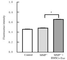
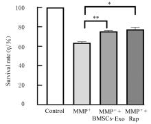
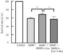
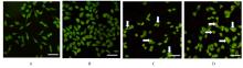
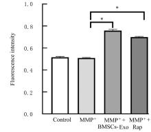
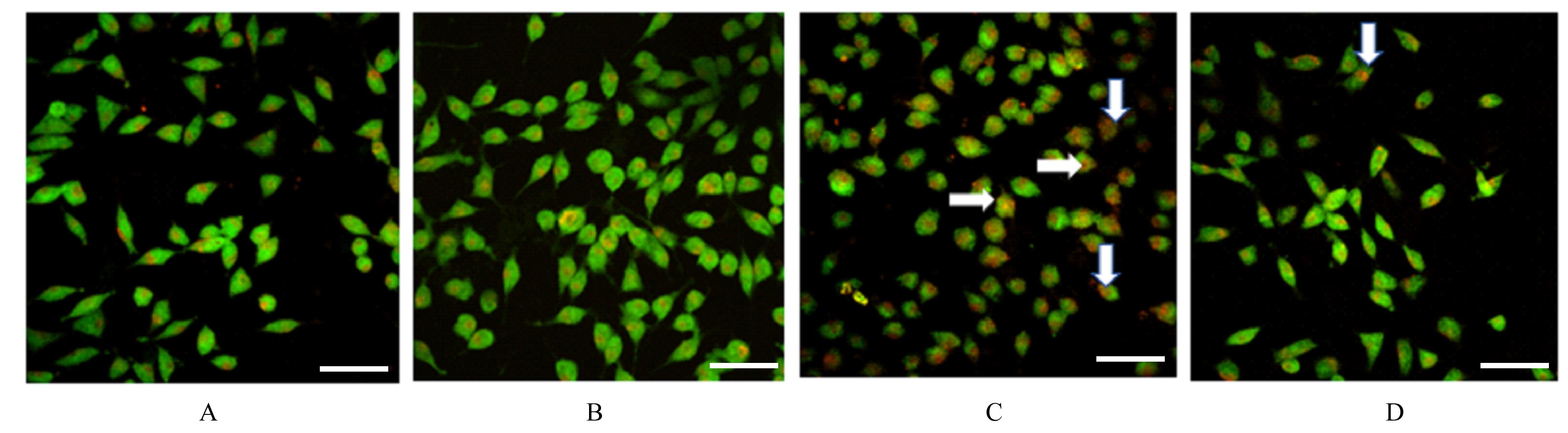
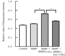
  • Received:2021-12-28 Online:2022-05-28 Published:2022-06-21
  • Contact: Fankai MENG E-mail:mfk200372019@126.com

摘要: 目的

探讨骨髓间充质干细胞(BMSCs)来源外泌体(Exo)诱导自噬对1-甲基-4-苯基吡啶离子(MPP+)抑制人神经母细胞瘤SH-SY5Y细胞存活的影响,阐明其作用机制。

方法

将人神经细胞肿瘤SH-SY5Y细胞分别给予不同浓度(0、0.25、0.50、1.00和2.00 mmol·L-1)MMP+作用24和48 h后检测各组细胞存活率。实验分为对照组[给予磷酸盐缓冲液(PBS)]、MPP+组(给予0.50 mmol·L-1 MPP+)、MPP++BMSCs的上清液(BMSCs-Sup)组(给予0.50 mmol·L-1 MPP+和BMSCs Sup)、MPP++BMSCs-Exo组(给予0.50 mmol·L-1 MPP+和100 mg·L-1 BMSCs Exo)、MPP++雷帕霉素(Rap)组(给予0.50 mmol·L-1 MPP+和2 mmol·L-1 Rap)和MPP++BMSCs-Exo+3-甲基腺嘌呤(3-MA)组(给予0.50 mmol·L-1 MPP+、100 mg·L-1 BMSCs-Exo和1 mmol·L-1 3-MA)。MTT法检测各组SH-SY5Y细胞存活率,共聚焦显微镜下观察吖啶橙染色后各组SH-SY5Y细胞中自噬酸性囊泡荧光强度,Western blotting法检测各组细胞中CD63、TGS101和Calnexin蛋白表达情况。

结果

MTT检测,SH-SY5Y细胞存活率随着MPP+浓度的升高或处理时间的延长逐渐降低(P<0.05),且呈浓度和时间依赖性。与MPP+组比较,MPP++BMSCs-Sup组、MPP++BMSCs-Exo组和MPP++Rap组SH-SY5Y细胞存活率升高(P<0.05);与MPP++BMSCs-Exo组比较,MPP++BMSCs-Exo+3-MA组SH-SY5Y细胞存活率降低(P<0.05)。细胞吖啶橙染色,与MPP+组比较,MPP++BMSCs-Sup组、MPP++BMSCs-Exo组和MPP++Rap组SH-SY5Y细胞中自噬酸性囊泡荧光强度增加(P<0.05);与MPP++BMSCs-Exo组比较,MPP++BMSCs-Exo+3-MA组SH-SY5Y细胞中自噬酸性囊泡荧光强度降低(P<0.05)。Western blotting法检测,各组细胞中外泌体标志蛋白CD63和TGS101蛋白高表达,内质网标志蛋白Calnexin低表达。

结论

BMSCs-Exo通过激活细胞自噬抵抗MPP+诱导的SH-SY5Y细胞存活率下降。

关键词: 外泌体, 骨髓间充质干细胞, 自噬, 细胞存活率, SH-SY5Y细胞

Abstract: Objective

To investigate the effect of autophagy induced by exosomes (exo) derived from the bone marrow mesenchymal stem cells (BMSCs) on the survival of the human neuroblastoma SH-SY5Y cells inhibited by 1-methyl-4-phenylpyridine ion (MPP+), and to darify its mechanism.

Methods

The human neuroblastoma SH-SY5Y cells were given different concentrations(0,0.25,0.50,1.00,and 2.00 mmol·L-1)MMP+,and the survival rates of the SH-SY5Y cells were detected after 24 and 48 h.The experiment was divided into control group[given phosphate buffered saline(PBS)], 0.50 mmol·L-1 MPP+ group (given 0.50 mmol·L-1 MPP+),MPP++BMSCs supernatnt (BMSCs-Sup)group (given 0.50 mmol·L-1 MPP++BMSCs-Sup),0.50 mmol·L-1 MPP++BMSCs-Exo group (given 0.50 mmol·L-1 MPP+ +100 mg·L-1 BMSCs-Exo),MPP+ +rapamycin(Rap) group (given 0.50 mmol·L-1 MPP+ +2 mmol·L-1 Rap) and MPP+ +BMSCs-Exo+3-methyladenine(3-MA) group(given 0.50 mmol·L-1 MPP+ +100 mg·L-1 BMSCs-Exo +1 mmol·L-1 3-MA). The survival rates of the SH-SY5Y cells in various groups were detected by MTT assay.The fluorescence intensities of autophagic acid vesicles in the SH-SY5Y cells in various groups were detected by acridine orange staining under confocal microscope.The expression levels of CD63, TGS101,and Calnexin proteins in the SH-SY5Y cells in various groups were detected by Western blotting method.

Results

The MTT assay results showed that the survival rates of the SH-SY5Y cells were decreased gradually with the increasing of the MPP+ concentration or the extension of treatment time (P<0.05) in a concentration- and time-dependent manner. Compared with MPP+ group, the survival rates of the SH-SY5Y cells in MPP+ + BMSCs-Sup group, MPP+ +BMSCs-Exo group,and MPP+ + Rap group were increased (P<0.05). Compared with MPP+ +BMSCs-Exo group, the survival rate of the SH-SY5Y cells in MPP+ +BMSCs-Exo + 3-MA group was decreased (P<0.05).The acridine orange staining results showed that compared with control group, the fluorescence intensities of autophagic acid vesicles in the SH-SY5Y cells in MPP+ + BMSCs-Sup group, MPP+ +BMSCs-Exo group and MPP+ + Rap group were increased (P<0.05); compared with MPP+ +BMSCs-Exo group, the fluorescence intensity of autophagic acid vesicles in the SH-SY5Y cells in MPP+ +BMSCs-Exo + 3-MA group was decreased (P<0.05).The Western blotting results showed that the exosome marker proteins CD63 and TGS101 were highly expressed, while the endoplasmic reticulum marker protein Calnexin was lowly expressed.

Conclusion

BMSCs-Exo can resist the MPP+-induced decreasing of the survival of the SH-SY5Y cells by activating the autophagy.

Key words: Exosomes, Bone marrow mesenchymal stem cells, Autophagy, Cell viability, SH-SY5Y cells

中图分类号: 

  • R285.5