吉林大学学报(医学版) ›› 2025, Vol. 51 ›› Issue (6): 1542-1550.doi: 10.13481/j.1671-587X.20250610

• 基础研究 • 上一篇    下一篇

脂肪源性干细胞联合脱细胞支架对坐骨神经损伤大鼠脊神经节的保护作用及其机制

于晓敏1,朱清华2,王一伦2,任淼2,刘子嘉2,余泳仪2,杜元良3,刘东慧2,郭森2,付秀美2,4()   

  1. 1.承德医学院临床技能教学中心,河北 承德 067000
    2.承德医学院基础医学院人体解剖学 教研室,河北 承德 067000
    3.承德医学院附属医院骨外科,河北 承德 067000
    4.河北省神经损伤与修复重点实验室,河北 承德 067000
  • 收稿日期:2025-01-12 接受日期:2025-02-12 出版日期:2025-11-28 发布日期:2025-12-15
  • 通讯作者: 付秀美 E-mail:fuxiumei2012@163.com
  • 作者简介:于晓敏(1973-),女,河北省承德市人,副教授,医学硕士,主要从事神经损伤和修复方面的研究。
  • 基金资助:
    河北省科技厅自然科学基金项目(H2021406056);河北省教育厅高等学校科学技术研究项目(ZD2020178);承德医学院人体解剖与组织胚胎学优势学科资助项目(〔2023〕22号);承德医学院大学生创新创业训练计划项目(2022136);承德医学院大学生创新创业训练计划项目(2024074)

Protective effect of adipose-derived stem cells combined with acellular scaffolds on dorsal root ganglion in rats with sciatic nerve injury and its mechanism

Xiaomin YU1,Qinghua ZHU2,Yilun WANG2,Miao REN2,Zijia LIU2,Yongyi YU2,Yuanliang DU3,Donghui LIU2,Sen GUO2,Xiumei FU2,4()   

  1. 1.Clinical Skills Teaching Center,Chengde Medical University,Chengde 067000,China
    2.Department of Human Anatomy,School of Basic Medical Sciences,Chengde Medical University,Chengde 067000,China
    3.Department of Bone Surgery,Affiliated Hospital,Chengde Medical University,Chengde 067000,China
    4.Key Laboratory of Nerve Injury and Repair,Hebei Province,Chengde 067000,China
  • Received:2025-01-12 Accepted:2025-02-12 Online:2025-11-28 Published:2025-12-15
  • Contact: Xiumei FU E-mail:fuxiumei2012@163.com

摘要:

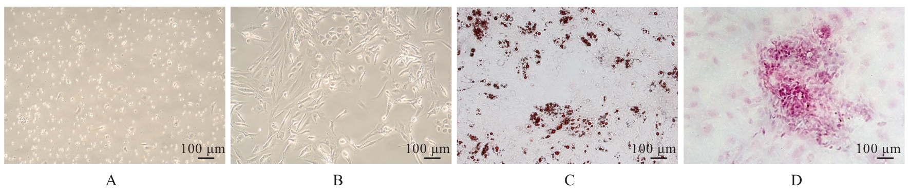
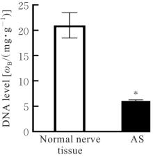
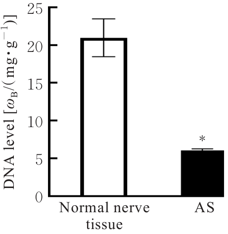
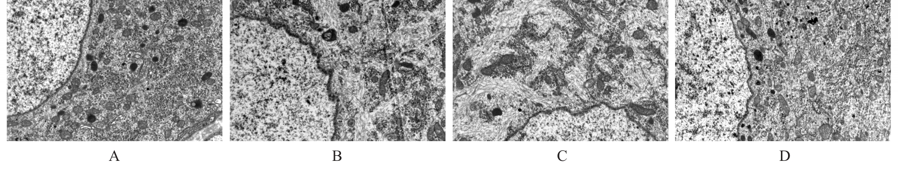
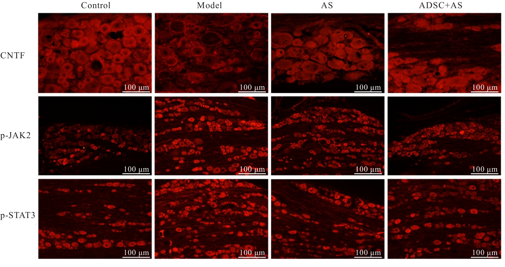
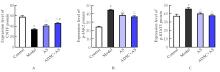
目的 观察脂肪源性干细胞(ADSC)联合脱细胞支架(AS)对坐骨神经损伤(SNI)大鼠脊神经节超微结构和睫状神经营养因子(CNTF)、Janus激酶2(JAK2)、磷酸化JAK2(p-JAK2)、信号转导与转录激活因子3(STAT3)和磷酸化STAT3(p-STAT3)蛋白及mRNA表达的影响,阐明ADSC联合AS对SNI大鼠脊神经节的保护作用及其可能机制。 方法 分离培养大鼠ADSC并检测其多向分化潜能。制备大鼠AS,将ADSC注入至AS中构建组织工程神经。36只大鼠随机分为对照组、模型组、AS组和ADSC+AS组。对照组大鼠常规饲养,不进行任何处理,其余各组大鼠采用切除右侧坐骨神经10 mm的方法建立SNI模型,模型组不再处理,AS组和ADSC+AS组大鼠分别将AS和构建的组织工程神经桥接于损伤神经的两断端处。术后6周,采用透射电镜观察各组大鼠脊神经节超微结构,免疫荧光法检测各组大鼠脊神经节中CNTF、p-JAK2和p-STAT3蛋白表达水平,实时荧光定量PCR(RT-qPCR)法检测各组大鼠脊髓神经节中CNTFJAK2STAT3 mRNA表达水平。 结果 原代ADSC培养7 d,倒置显微镜下可见数量较多、体积较大且呈长梭形的细胞,类似簇状或旋涡状排列;油红O染色镜下可见细胞中红色的脂滴,茜素红染色镜下可见钙化结节,表明分离培养的细胞具有多向分化能力。与正常神经组织比较,大鼠AS中DNA水平明显出降低(P<0.05)。与对照组比较,模型组大鼠脊神经节中细胞核膜凹凸不平,呈锯齿状改变,胞质内细胞器数目减少,线粒体肿胀、嵴断裂或缺失,结构不清晰;CNTF蛋白和mRNA表达水平均明显降低(P<0.01),p-JAK2和p-STAT3蛋白表达水平均明显升高(P<0.01),JAK2STAT3 mRNA表达水平均明显升高(P<0.01)。与模型组比较,AS组大鼠脊神经节中细胞核膜锯齿状改变明显减轻,胞质中细胞器数量增加,线粒体肿胀减轻;ADSC+AS组大鼠脊神经节中细胞核膜趋向完整,细胞器数量增加,线粒体肿胀和空泡化明显减轻;AS组和ADSC+AS组大鼠脊神经节中CNTF蛋白和mRNA表达水平均明显升高(P<0.01),p-JAK2和p-STAT3蛋白表达水平均明显降低(P<0.01),JAK2STAT3 mRNA表达水平明显降低(P<0.01)。与AS组比较,ADSC+AS组大鼠脊神经节中CNTF蛋白和mRNA表达水平均明显升高(P<0.05或P<0.01),p-JAK2和p-STAT3蛋白表达水平均明显降低(P<0.01),JAK2STAT3 mRNA表达水平明显降低(P<0.01)。 结论 ADSC联合AS应用可改善SNI大鼠脊神经节超微结构,其机制可能与ADSC联合AS应用可增加脊神经节中CNTF表达、降低JAK2/STAT3信号通路活化有关。

关键词: 脂肪源性干细胞, 脱细胞支架, 睫状神经营养因子, Janus激酶2, 信号转导与转录激活因子3信号通路, 脊神经节

Abstract:

Objective To observe the effects of adipose-derived stem cells (ADSC) combined with acellular scaffold (AS) on the ultrastructure of dorsal root ganglion and the protein and mRNA expression levels of ciliary neurotrophic factor (CNTF), Janus kinase 2 (JAK2), phosphorylated JAK2 (p-JAK2), signal transducer and activator of transcription 3(STAT3) and phosphorylated STAT3(p-STAT3) in the rats with sciatic nerve injury (SNI), and to clarify the protective effect of ADSC combined with AS on dorsal root ganglion in the SNI rats and its possible mechanism. Methods The rat ADSCs were isolated and cultured and their multidirectional differentiation potential was detected. The AS of rats was prepared, and ADSCs were injected into the AS to construct tissue-engineered nerve. A total of 36 rats were randomly divided into control group, model group, AS group, and ADSC+AS group. The rats in control group were routinely fed, and the rats in other groups were used to establish the SNI models by resecting 10 mm of right sciatic nerve. The rats in model group received no further treatment, while the rats in AS group and ADSC+AS group were bridged with AS and the constructed tissue-engineered nerve at the two ends of the injured nerve, respectively. At 6 weeks after surgery, transmission electron microscope was used to observe the ultrastructure of dorsal root ganglion of the rats in various groups; immunofluorescence method was used to detect the protein expression levels of CNTF, p-JAK2, and p-STAT3 in dorsal root ganglion of the rats; real-time fluorescence quantitative PCR (RT-qPCR) method was used to detect the mRNA expression levels of CNTFJAK2, and STAT3 in dorsal root ganglion of the rats in various groups. Results After 7 d of primary ADSC culture, a large number of large and long spindle-shaped cells were observed under the inverted microscope, arranged in clusters or whirlpools; red lipid droplets were observed with oil red O staining under microscope, and calcified nodules were observed with Alizarin red staining under microscope, indicating that the isolated and cultured cells had multidirectional differentiation ability.Compared with normal nerve tissue, the level of DNA in AS of rats was significantly decreased (P<0.05). Compared with control group, the nuclear membrane of dorsal root ganglion cells in model group was uneven and serrated, the number of organelles in the cytoplasm was decreased, mitochondria were swollen with broken or missing cristae and unclear structure; the CNTF protein and mRNA expression levels were significantly decreased (P<0.01), the p-JAK2 and p-STAT3 protein expression levels were significantly increased (P<0.01), and the JAK2 and STAT3 mRNA expression levels were significantly increased (P<0.01). Compared with model group, the serrated change of nuclear membrane of the dorsal root ganglion cells in AS group was significantly alleviated, the number of organelles in the cytoplasm was increased, and mitochondrial swelling was reduced; in ADSC+AS group, the nuclear membrane of dorsal root ganglion cells tended to be intact, the number of organelles was increased, and mitochondrial swelling and vacuolization were significantly reduced; the CNTF protein and mRNA expression levels in the dorsal root ganglion in AS group and ADSC+AS group were significantly increased (P<0.01), the p-JAK2 and p-STAT3 protein expression levels were significantly decreased (P<0.01), and the JAK2 and STAT3 mRNA expression levels were significantly decreased (P<0.01). Compared with AS group, the CNTF protein and mRNA expression levels in ADSC+AS group were significantly increased (P<0.05 or P<0.01), the p-JAK2 and p-STAT3 protein expression levels were significantly decreased (P<0.01), and the JAK2 and STAT3 mRNA expression levels were significantly decreased (P<0.01). Conclusion The application of ADSC combined with AS can improve the ultrastructure of dorsal root ganglion in the SNI rats, and the mechanism may be related to the increased CNTF expression and decreased activation of the JAK2/STAT3 signaling pathway in the dorsal root ganglion by ADSC combined with AS application.

Key words: Adipose-derived stem cells, Acellular scaffold, Ciliary neurotrophic factor, Janus kinase 2, Signal transducer and activator of transcription 3 signaling pathway, Dorsal root ganglion

中图分类号: 

  • R322.85