吉林大学学报(医学版) ›› 2025, Vol. 51 ›› Issue (6): 1532-1541.doi: 10.13481/j.1671-587X.20250609

• 基础研究 • 上一篇    

敲减血红素结合蛋白1基因对小胶质细胞BV2增殖、迁移及炎症反应的影响及其机制

冯思凡1,李昀峰2,王嘉营1,马福槟1,王岩1()   

  1. 1.广东医科大学附属医院神经病学研究所 广东省衰老相关心脑疾病重点实验室,广东 湛江 524001
    2.广东省高州市人民医院神经内科,广东 高州 525200
  • 收稿日期:2025-01-20 接受日期:2025-02-22 出版日期:2025-11-28 发布日期:2025-12-15
  • 通讯作者: 王岩 E-mail:jwangyan@gdmu.edu.cn
  • 作者简介:冯思凡(1999-),女,陕西省咸阳市人,在读硕士研究生,主要从事阿尔茨海默病中小胶质细胞作用及其机制方面的的研究。
  • 基金资助:
    广东省科技厅自然科学基金基础与应用基础研究面上项目(2024A1515011464);广东省科技厅自然科学基金基础与应用基础研究面上项目(2022A1515010593);广东医科大学附属医院高层次人才科研启动项目(GCC2021010)

Effects of heme-binding protein 1 gene knockdown on proliferation, migration, and inflammatory response of microglia BV2 and their mechanisms

Sifan FENG1,Yunfeng LI2,Jiaying WANG1,Fubin MA1,Yan WANG1()   

  1. 1.Institute of Neurology,Affiliated Hospital,Guangdong Medical University,Guangdong Provincial Key Laboratory of Age-Related Cardiac and Cerebral Diseases,Zhanjiang 524001,China
    2.Department of Neurology,People’s Hospital,Gaozhou City,Guangdong Province,Gaozhou 525200,China
  • Received:2025-01-20 Accepted:2025-02-22 Online:2025-11-28 Published:2025-12-15
  • Contact: Yan WANG E-mail:jwangyan@gdmu.edu.cn

摘要:

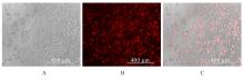
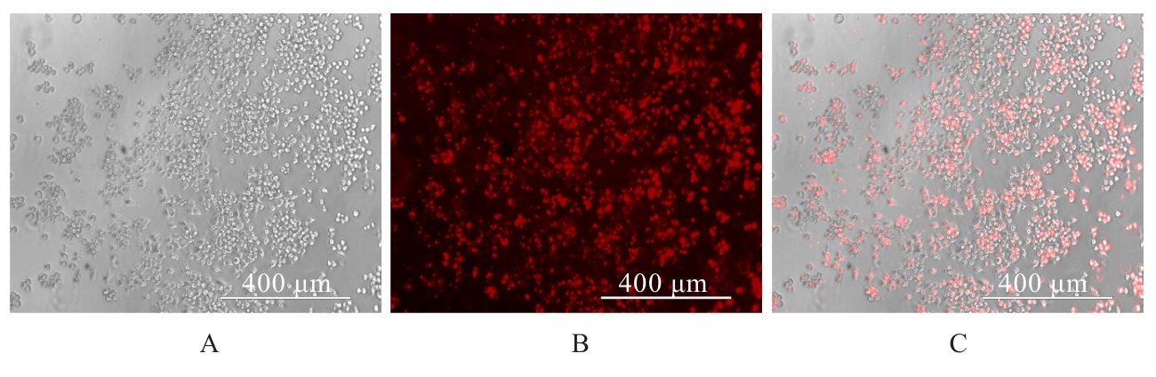
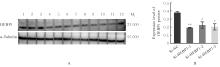
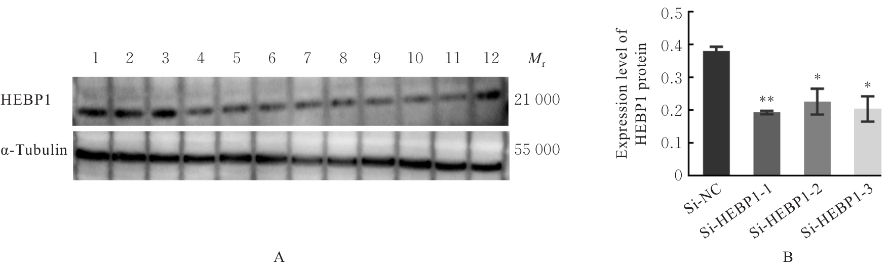
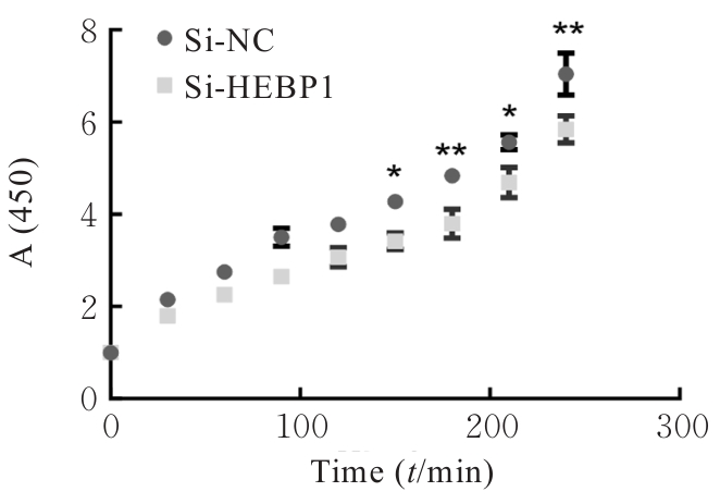
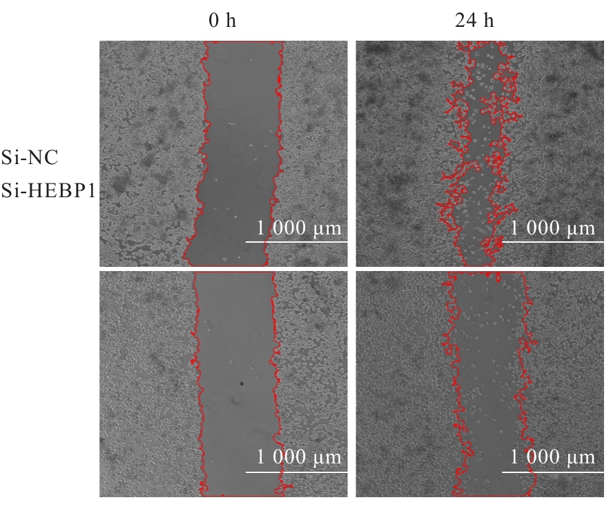
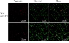
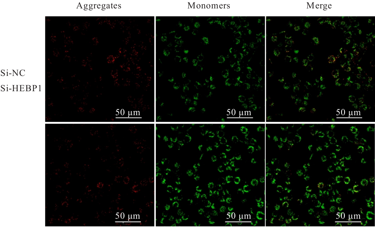
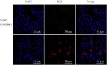
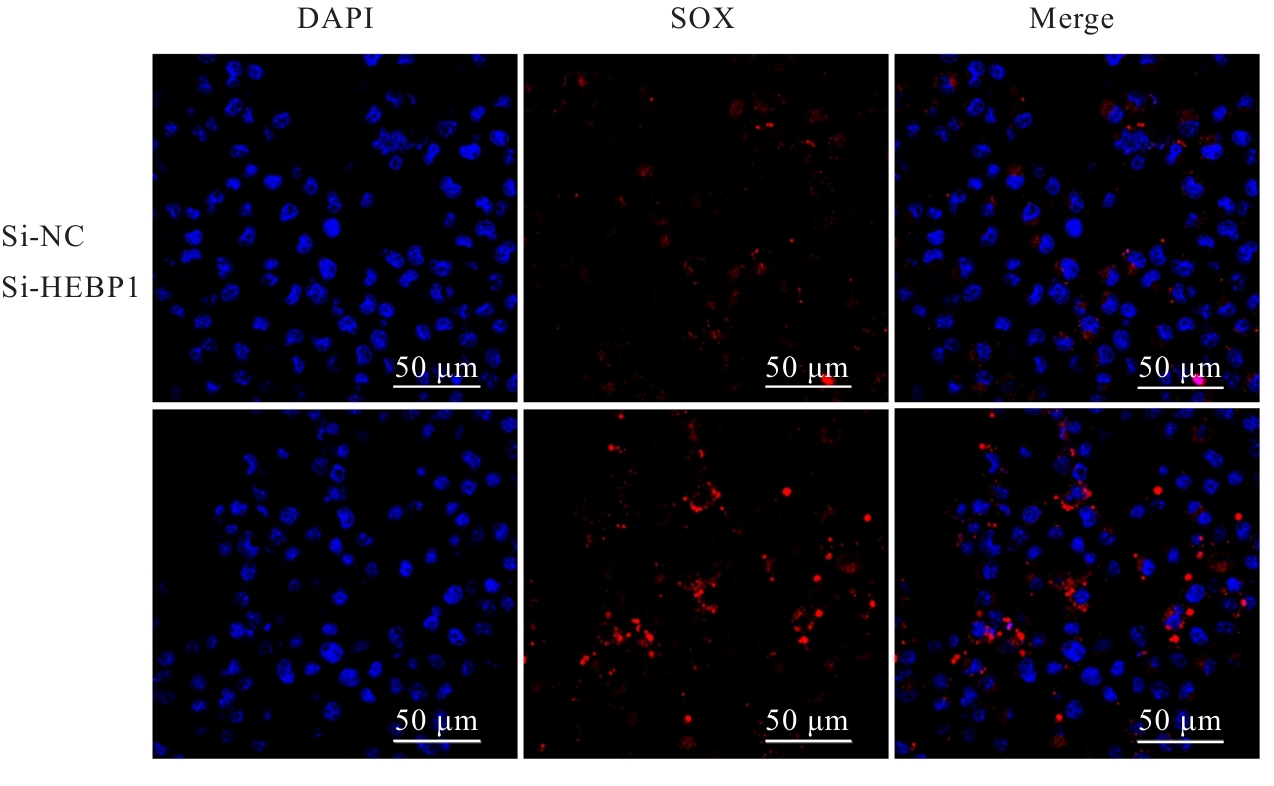
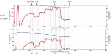
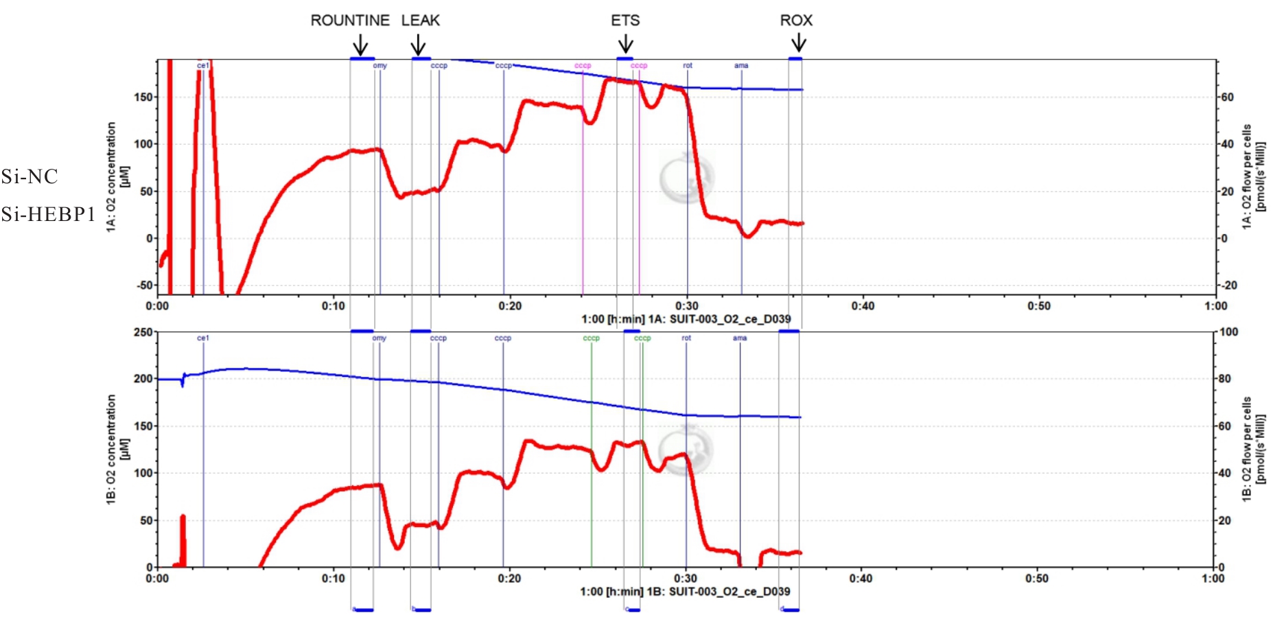
目的 探讨血红素结合蛋白1(HEBP1)基因下调对小胶质细胞BV2功能的影响,分析HEBP1在小胶质细胞中发挥的关键作用。 方法 构建阴性对照和HEBP1敲减的小干扰RNA(siRNA),敲减鼠源的小胶质细胞BV2中的HEBP1基因,获得HEBP1敲减BV2细胞模型。BV2细胞分为si-NC组、si-HEBP1-1组、si-HEBP1-2组和si-HEBP1-3组,采用实时荧光定量PCR法(RT-qPCR)法和Western blotting法检测细胞敲减后HEBP1 mRNA和蛋白表达水平,筛选出敲减效果最显著的siRNA用于后续实验。采用细胞计数试剂盒8(CCK-8)法检测si-NC组和si-HEBP1组BV2细胞增殖活性,细胞划痕实验检测2组BV2细胞迁移率,试剂盒检测2组BV2细胞中线粒体膜电位和活性氧(ROS)水平,线粒体呼吸功能测定仪检测2组BV2细胞线粒体呼吸功能。BV2细胞分为si-NC组、si-NC+脂多糖(LPS)组、si-HEBP1组和si-HEBP1+LPS组,RT-qPCR法检测各组BV2细胞中HEBP1、白细胞介素1β(IL-)、肿瘤坏死因子α(TNF-α)和白细胞介素6(IL-6) mRNA表达水平,Western blotting法检测各组BV2细胞中HEBP1蛋白表达水平。 结果 携带红色荧光标签CY3的siRNA转染BV2细胞,转染效率可达90%以上;与si-NC组比较,si-HEBP1-1组、si-HEBP1-2组和si-HEBP1-3组BV2细胞中HEBP1蛋白表达水平均明显降低(P<0.05或P<0.01),其中si-HEBP1-1组降低最明显;与si-NC组比较,si-HEBP1-1组、si-HEBP1-2组和si-HEBP1-3组BV2细胞中HEBP1 mRNA表达水平均明显降低(P<0.01),其中si-HEBP1-1组降低最明显,提示si-HEBP1-1是HEBP1敲减效果最好的siRNA,HEBP1敲减的BV2细胞模型构建成功。CCK-8法检测,与si-NC组比较,si-HEBP1组BV2细胞增殖活性明显降低(P<0.05或P<0.01);从90 min开始,2组细胞增殖活性差异更加明显。细胞划痕实验,与si-NC组比较,si-HEBP1组细胞迁移率明显降低(P<0.05)。荧光显微镜观察,与si-NC组比较,si-HEBP1组细胞线粒体膜电位明显降低(P<0.05);与si-NC组比较,si-HEBP1组BV2细胞中ROS水平明显升高(P<0.05)。细胞线粒体呼吸功能测定,与si-NC组比较,si-HEBP1组BV2细胞基础呼吸值(ROUNTINE)和细胞质子漏耗氧量(LEAK)均明显降低(P<0.05或P<0.01),2组细胞电子传递呼吸值(ETS)和剩余耗氧量(ROX)差异均无统计学意义(P>0.05);与si-NC组比较,si-HEBP1组BV2细胞三磷酸腺苷(ATP)生成量明显减少(P<0.05)。RT-qPCR法检测,与si-NC组比较,si-NC+LPS组BV2细胞中IL-TNF-αIL-6 mRNA水平均明显升高(P<0.01);与si-HEBP1组比较,si-HEBP1+LPS组BV2细胞中IL-TNF-αIL-6 mRNA水平均明显升高(P<0.01);与si-NC+LPS组比较,si-HEBP1+LPS组BV2细胞中IL-TNF-αIL-6 mRNA表达水平均明显升高(P<0.01)。 结论 敲减HEBP1基因可降低小胶质细胞BV2增殖和迁移能力,并增强对LPS刺激的炎症反应,其机制可能与BV2细胞线粒体功能受损和ATP产生减少有关。

关键词: 血红素结合蛋白1, 小胶质细胞, 线粒体功能, 神经炎症, 活性氧, 细胞增殖, 细胞迁移

Abstract:

Objective To explore the effect of heme-binding protein 1(HEBP1) down-regulation on the function of microglia BV2, and to clarify the key role of HEBP1 in the microglia. Methods Negative control and HEBP1 knockdown small interfering RNA (siRNA) were constructed to knockdown HEBP1 gene in mouse-derived microglial BV2, and the HEBP1 knockdown BV2 cell models were obtained. The BV2 cells were divided into si-NC group, si-HEBP1-1 group, si-HEBP1-2 group, and si-HEBP1-3 group. Real-time fluorescence quantitative PCR(RT-qPCR) and Western blotting methods were used to detect the expression levels of HEBP1 mRNA and protein in the BV2 cells after knockdown; the siRNA with the most significart knockdown effect was selected for stlbsequent expreriments.The proliferation abilities of the cells in si-NC group and si-HEBP1 group were detected by cell counting kit-8(CCK-8) assay, and the cell migration rates were assessed by scratch assay; the cellular mitochondrial membrane potential and reactive oxygen species (ROS) levels were detected by kits; the cellular mitochondrial respiratory function was detected by mitochondrial respirometer. The BV2 cells were divided into si-NC group, si-NC+lipopolysacch aride(LPS) group, si-HEBP1 group, and si-HEBP1+LPS group. RT-qPCR method was used to detect the expression levels of HEBP1, interleukin-1β(IL-),tumor necrosis factor-α(TNF-α),and interleukin-6(IL-6) mRNA in the BV2 cells in various groups, and Western blotting method was used to detect the expression levels of HEBP1 protein in the BV2 cells in various groups. Results When the BV2 cells were transfected with siRNA carrying with red fluorescence tag CY3, the transfect effricacy was above 90%; compared with si-NC group, the expression levels of HEBP1 protein in the BV2 cells in si-HEBP1-1 group, si-HEBP1-2 group, and si-HEBP1-3 group were significantly decreased (P<0.05 or P<0.01), especially in si-HEBP1-1 group. Compared with si-NC group, the expression levels of HEBP1 mRNA in the BV2 cells in si-HEBP1-1 group, si-HEBP1-2 group, and si-HEBP1-3 group were significantly decreased (P<0.01), especially in si-HEBP1-1 group; indicating that si-HEBP1-1 was the siRNA with best HEBP1 knowdown effect, and the HEBP1 knockdown BV2 cell model was successfully constructed. The CCK-8 resuts showed that compared with si-NC group, the proliferation activities of the BV2 cells in si-HEBP1 group were decreased (P<0.05 or P<0.01); from 90 min, the differences in proliferation activities of the BV2 cells in two groups were obvious. The cell scratch experiment results showed that compared with si-NC group, the cell migration rate in si-HEBP1 group was significantly decreased (P<0.05). The fluorescence microscope results showed that compared with si-NC group, the mitochondrial membrane potential of the BV2 cells in si-HEBP1 group was significantly decreased (P<0.05); compared with si-NC group, the ROS level in the BV2 cells in si-HEBP1 group was significantly increased(P<0.05). The mitochondrial respiration function testing results showed that compared with si-NC group, routine respiration(ROUNTINE) and leak respiration(LEAK) in si-HEBP1 group were significautly decreased (P<0.05 or P<0.01), and electron transfer system capacity(ETS) and residual oxygen consumption(ROX) had no significant differences (P>0.05); the ATP amount was decreased (P<0.05). The RT-qPCR results showed that compared with si-NC group, the expression levels of IL-TNF-α, and IL-6 mRNA in the BV2 cells in si-NC+LPS group were significantly decreased (P<0.01); compared with si-HEBP1 group, the expression levels of IL-TNF-α, and IL-6 mRNA in the BV2 cells in si-HEBP1+LPS group were significantly decreased (P<0.01); compared with si-NC+LPS group, the expression levels of IL-TNF-α, and IL-6 mRNA in the BV2 cells in si-HEBP1+LPS group were significantly increased (P<0.01). Conclusion Knockdown of HEBP1 gene can decrease the proliferation and migration abilities of the microglia BV2 and increase inflammatory response to LPS stimulus, and their mechanisms may be related to mitochondrial function damage and decreased ATP production of the BV2 cells.

Key words: Heme-binding protein 1, Microglia, Mitochondrial function, Neuroinflammation, Ractive oxygen species, Cell proliferation, Cell migration

中图分类号: 

  • R741.02