吉林大学学报(医学版) ›› 2021, Vol. 47 ›› Issue (1): 118-124.doi: 10.13481/j.1671-587x.20210116

• 基础研究 • 上一篇    下一篇

丹参酮ⅡA对宫腔黏连模型大鼠子宫内膜容受性的改善作用及其机制

刘静乔1,孟亚丽1(),徐淑稳1,王云灿2,王娜1,王玉静1   

  1. 1.河北医科大学第一医院妇科,河北 石家庄 050031
    2.河北医科大学第一医院超声科,河北 石家庄 050031
  • 收稿日期:2020-05-08 出版日期:2021-01-28 发布日期:2021-01-27
  • 通讯作者: 孟亚丽 E-mail:jingqiao8383@163.com
  • 作者简介:刘静乔(1983-), 女, 河北省晋州市人,主治医师,医学硕士,主要从事妇产科基础和临床方面的研究。
  • 基金资助:
    河北省卫健委医学科学研究项目(20190486)

Improvement effect of tanshinone ⅡA on endometrial receptivity of intrauterine adhesion model rats and its mechanism

Jingqiao LIU1,Yali MENG1(),Shuwen XU1,Yuncan WANG2,Na WANG1,Yujing WANG1   

  1. 1.Department of Gynecology,First Hospital,Hebei Medical University,Shijiazhuang 050031,China
    2.Department of Ultrasound,First Hospital,Hebei Medical University,Shijiazhuang 050031,China
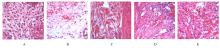
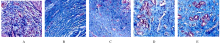
  • Received:2020-05-08 Online:2021-01-28 Published:2021-01-27
  • Contact: Yali MENG E-mail:jingqiao8383@163.com

摘要: 目的

观察宫腔黏连(IUA)模型大鼠子宫内膜中整合素αVβ3和白血病抑制因子(LIF)表达水平,探讨丹参酮ⅡA对IUA大鼠子宫内膜容受性的影响。

方法

50只雌性SD大鼠随机分为对照组、模型组、低剂量丹参酮ⅡA组、中剂量丹参酮ⅡA组和高剂量丹参酮ⅡA组。采用刮宫法建立大鼠IUA模型。造模7 d后低、中和高剂量丹参酮ⅡA组大鼠分别灌胃给予5、10和20 mg·kg-1丹参酮ⅡA,对照组和模型组大鼠给予等体积生理盐水,每天1次,连续给药14 d。HE染色观察各组大鼠子宫内膜组织病理形态表现,Masson染色检测各组大鼠子宫内膜纤维化面积比,免疫组织化学法检测各组大鼠子宫内膜组织中整合素αVβ3和LIF蛋白表达水平,实时荧光定量PCR(RT-qPCR)法检测各组大鼠子宫内膜组织中整合素αVβ3和LIF mRNA表达水平。

结果

HE染色,对照组大鼠宫腔结构完整,子宫内膜较厚,腺体丰富,细胞排列整齐;与对照组比较,模型组大鼠子宫内膜组织中细胞排列紊乱,纤维化增生;与模型组比较,低、中和高剂量丹参酮ⅡA组大鼠宫腔结构明显改善,子宫内膜增厚,细胞排列较整齐,纤维细胞减少。与对照组比较,模型组大鼠子宫内膜组织中腺体数明显减少(P<0.05),纤维化面积比明显升高(P<0.05),整合素αVβ3和LIF蛋白及mRNA表达水平明显降低(P<0.05);与模型组比较,低、中和高剂量丹参酮ⅡA组大鼠子宫内膜组织中腺体数明显增加(P<0.05),纤维化面积比明显降低(P<0.05),整合素αVβ3和LIF蛋白及mRNA表达水平明显升高(P<0.05);与低剂量丹参酮ⅡA组比较,中和高剂量丹参酮ⅡA组大鼠子宫内膜组织中腺体数明显增加(P<0.05),纤维化面积比明显降低(P<0.05),整合素αVβ3和LIF蛋白及mRNA表达水平明显升高(P<0.05);与中剂量丹参酮ⅡA组比较,高剂量丹参酮ⅡA组大鼠子宫内膜纤维化面积比明显降低(P<0.05),整合素αVβ3和LIF蛋白及mRNA表达水平明显升高(P<0.05)。

结论

丹参酮ⅡA可通过上调子宫内膜组织中整合素αVβ3和LIF的表达,改善IUA大鼠子宫内膜容受性,且呈剂量-效应依赖性。

关键词: 宫腔黏连, 丹参酮ⅡA, 子宫内膜, 容受性

Abstract: Objective

To observe the expression levels of integrin αVβ3 and leukemia inhibitory factor(LIF) in endometrium of the intrauterine adhesion(IUA) model rats, and to explore the effect of tanshinone ⅡA on endometrial receptivity of the IUA rats.

Methods

Fifty female SD rats were randomly divided into control group, model group, low dose of tanshinone ⅡA group, medium dose of tanshinone ⅡA group and high dose of tanshinone ⅡA group. The IUA rat models were established by curettage method. Seven days after molding, the rats in low, medium and high doses of tanshinone ⅡA groups were respectively given 5, 10 and 20 mg·kg-1 tanshinone ⅡA, while the rats in control group and model group were given equal volume saline once a day for 14 d. HE staining was used to observe the pathomorphology of endometrium tissue of the rats in various groups. Masson staining was used to detect the fibrotsis area ratios of endometrium of the rats in various groups. Immunohistochemistry method was used to observe the expression levels of integrin αVβ3 and LIF proteins in the endometrium tissue of the rats in various groups. Real-time quantitative PCR(RT-qPCR) was used to detect the expression levels of integrin αVβ3 and LIF mRNA in the endometrium tissue of the rats in various groups.

Results

The HE staining results showed the complete structure of uterine cavity, thick endometrium, abundant glands and orderly cell arrangement of the rats in control group; compared with control group, the cells in endometrium tissue of of the rats in model group were disorganized with fibrotic hyperplasia;compared with model group, the structures of uterine cavities of the rats in low, medium and high does of tanshinone ⅡA groups were obviously improved, the endometrium was thickened, the cell arrangement was in better order, and the fiber cells were decreased. Compared with control group, the number of endometrial glands of the rats in model group was significantly decreased (P<0.05), while the fibrosis area ratio was significantly increased (P<0.05), and the expression levels of integrin αVβ3 and LIF protein and mRNA were significantly decreased (P<0.05). Compared with model group, the number of endometrial glands in low, medium and high doses of tanshinone ⅡA groups were significantly increased (P<0.05), while the fibrosis area ratios were significantly decreased (P<0.05), and the expression levels of integrin αVβ3 and LIF protein and mRNA were significantly increased (P<0.05). Compared with low dose of tanshinone ⅡA group, the number of endometrial glands of the rats in medium and high doses of tanshinone ⅡA groups was significantly increased (P<0.05), while the fibrosis area ratios were significantly decreased (P<0.05), and the expression levels of integrin αVβ3 and LIF proteins and mRNA were significantly increased (P<0.05);compared with medium dose of tanshinone ⅡA group, the number of endometrial glands of the rats in high dose of tanshinone ⅡA group was significantly increased (P<0.05), while the fibrosis area ratios was significantly decreased (P<0.05), and the expression levels of integrin αVβ3 and LIF protein and mRNA were significantly increased (P<0.05).

Conclusion

Tanshinone ⅡA can improve the endometrial receptivity of the IUA rats by upregulating the expressions of integrin αVβ3 and LIF in endometrium tissue of the rats in a dose-dependent manner.

Key words: intrauterine adhesion, tanshinone ⅡA, endometrium, receptive

中图分类号: 

  • R711.74