吉林大学学报(医学版) ›› 2022, Vol. 48 ›› Issue (3): 591-599.doi: 10.13481/j.1671-587X.20220306

• 基础研究 • 上一篇    

miR-152降低低密度脂蛋白受体表达对子宫内膜癌细胞增殖和侵袭的抑制作用

汪国武1,2,姚远1,张雨3,徐娜1,刘芳2,4()   

  1. 1.石河子大学医学院第一附属医院妇产科,新疆 石河子 832000
    2.四川省遂宁市中心医院妇科,四川 遂宁 629000
    3.新疆维吾尔自治区石河子市人民医院妇科,新疆 石河子 832000
    4.南方医科大学第五附属医院妇科,广东 广州 510900
  • 收稿日期:2021-09-25 出版日期:2022-05-28 发布日期:2022-06-21
  • 通讯作者: 刘芳 E-mail:lfdzzj@163.com
  • 作者简介:汪国武(1987-),男,四川省宜宾市人,住院医师,医学硕士,主要从事妇科肿瘤基础和临床方面的研究。
  • 基金资助:
    国家自然科学基金项目(81460225);四川省卫健委科研项目(19PJ147);四川省遂宁市中心医院自筹项目(ZHYX201812010)

Inhibitory effect of miR-152 on proliferation and invasion of endometrial carcinoma cells by reducing low-density lipoprotein receptor expression

Guowu WANG1,2,Yuan YAO1,Yu ZHANG3,Na XU1,Fang LIU2,4()   

  1. 1.Department of Obstetrics and Gynecology,First Affiliated Hospital,School of Medical Sciences,Shihezi University,Shihezi 832000,China
    2.Department of Gynecology,Central Hospital,Suining City,Sichuan Province,Suining 629000,China
    3.Department of Gynecology,People’s Hospital,Shihezi City,Xinjiang Uygur Autonomous Region,Shihezi 832000,China
    4.Department of Gynecology,Fifth Affiliated Hospital,Southern Medical University,Guangzhou 510900,China
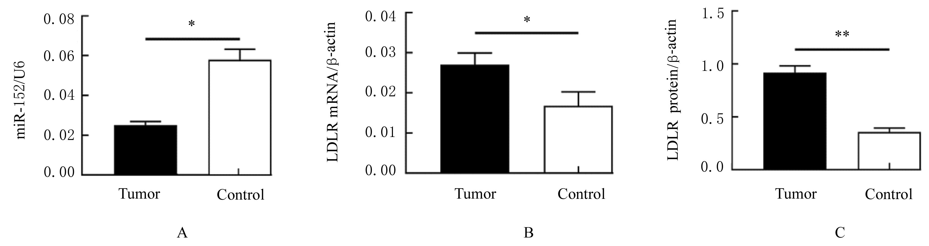
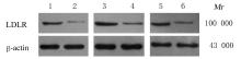
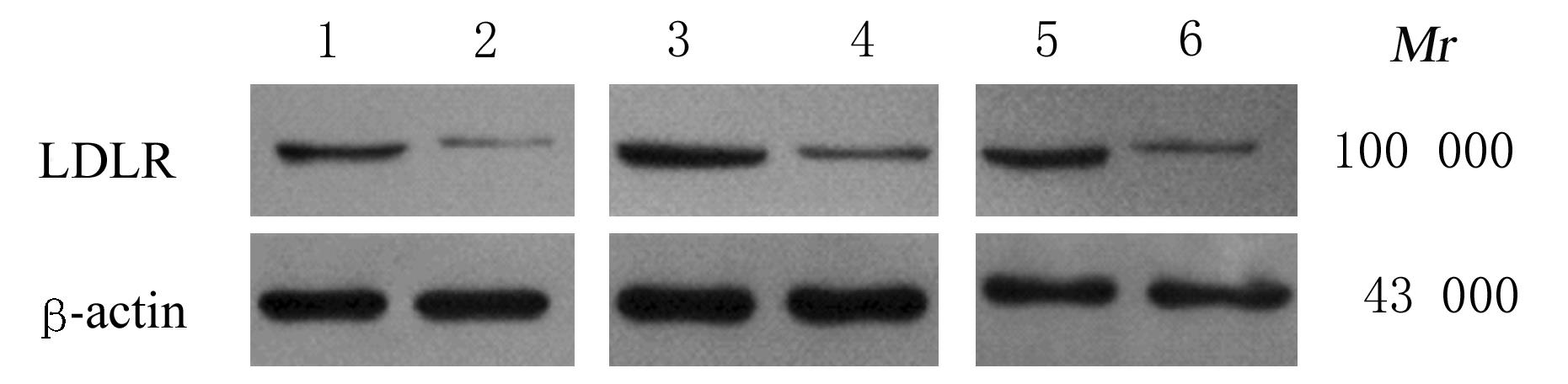
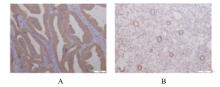
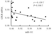
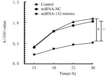
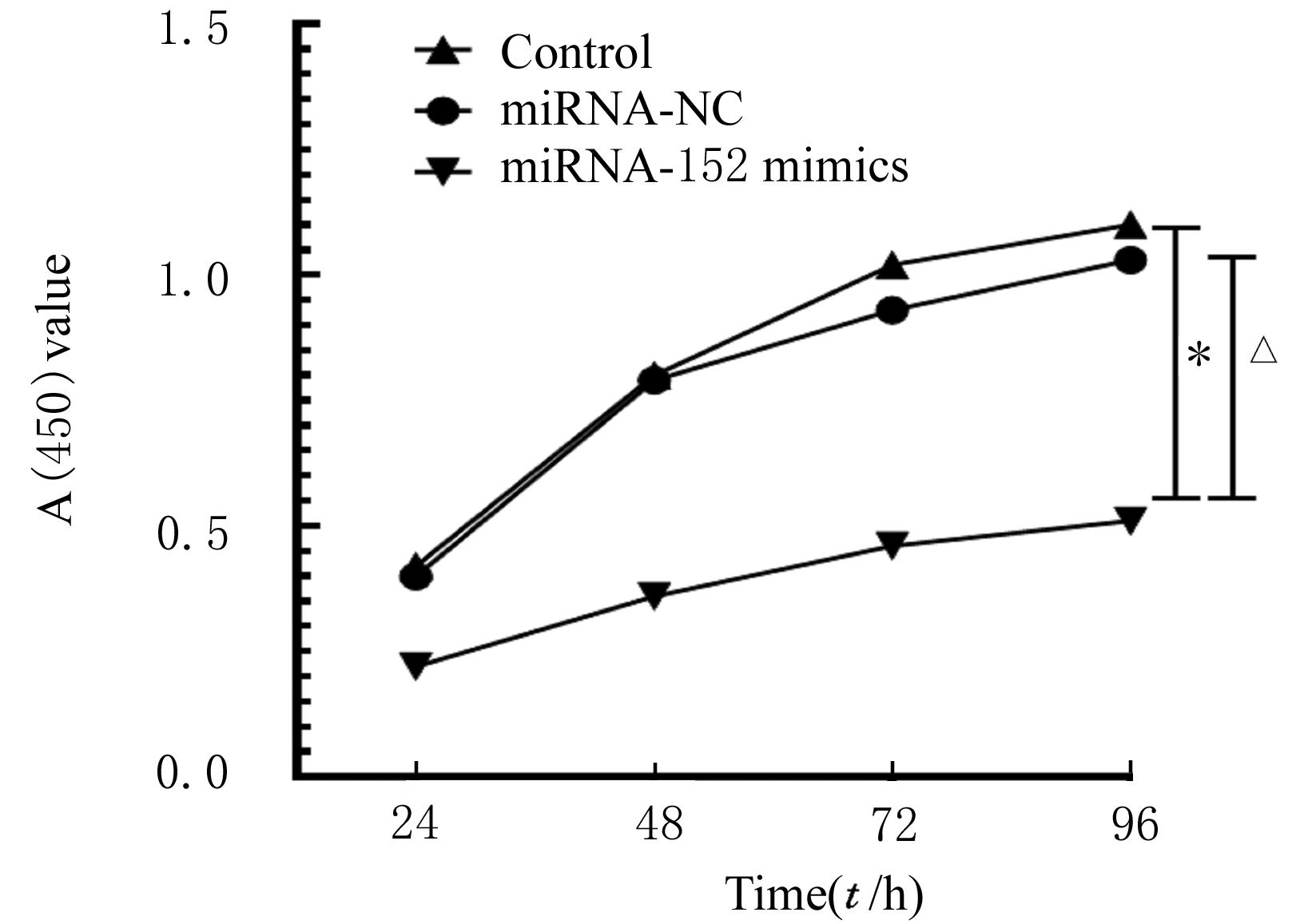
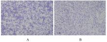
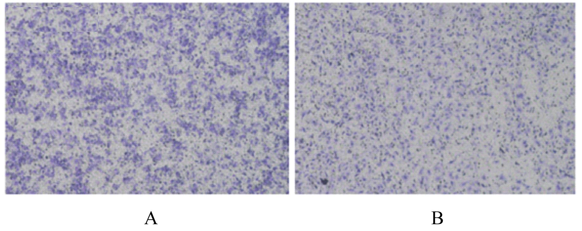
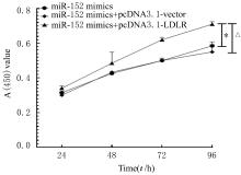
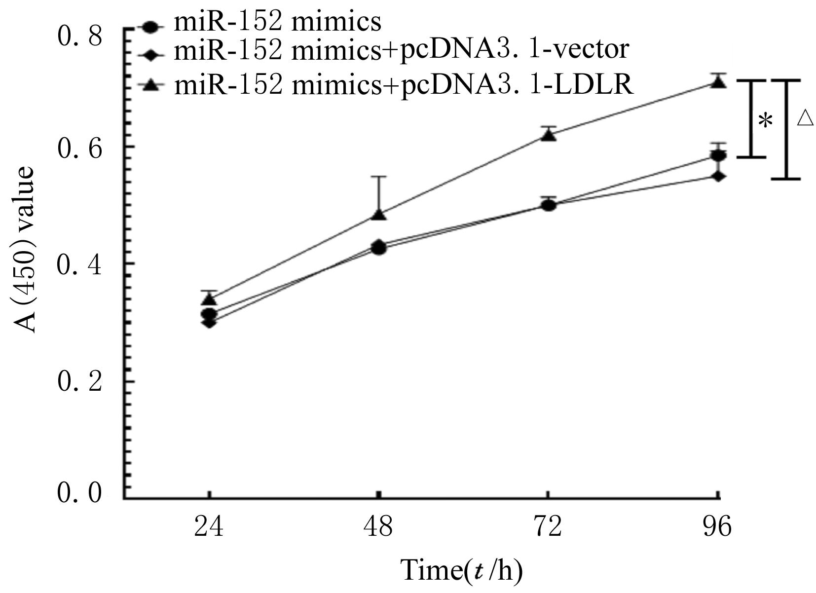
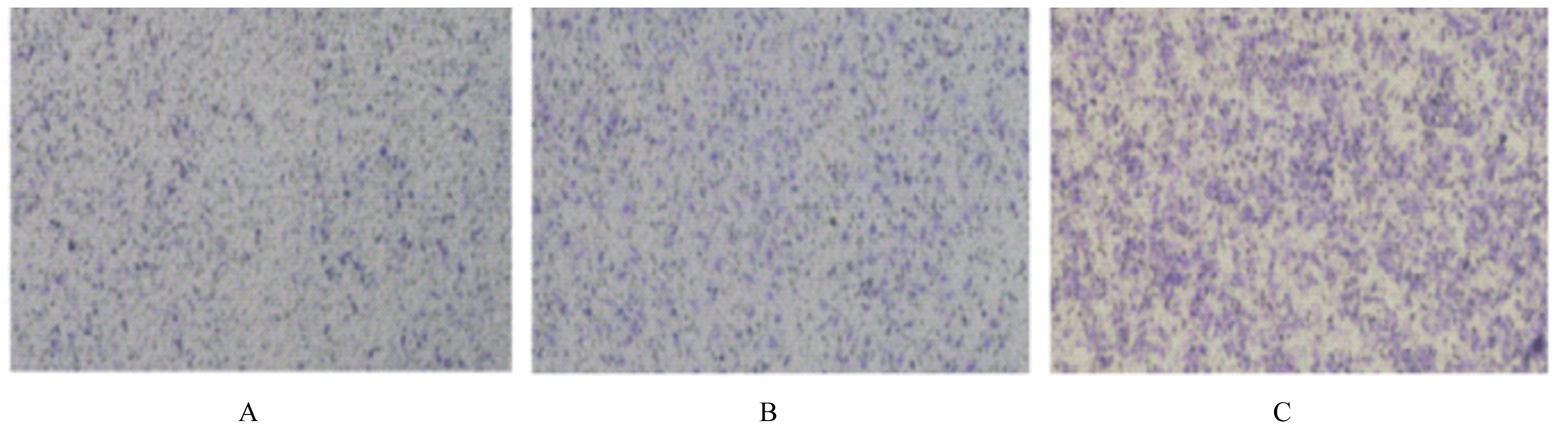
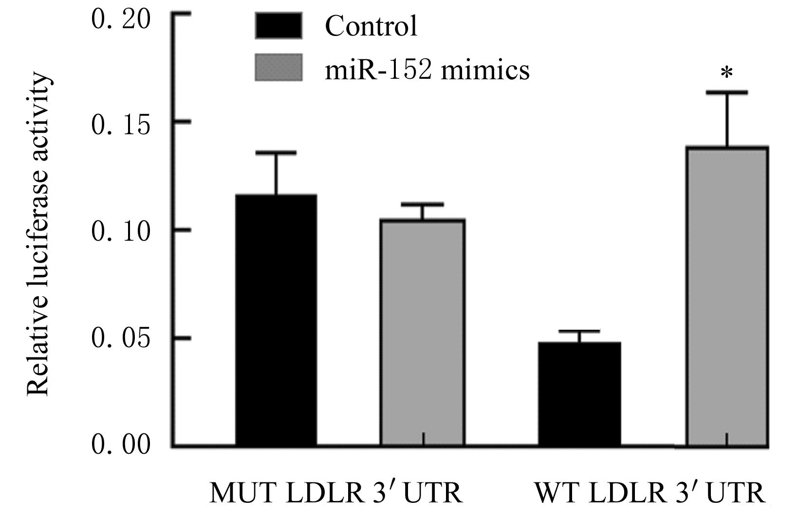
  • Received:2021-09-25 Online:2022-05-28 Published:2022-06-21
  • Contact: Fang LIU E-mail:lfdzzj@163.com

摘要: 目的

探讨微小RNA-152(miR-152)在子宫内膜癌(EC)组织中的表达和作用,为研究EC的发病机制和靶向治疗方案提供依据。

方法

选取21例EC患者癌组织(肿瘤组)和39例非EC患者内膜组织(对照组)及人EC RL95-2细胞和293T细胞为研究对象。收集肿瘤组和对照组患者的年龄、身高、体质量(BM)、体质量指数(BMI)、收缩压(SBP)、舒张压(DBP)、腰围(WC)和腹围(AC)等临床资料,检测2组患者甘油三酯(TG)、总胆固醇(TC)、低密度脂蛋白(LDL)、高密度脂蛋白(HDL)、Ki-67和低密度脂蛋白受体(LDLR)水平,生物信息学方法预测miR-152下游与增殖和侵袭相关的靶基因LDLR表达,实时荧光定量PCR(RT-qPCR)法检测肿瘤组和对照组患者子宫内膜组织中miR-152表达水平,Western blotting法和免疫组织化学检测法检测肿瘤组和对照组患者子宫内膜组织中LDLR 蛋白表达水平,Person相关分析法分析LDLR mRNA表达与患者一般资料、生化指标及miR-152表达的相关性。将RL95-2细胞分为空质粒组(转染miR-NC 空质粒)、miR-152组(转染miR-152-mimics)、miR-152-mimics+pcDNA3.1-vector组(同时转染miR-152-mimics和pcDNA3.1-vector) 和miR-152-mimics+pcDNA3.1-LDLR 组(同时转染miR-152-mimics和 pcDNA3.1-LDLR)。CCK-8法检测各组细胞增殖率,Transwell法检测各组细胞的侵袭细胞数,双荧光素酶报告基因实验检测各组293T细胞中荧光素酶活性。

结果

肿瘤组患者子宫内膜组织中miR-152表达水平明显低于对照组(P<0.05),LDLR mRNA和蛋白表达水平均高于对照组(P<0.05 或P<0.01)。双荧光素酶报告基因检测,LDLR为 miR-152的靶基因。Person相关分析,肿瘤组患者子宫内膜组织中LDLR mRNA表达与Ki-67、BMI、TC、TG、BM和WC呈正相关关系(r=0.4490, r=0.4377, r=0.4472, r=0.4706, r=0.5882, r=0.5130, P<0.05),与miR-152的表达呈负相关关系(r=-0.4378,P<0.05)。与空质粒组比较,miR-152组RL95-2细胞增殖率降低(P<0.05),侵袭细胞数减少(P<0.05);与miR-NC组比较,miR-152组RL95-2细胞增殖率降低(P<0.05),侵袭细胞数减少(P<0.05);与miR-152-mimics+pcDNA3.1-vector组比较,miR-152-mimics+pcDNA3.1-LDLR组细胞增殖率升高(P<0.01),侵袭细胞数增多(P<0.01)。

结论

miR-152通过降低LDLR的表达抑制EC细胞增殖和侵袭。

关键词: 微小RNA-152, 子宫内膜癌, 低密度脂蛋白受体, 细胞增殖, 细胞侵袭

Abstract:

Objective: To investigate the expression and role of microRNA-152 (miR-152) in endometrial carcinoma (EC) tissue, and to provide the basis for study of pathogenesis and targeted treatment of EC.

Methods

The cancer tissues of 21 patients with EC (tumor group) and 39 patients with non-EC (control group) and human endometrial cancer RL95-2 cells and 293T cells were selected as the subjects. The clinical data such as age, height, body mass (BM), body mass index (BMI), systolic blood pressure (SBP), diastolic blood pressure (DBP), waist circumference (WC) and abdominal circumference (AC) of the patients in tumor group and control group were collected. The levels of triglyceride(TG),total cholesterol (TC), low-density lipoprotein (LDL), high density lipoprotein (HDL), Ki-67 and low-density lipoprotein receptor (LDLR) of the patients in two groups were measured.Bioinformatics method was used to predict the expression of target gene LDLR related to the proliferation and invasion downstream of miR-152. Real-time fluorescence quantitative PCR (RT-qPCR) method was used to detect the expression level of miR-152 in endometrial tissue of the patients in tumor group and control group, Western blotting method and immunohistochemistry were used to detect the expression levels of LDLR protein in endometrial tissue of the patients in tumor group and control group, and Person correlation analysis was used to analyze the correlations between the expression of LDLR mRNA and the general data, biochemical indexes of the patients and miR-152 expression. The RL95-2 cells were divided into empty plasmid group (transfected with miR-NC empty plasmid), miR-152 group (transfected with miR-152-mimics), miR-152-mimics+pcDNA3.1-vector group (simultaneously transfected with miR-152-mimics and pcDNA3.1-vector), and miR-152-mimics+pcDNA3.1-LDLR group (simultaneously transfected with miR-152-mimics+pcDNA3.1-LDLR). The proliferation rates of the EC cells in various groups were detected by CCK-8 method, and the number of invasive EC cells in various groups was detected by Transwell assay. The luciferase activities of the 293T cells in various groups were detected by double luciferase reporter gene experiment.

Results

The expression level of miR-152 in endometrial tissue of the patients in tumor group was significantly lower than that in control group (P<0.05), and the expression levels of LDLR mRNA and protein were higher than those in control group (P<0.05 or P<0.01). The results of dual-luciferase reporter gene assay showed that LDLR was the target gene of miR-152.The Person correlation analysis results showed that the expression of LDLR mRNA in endometrial tissue of the patients in tumor group had a positive correlation with Ki-67, BMI, TC, TG, BM,and WC (r=0.4490, r=0.4377, r=0.4472, r=0.4706, r=0.5882, r=0.5130, P<0.05), and it was negatively correlated with the expression of miR-152(r=-0.4378, P<0.05). Compared with empty plasmid group, the proliferation rate of the RL95-2 cells in miR-152 group was decreased (P<0.05), and the number of invasive cells was decreased (P<0.05); compared with miR-152-mimics+pcDNA3.1-vector group, the proliferation rate of the cells in miR-152-mimics+pcDNA3.1-LDLR group was increased (P<0.01), and the number of invasive cells was increased (P<0.01).

Conclusion

MiR-152 inhibits the proliferation and invasion of the EC cells by decreasing the expression of LDLR.

Key words: MicroRNA-152, Endometrial carcinoma, Low-density lipoprotein receptor, Cell proliferation, Cell invasion

中图分类号: 

  • R737.33