吉林大学学报(医学版) ›› 2022, Vol. 48 ›› Issue (4): 954-961.doi: 10.13481/j.1671-587X.20220415

• 基础研究 • 上一篇    下一篇

载TGF-β3甲基丙烯酰化肝素对牙髓干细胞成骨分化能力的影响及其机制

邹馨颖,高爽,赵红,刘新,赵远航,宋嘉卓,闫琳琳,张志民()   

  1. 吉林大学口腔医院牙体牙髓科,吉林 长春 130021
  • 收稿日期:2021-10-26 出版日期:2022-07-28 发布日期:2022-07-26
  • 通讯作者: 张志民 E-mail:zhangzhim@jlu.edu.cn
  • 作者简介:邹馨颖(1994-),女,安徽省涡阳县人,住院医师,医学硕士,主要从事口腔内科方面的研究。
  • 基金资助:
    吉林省科技厅科技发展计划项目(20160101003JC);吉林省发改委科研项目(2016C045-3);吉林省财政厅科技项目(JCSZ2020304-2);吉林省卫生厅科研项目(2016S025)

Effect of TGF-β3-loaded methacrylated heparin on osteogenic differentiation of dental pulp stem cells and its mechanism

Xinying ZOU,Shuang GAO,Hong ZHAO,Xin LIU,Yuanhang ZHAO,Jiazhuo SONG,Linlin YAN,Zhimin ZHANG()   

  1. Department of Endodontics,Stomatology Hospital,Jilin University,Changchun 130021,China
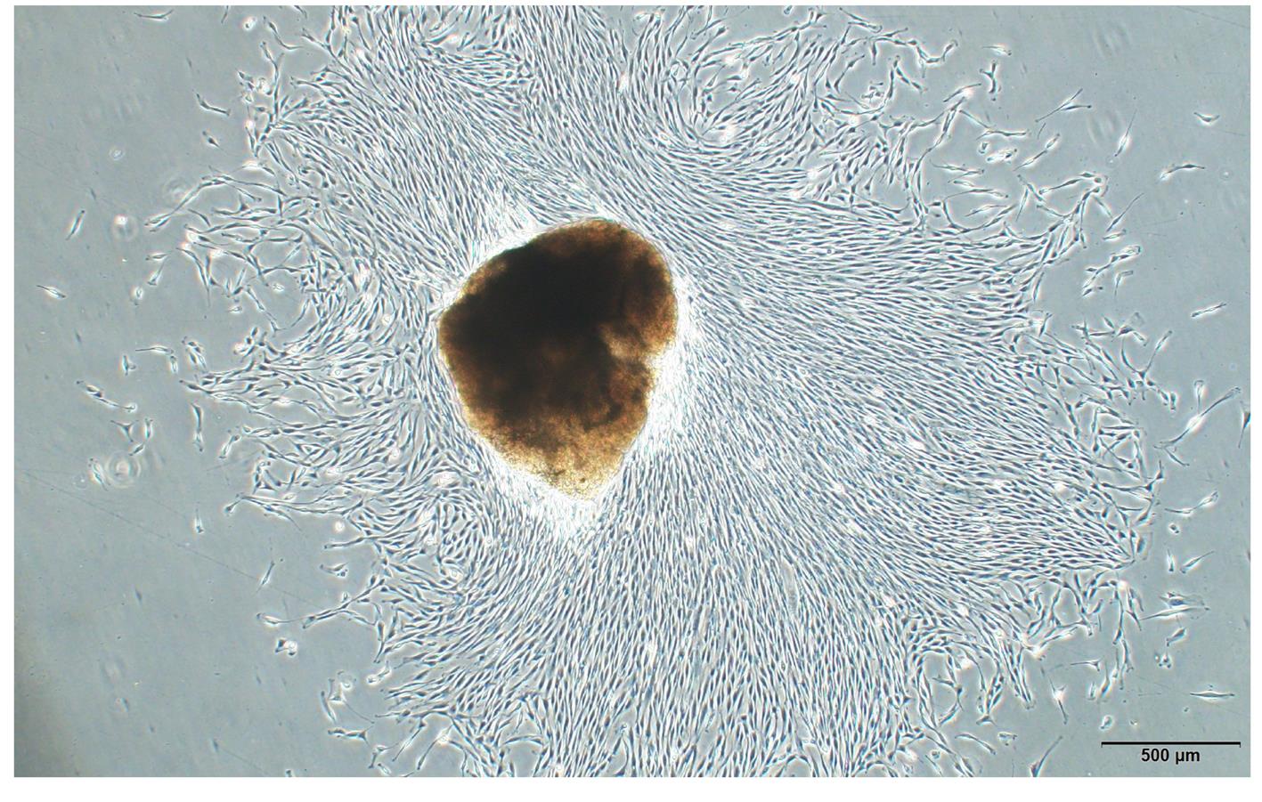
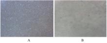
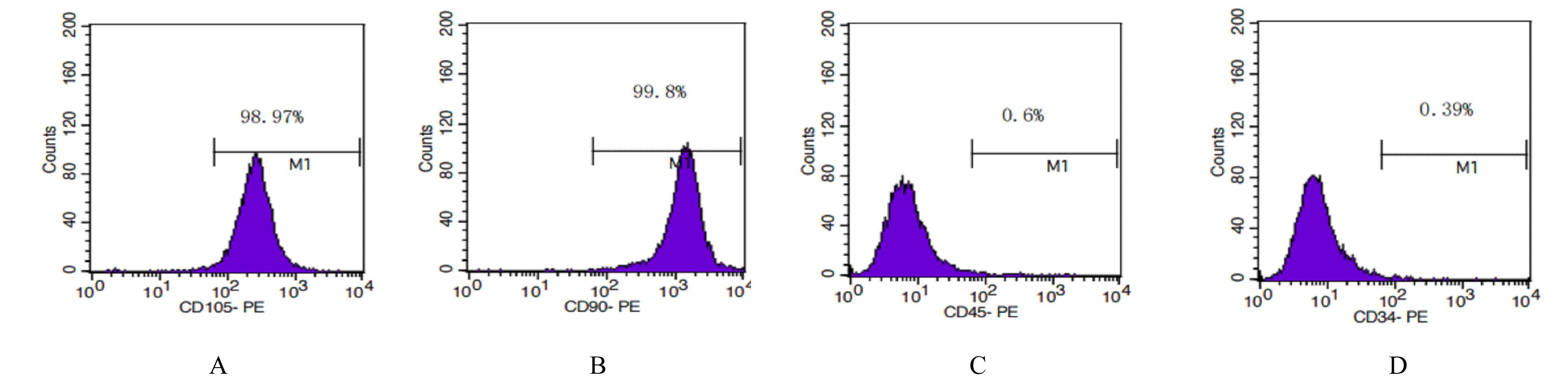
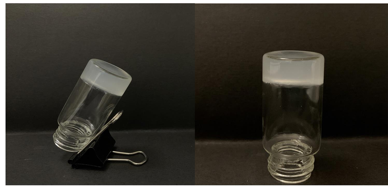
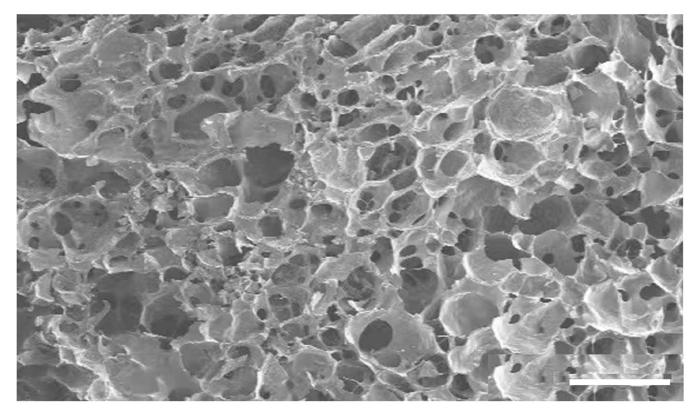
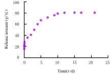
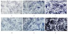
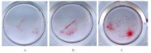
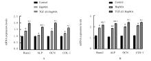
  • Received:2021-10-26 Online:2022-07-28 Published:2022-07-26
  • Contact: Zhimin ZHANG E-mail:zhangzhim@jlu.edu.cn

摘要: 目的

探讨载转化生长因子β3(TGF-β3)甲基丙烯酰化肝素(HepMA)对牙髓干细胞(DPSCs)成骨分化能力的促进作用,阐明其可能的作用机制。

方法

体外分离培养人牙髓干细胞(hDPSCs)并通过流式细胞术进行细胞鉴定。合成光固化HepMA水凝胶,利用扫描电子显微镜观察材料表面形态特征。合成载不同浓度(20、40、60、80和100 μg?L-1)TGF-β3的HepMA(TGF-β3-HepMA),采用其浸提液分别培养hDPSCs 1、3和5 d,同时设对照组,采用 CCK-8 法筛选出载TGF-β3的最适浓度。采用酶联免疫吸附测定(ELISA)法检测各时间点TGF-β3-HepMA中TGF-β3的累积释放量并绘制药物释放曲线。hDPSCs分为对照组、HepMA组和载最适浓度TGF-β3的 HepMA(TGF-β3-HepMA)组,加入含有相对应浸提液的培养基培养24 h,配制成骨诱导液分别诱导hDPSCs 7、14和21 d,采用碱性磷酸酶(ALP)染色和茜素红染色法检测各组细胞ALP染色及钙结节形成情况,实时荧光定量PCR(RT-qPCR)法检测各组细胞中成骨相关因子mRNA表达水平。

结果

分离培养的hDPSCs在光学显微镜下观察呈长梭形;流式细胞术检测,培养的hDPSCs中 CD105和CD90呈阳性表达,CD34和CD45呈阴性表达,验证本研究中分离培养的细胞为hDPSCs。扫描电子显微镜(SEM)观察,HepMA平均孔径为50~70 μm。ELISA法检测,TGF-β3在7 d内呈突释阶段后缓慢释放,21 d左右达到平衡。CCK-8法检测,与0 μg?L-1 TGF-β3-HepMA组比较,20、40和60 μg?L-1 TGF-β3-HepMA组hDPSCs增殖率呈剂量依赖性升高(P<0.05),故载TGF-β3的最适浓度为60 μg?L-1。成骨诱导7和14 d时,与对照组和HepMA组比较,TGF-β3-HepMA组细胞中ALP染色面积最大且染色颜色最深,14 d时细胞中ALP染色较7 d时更加明显;成骨诱导21 d时,与对照组和HepMA组比较,TGF-β3-HepMA组细胞中茜素红染色的矿化钙结节面积最大。RT-qPCR法检测,培养7和14 d时,与对照组比较,HepMA组和TGF-β3-HepMA组细胞中Runt相关转录因子2(Runx2)、ALP、骨钙桥素(OCN)和Ⅰ型胶原蛋白(COL-Ⅰ) mRNA表达水平明显升高(P<0.05或P<0.01);与HepMA组比较,TGF-β3-HepMA组细胞中 Runx2、ALP、OCN和COL-Ⅰ mRNA表达水平明显升高(P<0.05)。

结论

载TGF-β3的HepMA能够提高hDPSCs成骨分化能力,其机制可能与上调成骨相关因子的表达有关。

关键词: 转化生长因子β3, 牙髓干细胞, 甲基丙烯酰化肝素, 成骨分化

Abstract:

Objective: To investigate the promotion effect of transforming growth factor β3(TGF-β3)-loaded methacrylamide heparin(HepMA) on the osteogenic differentiation of dental pulp stem cells(DPSCs), and to elucidate its possible mechanism.

Methods

The hDPSCs were extracted and cultured in vitro and identified by flow cytometry. A light-cured HepMA hydrogel was synthesized, and the surface morphology of the material was observed by scanning electron microscope.The HepMA loaded with TGF-β3 (TGF-β-HepMA) at different concentrations (20, 40, 60, 80 and 100 μg?L-1) was synthesized and its extract was prepared to culture the hDPSCs for 1, 3 and 5 d; meanwhile, control group was established, and the optimal concentration of TGF-β3 loaded was screened out by CCK-8 method. Enzyme-linked immunosorbent assay (ELISA) was used to detect the cumulative release amounts of TGF-β3 in TGF-β-HepMA at different time points and the drug release curve was drawn.The HDPSCs were divided into control group, HepMA group and HE PMA group loaded with the optimal concentration of TGF-β3 (TGF-β3-HepMA). After being cultured for 24 h in the medium containing the corresponding extracts, the osteogenic induction solution was prepared to induce hHDPSCs for 7, 14 and 21 d, respectively. ALP staining and Alizarin red staining were used to detect the cell ALP staining and calcium nodule formation in each group. Real-time fluorescentce quantitative PCR(RT-qPCR) was used to detect the mRNA expression levels of osteogenic related factors in the cells in each group.

Results

The hDPSCs isolated and cultured were spindle-shaped under light microscope.The flow cytometry results showed positive expressions of CD105 and CD90 and negative expressions of CD34 and CD45 in the cultured hDPSCs, confirming that the cells isolated and cultured in this experiment were the hDPSCs. The scanning electron microscope (SEM) observation showed that the average pore size of HepMA was 50-70 μm. The ELISA results showed that TGF-β3 released slowly after a sudden release phase within 7 d and reached the equilibrium around 21 d.The CCK8 method results showed that compared with 0 μg?L-1 TGF-β3-HepMA group, the proliferation rate of hDPSCs in 60 μg?L-1 TGF-β 3-HEPMA group was increased(P<0.05), so the optimal concentration of TGF-β3 loaded was 60 μg?L-1. Compared with control group and HepMA group, the ALP staining area in tTGF-β3-HepMA group was the largest and the most stained in color when osteogenesis induction for 7 and 14 d, and the ALP staining in cells on the 14th day was more significant than that on the 7th day. Compared with control group and the HepMA group, the mineralized calcium nodule area stained with Alizarin red was the largest in TGF-β3-HepMA group at 21 d of osteogenic induction. The RT-qPCR results showed that compared with control group, the mRNA expression levels of Runx2, ALP, OCN and COL-Ⅰ in the cells in HepMA and TGF-β3-HepMA groups were significantly increased after 7 and 14 d of culture(P<0.05 or P<0.01); compared with HepMA group, the mRNA expression levels of Runx2, ALP, OCN and COL-Ⅰ in the cells in TGF-β3-HepMA group were significantly increased (P<0.05).

Conclusion

HepMA loading TGF-β3 can improve the osteogenic differentiation of hDPSCs, and its mechanism may be related to the up-regulation of the expressions of osteogenic related factors.

Key words: Transforming growth factor-beta 3, Dental pulp stem cells, Methyl acryloyl heparin, Osteogenic differentiation

中图分类号: 

  • R782