吉林大学学报(医学版) ›› 2022, Vol. 48 ›› Issue (5): 1167-1174.doi: 10.13481/j.1671-587X.20220509

• 基础研究 • 上一篇    

桑黄酸性多糖对胆管结扎所致小鼠肝纤维化的改善作用及其机制

谷明柳1,林逢源1,吴雪峰1,周嘉宁1,卢学春2,杜培革1,安丽萍1()   

  1. 1.北华大学药学院微生物与生化药学教研室,吉林 吉林 132013
    2.解放军总医院第二医学中心 血液科 国家老年疾病临床研究中心,北京 100853
  • 收稿日期:2021-12-18 出版日期:2022-09-28 发布日期:2022-11-15
  • 通讯作者: 安丽萍 E-mail:630114130@qq.com
  • 作者简介:谷明柳(1996-),女,吉林省松原市人,在读硕士研究生,主要从事肝纤维化基础方面的研究。
  • 基金资助:
    吉林省发改委产业创新专项资金项目(2018C046-3)

Improvement effect of Phellinus igniarius acidic polysaccharide on liver fibrosis induced by bile duct ligation in mice and its mechanism

Mingliu GU1,Fengyuan LIN1,Xuefeng WU1,Jianing ZHOU1,Xuechun LU2,Peige DU1,Liping AN1()   

  1. 1.Department of Microbiology and Biochemical Pharmacy,School of Pharmacy,Beihua University,Jilin 132013,China
    2.Department of Hematology,Second Medical Center&National Clinical Research Center for Geriatric Diseases,Chinese PLA General Hospital,Beijing 100853,China
  • Received:2021-12-18 Online:2022-09-28 Published:2022-11-15
  • Contact: Liping AN E-mail:630114130@qq.com

摘要:

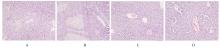
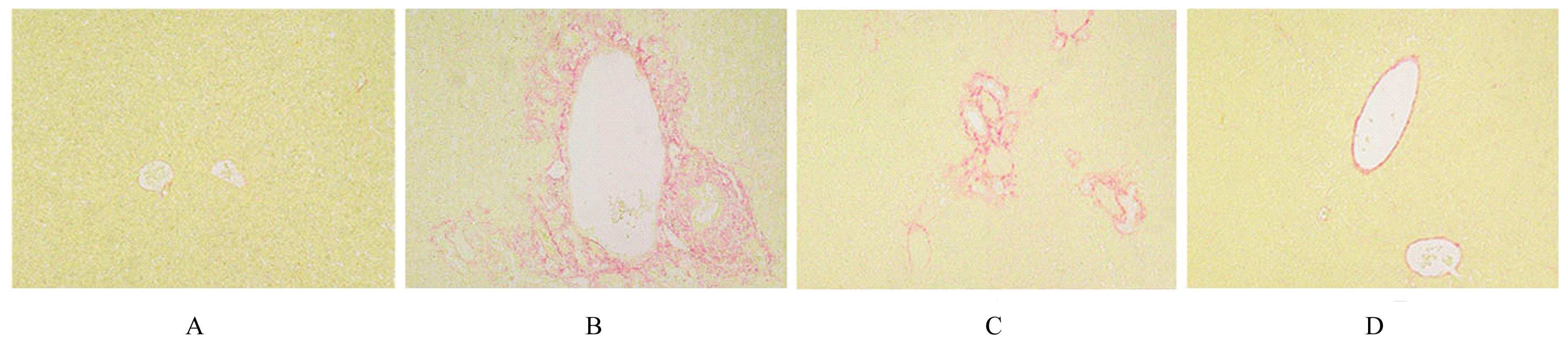
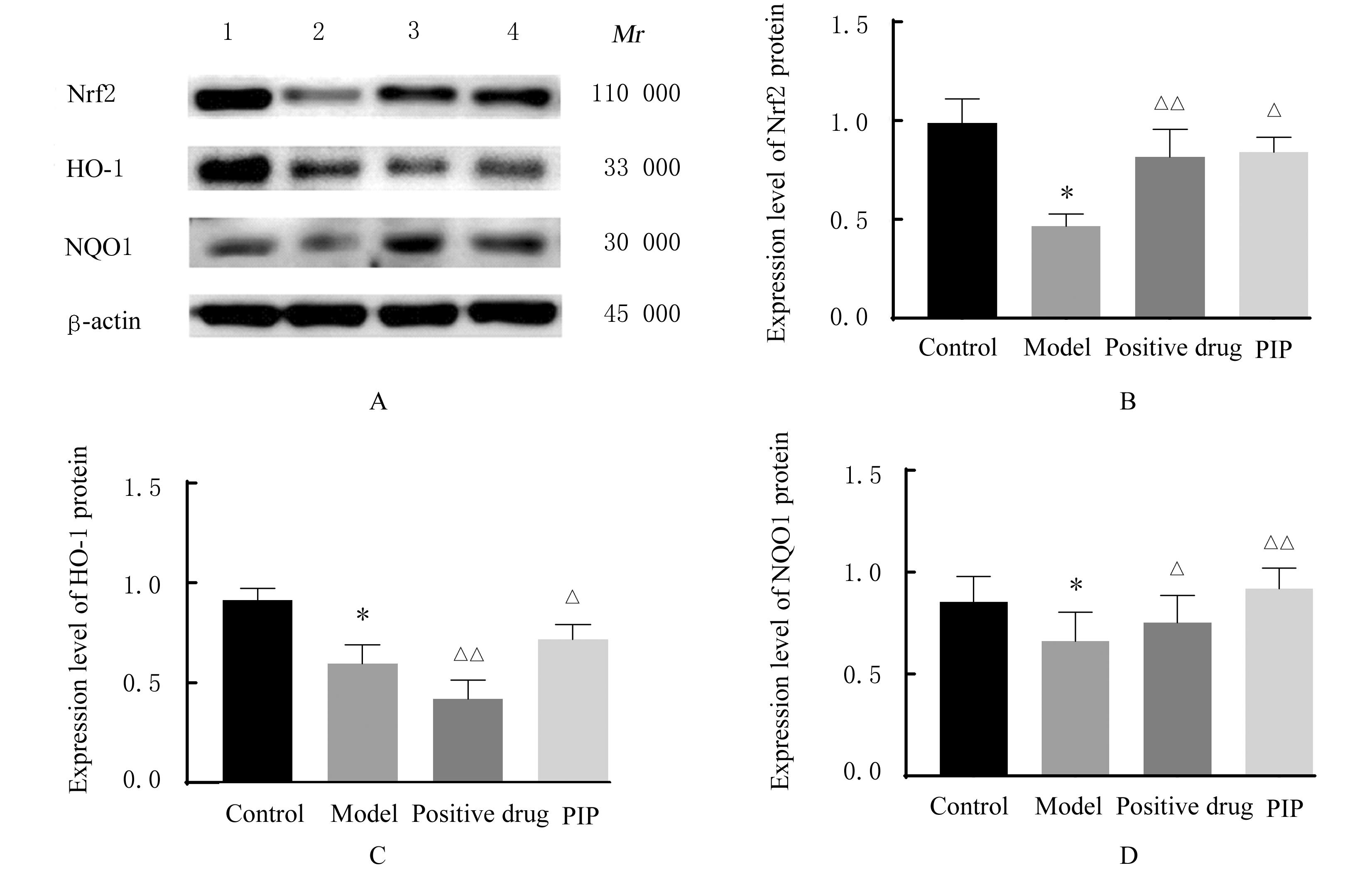
目的 探讨桑黄酸性多糖(PIP)对胆管结扎(BDL)所致小鼠肝纤维化的改善作用,阐明其可能的作用机制。 方法 60只4周龄雄性C57/BL6小鼠随机分为对照组、模型组、阳性药组和PIP给药组(PIP组),每组15只。对照组小鼠只绕胆总管穿过手术线但不结扎,模型组、阳性药组和PIP组小鼠结扎胆管。术后给药3周,末次给药12 h后眼球取血,处死小鼠。称量小鼠体质量和肝脏质量,并计算肝脏指数;微板法检测各组小鼠血清中丙氨酸氨基转移酶(ALT)和天门冬氨酸氨基转移酶(AST)水平;双抗体夹心法检测各组小鼠血清中Ⅲ型前胶原(PC Ⅲ)和Ⅳ型胶原(Col Ⅳ)水平;HE染色和天狼猩红染色观察各组小鼠肝组织病理形态表现和肝纤维化程度;检测各组小鼠肝组织中超氧化物歧化酶(SOD)和谷胱甘肽过氧化物酶(GSH-Px)活性及丙二醛(MDA)和谷胱甘肽(GSH)水平。Western blotting法检测各组小鼠肝组织中核因子E2相关因子(Nrf2)、血红素加氧酶1(HO-1)和醌氧化还原酶1(NQO1)蛋白表达水平。 结果 与对照组比较,模型组小鼠体质量差异无统计学意义(P>0.05),肝脏质量和肝脏指数均升高(P<0.05);与模型组比较,PIP组小鼠体质量无明显变化(P>0.05),肝脏质量和肝脏指数降低(P<0.05)。HE染色,对照组小鼠肝组织无异常表现;模型组小鼠肝组织中肝小叶结构破坏,可见灶状液化性肝坏死和较多炎症细胞浸润;与模型组比较,阳性药组和PIP组小鼠肝组织坏死程度明显减轻,炎症细胞浸润减少。天狼猩红染色,与对照组比较,模型组小鼠肝组织中胶原纤维含量明显增加;与模型组比较,PIP组小鼠肝组织胶原纤维含量明显减少。与对照组比较,模型组小鼠血清中ALT和AST水平明显升高(P<0.01),PC Ⅲ和Col Ⅳ水平明显升高(P<0.05),小鼠肝组织中SOD和GSH-Px活性明显降低(P<0.01),MDA和GSH水平明显升高(P<0.05或P<0.01);与模型组比较,阳性药组和PIP组小鼠血清中ALT和AST水平明显降低(P<0.01),PC Ⅲ和Col Ⅳ水平明显降低(P<0.05或P<0.01),小鼠肝组织中SOD和GSH-Px活性升高(P<0.05),GSH和MDA水平降低(P<0.05)。Western blotting法检测,与对照组比较,模型组小鼠肝组织中Nrf2、HO-1和NQO1蛋白表达水平均降低(P<0.05);与模型组比较,PIP组小鼠肝组织中Nrf2、HO-1和NQO1蛋白表达水平明显升高(P<0.05或P<0.01)。 结论 PIP能改善BDL导致的小鼠肝纤维化,其作用机制可能与降低肝脏氧化应激水平,调节Nrf2/HO-1信号通路中关键因子Nrf2、HO-1和NQO1蛋白表达水平有关。

关键词: 桑黄酸性多糖, 肝纤维化, 胆管结扎, 核因子E2相关因子, 血红素加氧酶1

Abstract:

Objective To investigate the improvement effect of Phellinus igniarius acidic polysaccharide(PIP) on liver fibrosis in the mice induced by bile duct ligation (BDL), and to clarify its possible mechanism. Methods A total of sixty 4-week-old male C57/BL6 mice were randomly divided into control group, model group, positive drug group,and PIP group,with 15 mice in each group. The mice in control group only received the passed surgical line without ligation around the common bile duct, and the bile duct of mice in model group, postive drug group and PIP group were ligated. After administration for 3 weeks, the blood was taken from eyeball 12 h after the last administration, and the mice were sacrificed. The body weights and liver weights of mice were weighed, and the liver index was calculated. Microplate method was used to determine the levels of alanine aminotransferase (ALT) and aspartate aminotransferase (AST) in serum of the mice in various goups; the levels of type Ⅲ procollagen (PC Ⅲ) and cellagen type Ⅳ(Col Ⅳ) in serum were determined by double antibody sandwich method. HE staining and Sirius red staining were used to observe the pathomorphology and fibrosis degrees of liver tissue of the mice in various groups; the activities of superoxide dismutase (SOD) and glutathione peroxidase (GSH-Px) and the levels of malondialdehyde (MDA)and glutathione (GSH) in liver tissue of the mice in various groups were determined. The expression levels of nuclear factor-E2 related factor (Nrf2), heme oxygenase-1 (HO-1) and quinone oxidoreductase 1 (NQO1) proteins in liver tissue of the mice in various groups were detected by Western blotting method. Results Compared with control group,the body weight of the mice in model group had no significant difference(P>0.05),and the liver weight and liver index were increased(P<0.05);compared with model group, the body weight of the mice in PIP group had no significant change(P>0.05), and the liver weight and liver index were decreased(P<0.05).The HE staining results showed no abnormalities in liver tissue of the mice in control group;the liver lobular structure was destroyed and focal liquefied hepatic necrosis and more inflammatory cell infiltration were seen in liver tissue of the mice in model group;compared with model group, the necrosis degrees of liver tissue of the mice in positive drug group and PIP group were significantly reduced, and the infiltration of inflammatory cells was reduced. The Sirius red staining results showed that compared with control group,the content of collagen fibers in liver tissue of the mice in model group was significantly increased;compared with model group,the content of collagen fibers in liver tissue of the mice in PIP group was significantly decreased.Compared with control group,the ALT and AST levels in serum of the mice in model group were increased(P<0.01),PC Ⅲ and Col Ⅳ levels were increased(P<0.05), the activities of SOD and GSH-Px were significantly decreased(P<0.01),and the levels of MDA and GSH were significantly increased(P<0.05 or P<0.01); compared with model group, the levels of ALT and AST in serum of the mice in positive drug group and PIP group were significantly decreased (P<0.01), the levels of PC Ⅲ and Col Ⅳ were significantly decreased (P<0.05 or P<0.01), the activities of SOD and GSH-Px in liver tissue of the mice were increased (P<0.05), and the levels of GSH and MDA were decreased (P<0.05).The Western blotting results showed that compared with control group,the expression levels of Nrf2,HO-1,and NQO1 proteins in liver tissue of the mice in model group were decreased (P<0.05);compared with model group, the expression levels of Nrf2,HO-1,and NQO1 proteins in liver tissue of the mice in PIP group were significantly increased (P<0.05 or P<0.01). Conclusion PIP can improve the liver fibrosis in the mice caused by BDL, and its mechanism may be related to reducing the level of liver oxidative stress and regulating the expression levels of Nrf2, HO-1 and NQO1 proteins in Nrf2 / HO-1 signaling pathway.

Key words: Phellinus igniarius acid polysaccharides, Liver fibrosis, Bile duct ligation, Nuclear factor-E2 related factor, Heme oxygenase-1

中图分类号: 

  • R575.2