吉林大学学报(医学版) ›› 2021, Vol. 47 ›› Issue (5): 1124-1130.doi: 10.13481/j.1671-587X.20210507

• 基础研究 • 上一篇    下一篇

莪术醇对小鼠肝窦内皮细胞结构的影响及其对肝内血管新生的抑制作用

郑洋1,王佳慧1,彭岳2,原鲜玲2,汪磊1,赵铁建2()   

  1. 1.广西中医药大学赛恩斯新医药学院医学系,广西 南宁 530222
    2.广西中医药大学基础医学院 生理教研室,广西 南宁 530222
  • 收稿日期:2021-01-31 出版日期:2021-09-28 发布日期:2021-10-26
  • 通讯作者: 赵铁建 E-mail:570041687@qq.com
  • 作者简介:郑 洋(1991-),男,安徽省无为市人,助教,医学硕士,主要从事民族药防治慢性肝病等方面的研究。
  • 基金资助:
    国家自然科学基金项目(81960751);广西科技厅青年基金项目(2020GXNSFBA297094);广西教育厅中青年教师科研基础能力提升计划项目(2020KY59009);广西壮瑶药重点实验室项目(GXZYZZ2020-07);广西中医药大学青年基金项目(2020QN006)

Influence of curcumol in structure of liver sinusoidal endothelial cells of mice and its inhibitiory effect on intrahepatic angiogenesis

Yang ZHENG1,Jiahui WANG1,Yue PENG2,Xianling YUAN2,Lei WANG1,Tiejian ZHAO2()   

  1. 1.Department of Medicine,Faculty of Chinese Medicine Science,Guangxi University of Chinese Medicine,Nanning 530021,China
    2.Department of Physiology,College of Basic Medical Sciences,Guangxi University of Chinese Medicine,Nanning 530021,China
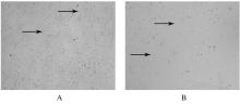
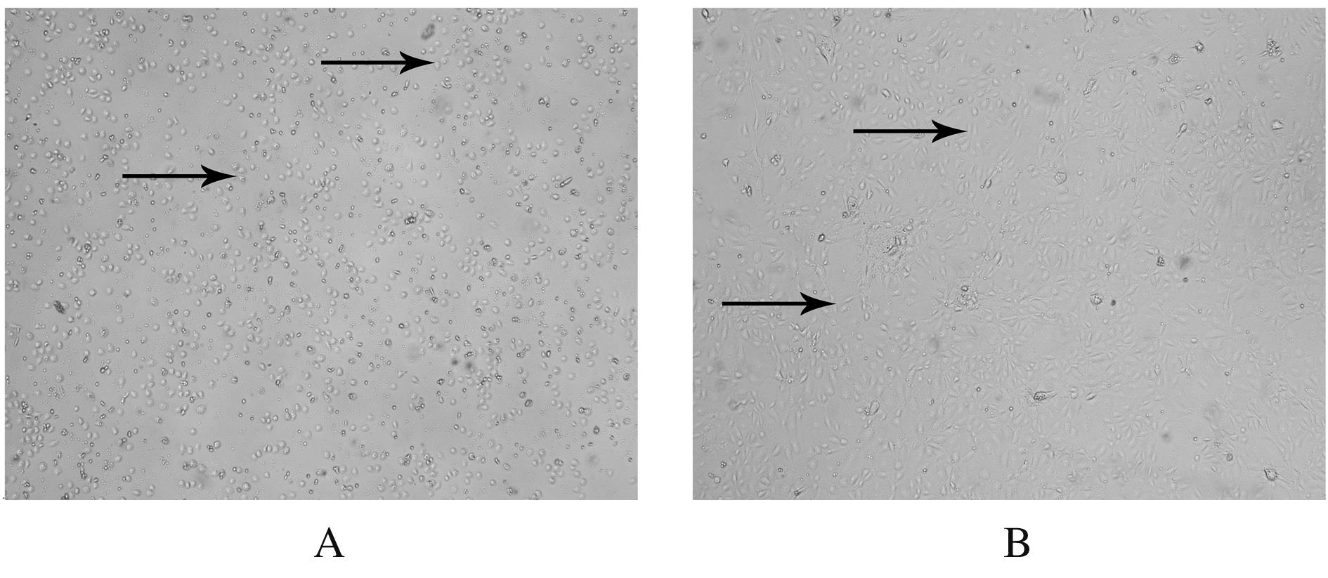
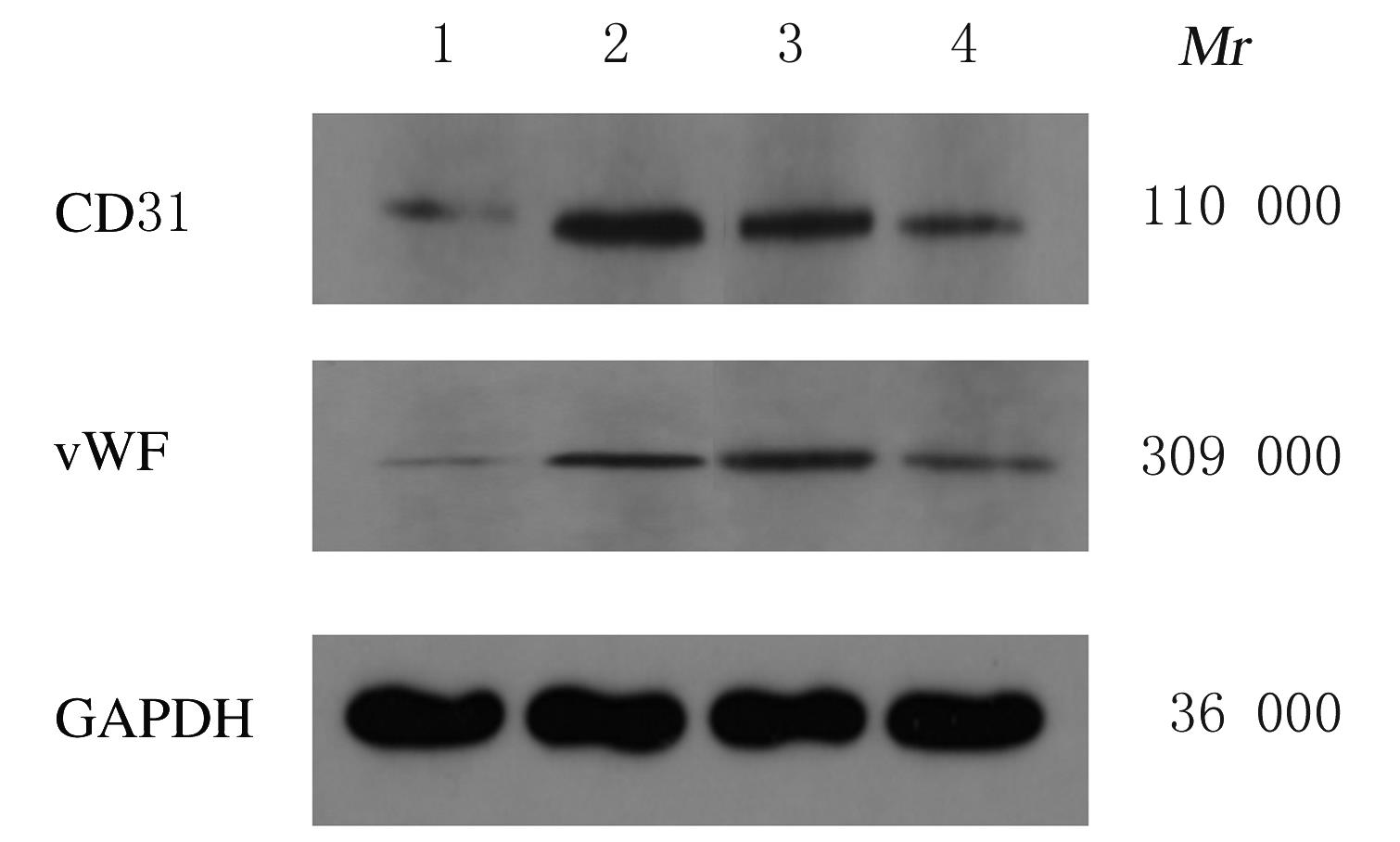
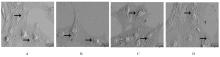
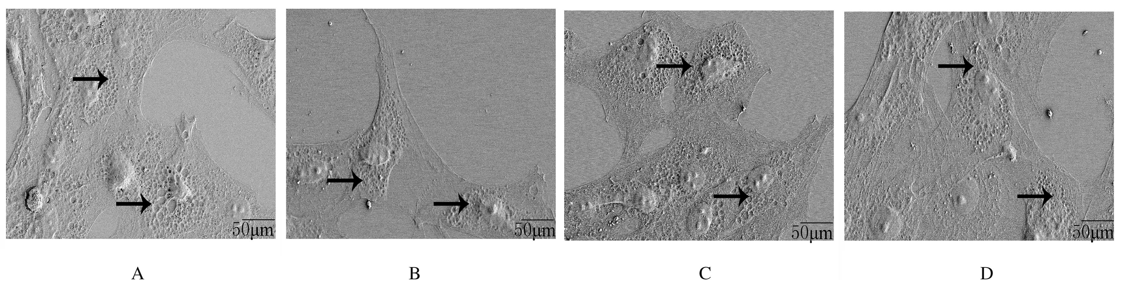
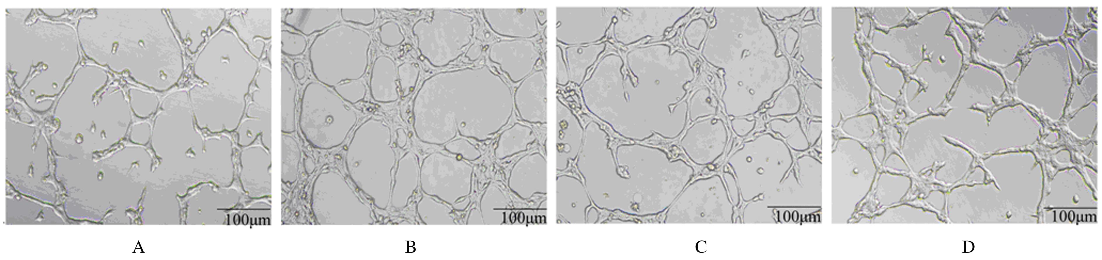
  • Received:2021-01-31 Online:2021-09-28 Published:2021-10-26
  • Contact: Tiejian ZHAO E-mail:570041687@qq.com

摘要: 目的

探讨莪术醇对小鼠肝窦内皮细胞(LSEC)结构的影响,阐明其对LSEC介导的肝内血管新生的作用。

方法

30只SPF级KM小鼠使用灌注及消化法分离原代LSEC,将其分为空白对照组、模型组、莪术醇组和丹参酚酸B组,空白对照组仅用普通培养基处理;模型组用100 μg·L-1廋素进行活化,培养细胞48 h;莪术醇组先用45 mg·L-1莪术醇处理30 min再用廋素培养细胞48 h;丹参酚酸B组先用1×10⁻⁶ mmol·L-1丹参酚酸B处理30 min再用廋素培养细胞48 h。采用MTT法检测各组细胞增殖率,实时荧光定量聚合酶链式反应(RT-qPCR)法检测各组细胞中分化抗原簇31(CD31)和血管性血友病因子(vWF) mRNA表达水平,免疫印迹法检测CD31和vWF蛋白表达水平,扫描电镜观察LSEC窗孔的结构,血管形成实验观察肝内血管新生情况。

结果

MTT法检测,模型组细胞增殖率较空白对照组明显降低(P<0.05),莪术醇组和丹参酚酸B组细胞增殖率较模型组明显下降(P<0.05),丹参酚酸B组与莪术醇组细胞增殖率比较差异无统计学意义(P>0.05)。RT-qPCR法检测,模型组细胞中CD31和vWF mRNA表达水平较空白对照组明显升高(P<0.05),莪术醇组和丹参酚酸B组细胞中CD31和vWF mRNA表达水平较模型组明显降低(P<0.05),丹参酚酸B组与莪术醇组细胞中CD31和vWF mRNA表达水平比较差异无统计学意义(P>0.05)。免疫印迹法检测,模型组细胞中CD31和vWF蛋白表达水平较空白对照组明显升高(P<0.05),莪术醇组和丹参酚酸B组细胞中CD31和vWF蛋白表达水平较模型组明显降低(P<0.05),莪术醇组与丹参酚酸B组细胞中CD31和vWF蛋白表达水平比较差异无统计学意义(P>0.05)。扫描电镜观察,空白对照组LSEC窗孔数量较多,与空白对照组比较,模型组窗孔数量减少;与模型组比较,莪术醇组和丹参酚酸B组LSEC窗孔数量有所增加。血管形成实验检测,模型组血管形成数量较空白对照组明显增加(P<0.05),莪术醇组和丹参酚酸B组血管形成数量较模型组明显减少(P<0.05),莪术醇组与丹参酚酸B组血管形成数量比较差异无统计学意义(P>0.05)。

结论

莪术醇通过调控小鼠LSEC的窗孔结构,从而抑制肝内血管新生。

关键词: 肝纤维化, 莪术醇, 肝窦内皮细胞, 血管新生

Abstract: Objective

To investigate the influence of curcumol on the structure of liver sinusoidal endothelial cells(LSEC) of the mice, and to clarify its effect on intrahepatic angiogenesis mediated by LSEC.

Methods

Perfusion and digestion methods were used in 30 SPF KM mice to isolate the primary LSEC. The cells were divided into blank control group, model group, curcumol group, and salvianolic acid B group. The cells in blank control group were treated with ordinary culture medium only; the cells in model group were activated with 100 μg·L-1 serotonin, and the cells were cultured for 48 h; the cells in curcumol group were treated with 45 mg·L-1 curcumol for 30 min and then the cells were cultured with serotonin for 48 h; the cells in salvianolic acid B group were first treated with 1×10⁻⁶ mmol·L-1 salvianolic acid B for 30 min and then cultured with alicin for 48 h. MTT assay was used to detect the proliferation rates of the cells in various groups; Real-time fluorescence quantitative polymerase chain reaction(RT-qPCR) method was used to determine the expression levels of CD31 and vWF mRNA in the cells in various groups; Western blotting method was used to detect the expression levels of CD31 and vWF proteins in the cells in various groups; scanning electron microscope was used to observe the structure of the LSEC; angiogenesis experiment was used to observe intrahepatic angiogenesis.

Results

The MTT test results showed that the proliferation rate of the cells in model group was significantly lower than that in blank control group (P<0.05); the cell proliferation rates in curcumol group and salvianolic acid B group were significantly lower than that in model group (P<0.05);there was no significant difference in the cell proliferation rate between salvianolic acid B group and curcumol group (P>0.05). The results of RT-qPCR showed that the expression levels of CD31 and vWF mRNA in the cells in model group was significantly higher than those in blank control group (P<0.05); the expression levels of CD31 and vWF mRNA in curcumol group and salvianolic acid B group were significantly lower than those in model group (P<0.05);there were no significant differences in the expression levels of CD31 and vWF mRNA between salvianolic acid B group and curcumol group(P>0.05). The results of Western blotting method showed that the expression levels of CD31 and vWF proteins in the cells in model group were significantly higher than those in blank control group (P<0.05); the expression levels of CD31 and vWF proteins in the cells in salvianolic acid B and curcumol group group were significantly lower than those in model group (P<0.05); there were no significant differences in the expression levels of CD31 and vWF proteins between salvianolic acid B group and curcumol group(P>0.05). The results of scanning electron microscope showed that the number of LSEC fenestrations of LSEC in blank control group was larger; compared with blank control group, the number of LSEC fenestrations in model group was decreased; compared with model group, the number of LSEC fenestrations in curcumol group and salvianolic acid B group was increased. The angiogenesis experiment results showed that the number of blood vessels in model group was significantly increased compared with blank group (P<0.05);the number of blood vessels in curcumol group and salvianolic acid B group was significantly lower than that in model group (P<0.05); and there was no statistically significant difference in the number of blood vessels between salvianolic acid B group and curcumol group (P>0.05).

Conclusion

Curcumol can inhibit the intrahepatic angiogenesis by regulating the fenestration structure of LSEC.

Key words: liver fibrosis, curcumol, sinusoidal endothelial cell, angiogenesis

中图分类号: 

  • R657.3