吉林大学学报(医学版) ›› 2026, Vol. 52 ›› Issue (2): 318-329.doi: 10.13481/j.1671-587X.20260203

• 基础研究 • 上一篇    

局部应用负载外泌体的DCC水凝胶对小鼠腹腔粘连的改善作用及其机制

马宁1,柳爽1,王成瑶2,卢嘉骏1,陈琳渝2,王梓祎2,赵伟钦2,王伟潼1,车鹏程2,孙红1()   

  1. 1.华北理工大学基础医学院 河北省慢性病重点实验室,河北 唐山 063210
    2.华北理工大学护理与 康复学院 河北省康复工程与再生医学重点实验室,河北 唐山 063000
  • 收稿日期:2025-04-16 接受日期:2025-06-12 出版日期:2026-03-28 发布日期:2026-04-15
  • 通讯作者: 孙红 E-mail:sunhong@ncst.edu.cn
  • 作者简介:马 宁(1998-),女,河北省秦皇岛市人,在读硕士研究生,主要从事组织修复和再生医学方面的研究。
  • 基金资助:
    河北省科技厅自然科学基金项目(H2023209021);河北省科技厅自然科学基金项目(H2024209013);河北省教育厅大学生创新创业训练计划项目(202410081057)

Improvement effect of topical application of exosome loaded hydrogel DCC on peritoneal adhesion in mice and its mechanism

Ning MA1,Shuang LIU1,Chengyao WANG2,Jiajun LU1,Linyu CHEN2,Ziyi WANG2,Weiqin ZHAO2,Weitong WANG1,Pengcheng CHE2,Hong SUN1()   

  1. 1.Hebei Provincial Key Laboratory of Chronic Diseases,School of Basic Medical Sciences,North China University of Science and Technology,Tangshan 063210,China
    2.Hebei Provincial Key Laboratory of Rehabilitation Engineering and Regenerative Medicine,School of Nursing and Rehabilitation,North China University of Science and Technology,Tangshan 063000,China
  • Received:2025-04-16 Accepted:2025-06-12 Online:2026-03-28 Published:2026-04-15
  • Contact: Hong SUN E-mail:sunhong@ncst.edu.cn

摘要:

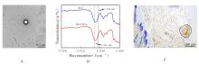
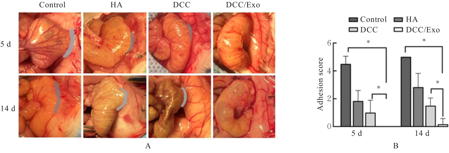
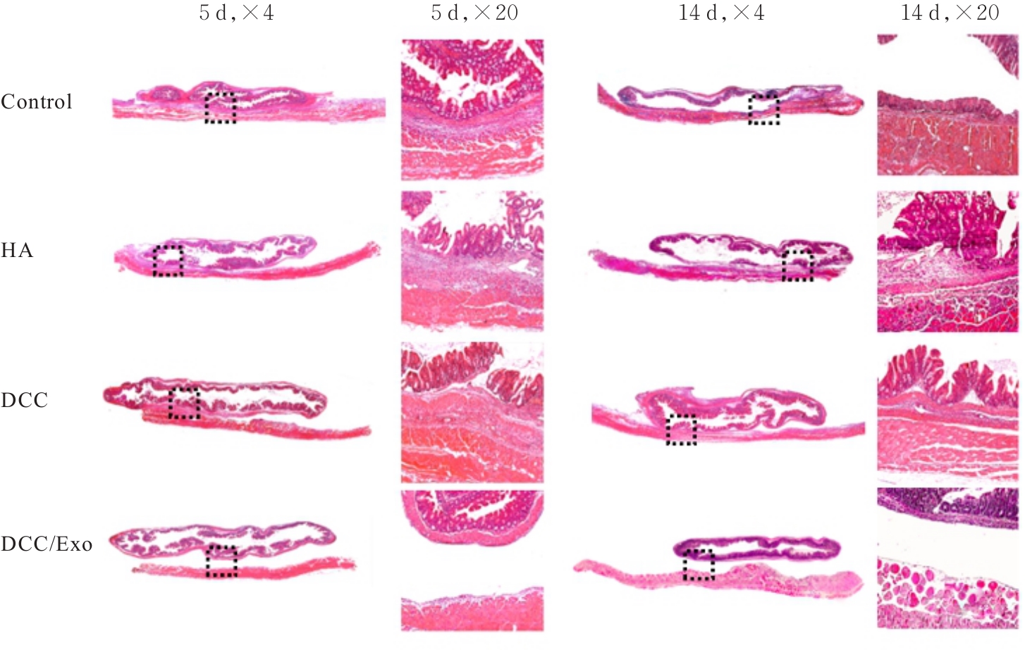
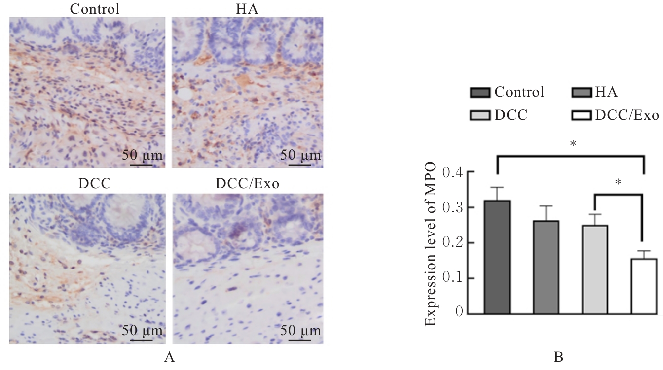
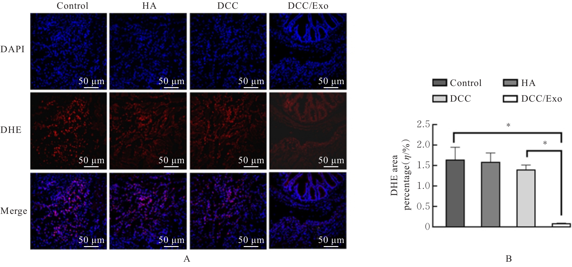
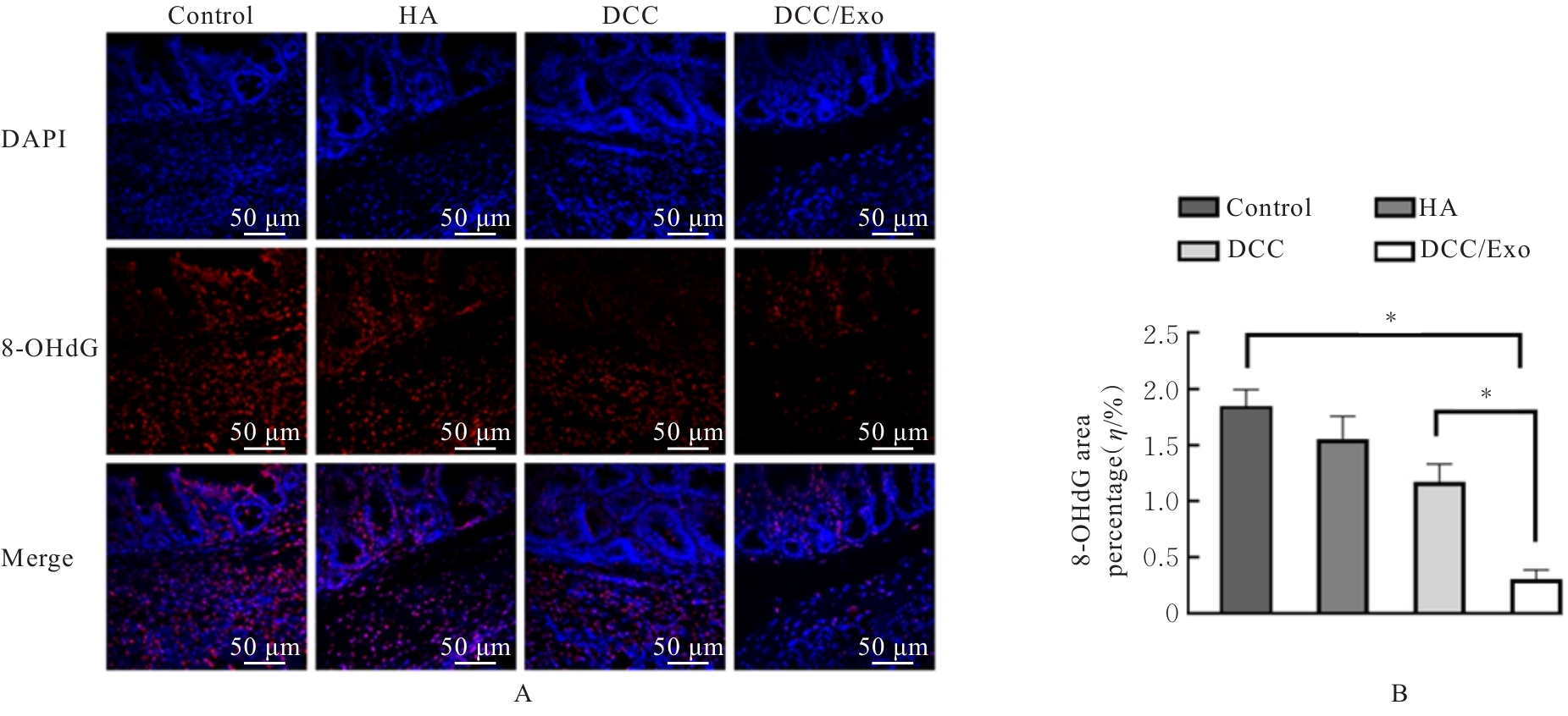
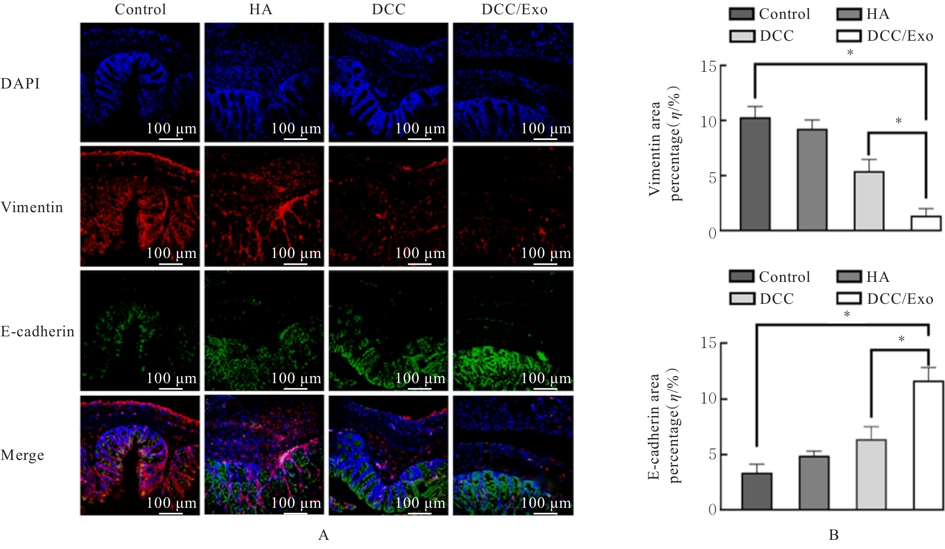
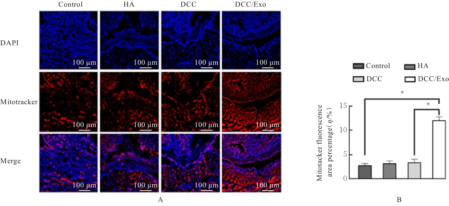
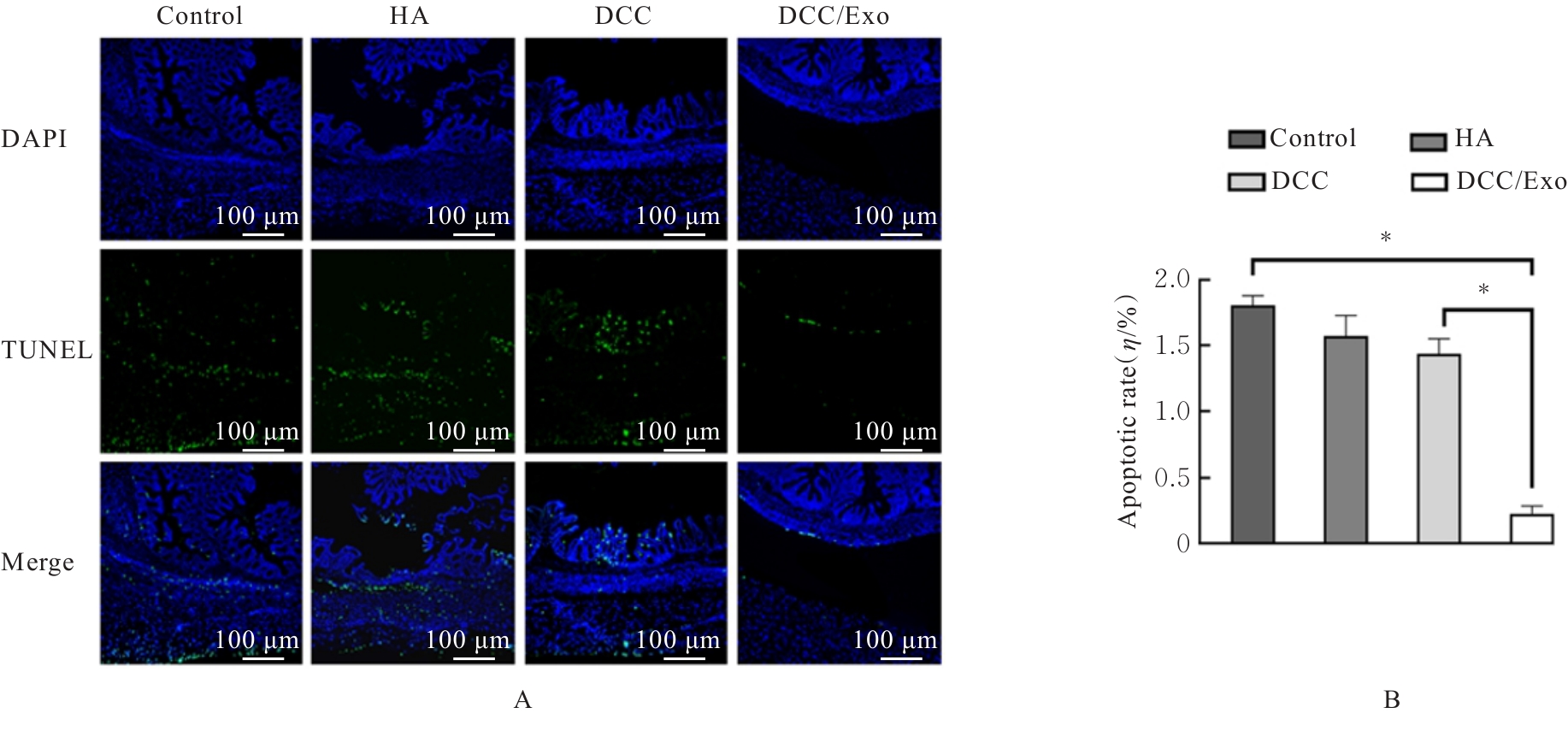
目的 探讨负载外泌体(Exo)的可注射氧化右旋糖酐(ODex)/羧甲基壳聚糖(CMCS)(DCC)水凝胶(DCC/Exo)对小鼠术后腹腔粘连(PA)的改善效果,并阐明其机制。 方法 提取大鼠Exo并制备DCC/Exo,检测其化学结构,采用免疫组织化学染色法检测注射DCC/Exo后小鼠背部皮肤组织中炎症因子Ly-6G的表达情况以评价其生物相容性。采用盲肠刮擦法建立小鼠术后PA模型。将KM小鼠随机分为对照组、透明质酸凝胶(HA)组、DCC组和DCC/Exo组。于术后5和14 d观察各组小鼠PA情况并进行评分,采用HE染色观察各组小鼠PA组织病理形态表现。于术后5 d,采用免疫组织化学染色法检测各组小鼠PA组织中髓过氧化物酶(MPO)表达水平,二氢乙锭(DHE)荧光探针法检测各组小鼠PA组织中活性氧(ROS)水平,免疫荧光染色法检测各组小鼠PA组织中氧化损伤标志物8-羟基脱氧鸟苷(8-OHdG)和间皮-间质转化(MMT)相关蛋白E-钙黏蛋白(E-cadherin)及波形蛋白(Vimentin)表达水平,Mitotracker荧光探针法检测各组小鼠PA组织中线粒体损伤程度,末端脱氧核苷酸转移酶dUTP缺口末端标记(TUNEL)染色法检测各组小鼠PA组织中细胞凋亡率。 结果 提取分离的Exo具有杯形双膜结构;DCC水凝胶负载Exo后,其特征吸收峰未发生明显位移。免疫组织化学染色法,小鼠背部皮肤组织中仅存在少量的中性粒细胞浸润。术后5和14 d,与对照组和DCC组比较,DCC/Exo组小鼠平均粘连评分明显降低(P<0.001)。HE染色,对照组小鼠PA严重且出现大量炎性细胞浸润,HA组和DCC组小鼠PA减轻,DCC/Exo组小鼠腹腔未见粘连,仅见少量炎症细胞。免疫组织化学染色,术后5 d,对照组小鼠PA组织表现出明显弥漫炎性浸润;与对照组和DCC组比较,DCC/Exo组小鼠PA组织中MPO表达水平明显降低(P<0.001)。DHE染色,与对照组和DCC组比较,DCC/Exo组小鼠PA组织中ROS水平明显降低(P<0.001)。免疫荧光染色,与对照组和DCC组比较,DCC/Exo组小鼠PA组织中8-OHdG和Vimentin表达水平明显降低(P<0.001),E-cadherin表达水平明显升高(P<0.001)。与对照组和DCC组比较,DCC/Exo组小鼠PA组织中Mitotracker荧光信号面积比例明显升高(P<0.001),线粒体损伤程度降低。与对照组和DCC组比较,DCC/Exo组小鼠PA组织中细胞凋亡率明显降低(P<0.001)。 结论 局部应用DCC/Exo复合水凝胶可有效减轻小鼠术后PA程度,其作用机制可能与抑制局部炎症浸润、降低组织氧化应激水平、减少细胞凋亡以及抑制MMT进程有关。

关键词: 术后腹腔粘连, 水凝胶, 外泌体, 氧化应激, 间皮-间质转化

Abstract:

Objective To investigate the ameliorative effect of the injectable oxidized dextran(ODex)/carboxymethyl chitosan(CMCS) hydrogel (DCC) loaded with exosomes(Exo) (DCC/Exo) on postoperative peritoneal adhesion (PA) in the mice,and to elucidate its mechanism. Methods The exosomes were extracted from the rats, and DCC/Exo was prepared and its chemical structure was characterized. Immunohistochemical staining method was used to detect the expression of the inflammatory factor Ly-6G in mouse dorsal skin tissue after injection of DCC/Exo to evaluate its biocompatibility. The mouse PA model was established by cecal abrasion. The KM mice were randomly divided into control group, hyaluronic acid (HA) group, DCC group, and DCC/Exo group. The PA condition was observed and scored in the mice in various groups at 5 and 14 d after operation. HE staining was used to observe the pathomorphology of PA tissue of the mice in various groups. At 5 d after operation, immunohistochemical staining method was used to detect the expression levels of myeloperoxidase (MPO) in PA tissue of the mice in various groups. The dihydroethidium (DHE) fluorescence probe assay was used to detect the levels of reactive oxygen species (ROS) in PA tissue of the mice in various groups; immunofluorescence staining was used to detect the expression levels of the oxidative damage marker 8-hydroxy-2'-deoxyguanosine (8-OHdG) and the mesothelial-mesenchymal transition (MMT)- related proteins E-cadherin and Vimentin in PA tissue of the mice in various groups. The Mitotracker fluorescence probe assay was used to detect the degree of mitochondrial damage in PA tissues of the mice in various groups. Terminal deoxynucleotidyl transferase dUTP nick end labeling (TUNEL) staining was used to detect the apoptotic rates of the cells in PA tissue of the mice in various groups. Results The isolated Exos exhibited a cup-shaped double-membrane structure. After loading with Exos, the characteristic absorption peaks of the DCC hydrogel showed no significant shift. The immunohistochemistry analysis results revealed only minimal neutrophil infiltration in mouse dorsal skin tissues. At 5 and 14 d after operation, compared with control group and DCC group, the mean adhesion score of the mice in DCC/Exo group was significantly decreased (P<0.001). The HE staining results showed severe PA with massive inflammatory cell infiltration of the mice in control group, mitigated PA in the mice in HA group and DCC group, and no adhesion with only a few inflammatory cells observed in the abdominal cavity of mice in DCC/Exo group. At 5 d after operation, the immunohistochemical staining results showed significant diffuse inflammatory infiltration in PA tissue of the mice in control group. Compared with control group and DCC group, the expression level of MPO in PA tissue of the mice in DCC/Exo group was significantly decreased (P<0.001). The DHE staining results showed that compared with control group and DCC group, the ROS level in PA tissue of the mice in DCC/Exo group was significantly decreased (P<0.001).The immunofluorescence staining results showed that compared with control group and DCC group, the expression levels of 8-OHdG and Vimentin in PA tissue of the mice in DCC/Exo group were significantly decreased (P<0.001), while the expression level of E-cadherin was significantly increased (P<0.001). Compared with control group and DCC group, the fraction of Mitotracker fluorescence area in PA tissue of the mice in DCC/Exo group was significantly increased (P<0.001), indicating a decreased degree of mitochondrial damage. Compared with control group and DCC group, the apoptotic rate of the cells in PA tissue of the mice in DCC/Exo group was significantly decreased (P<0.001). Conclusion Topical application of the DCC/Exo composite hydrogel may effectively attenuate postoperative PA in the mice, and its mechanism may be related to the suppression of local inflammatory infiltration, reduction of tissue oxidative stress, decrease in apoptosis, and inhibition of the MMT process.

Key words: Postoperative peritoneal adhesion, Hydrogel, Exosome, Oxidative stress, Mesothelial-mesenchymal transition

中图分类号: 

  • R318.08