吉林大学学报(医学版) ›› 2026, Vol. 52 ›› Issue (2): 330-339.doi: 10.13481/j.1671-587X.20260204

• 基础研究 • 上一篇    

芬戈莫德对2型糖尿病小鼠肝纤维化的改善作用及其机制

李舒1,2,郭迦期3,李宛凇4,甄艳凤2,翟红佳4,李捷4,房辉2()   

  1. 1.华北理工大学临床医学院内科教研室,河北 唐山 063000
    2.河北省唐山市工人医院内分泌科,河北 唐山 063000
    3.广东省深圳市龙华区中心医院重症医学科,广东 深圳 518000
    4.河北医科 大学临床医学院内科教研室,河北 石家庄 050000
  • 收稿日期:2025-05-14 接受日期:2025-07-06 出版日期:2026-03-28 发布日期:2026-04-15
  • 通讯作者: 房辉 E-mail:fanghui2818@126.com
  • 作者简介:李 舒(1996-),女,河北省唐山市人,在读硕士研究生,主要从事糖尿病认知功能障碍方面的研究。
  • 基金资助:
    河北省卫健委医学科学研究课题计划项目(20241681);河北省卫健委医学科学研究课题计划项目(20250216);河北省唐山市科技局自然科学基金资助项目(H2024105016)

Improvement effect of fingolimod on hepatic fibrosis in type 2 diabetes mellitus mice and its mechanism

Shu LI1,2,Jiaqi GUO3,Wansong LI4,Yanfeng ZHEN2,Hongjia ZHAI4,Jie LI4,Hui FANG2()   

  1. 1.Department of Internal Medicine,School of Clinical Medicine,North China University of Science and Technology,Tangshan 063000,China
    2.Department of Endocrinology,Tangshan Workers’ Hospital,Tangshan 063000,China
    3.Department of Critical Care Medicine,Central Hospital,Longhua District,Guangdong Province,Shenzhen 518000,China
    4.Department of Internal Medicine,School of Clinical Medicine,Hebei Medical University,Shijiazhuang 050000,China
  • Received:2025-05-14 Accepted:2025-07-06 Online:2026-03-28 Published:2026-04-15
  • Contact: Hui FANG E-mail:fanghui2818@126.com

摘要:

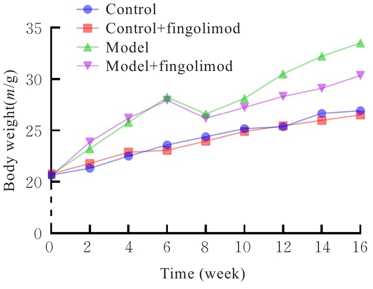
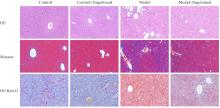
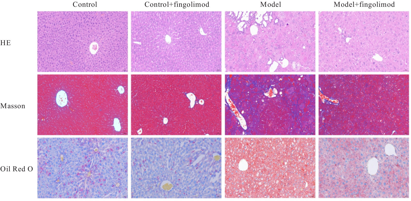
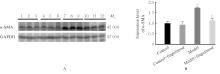
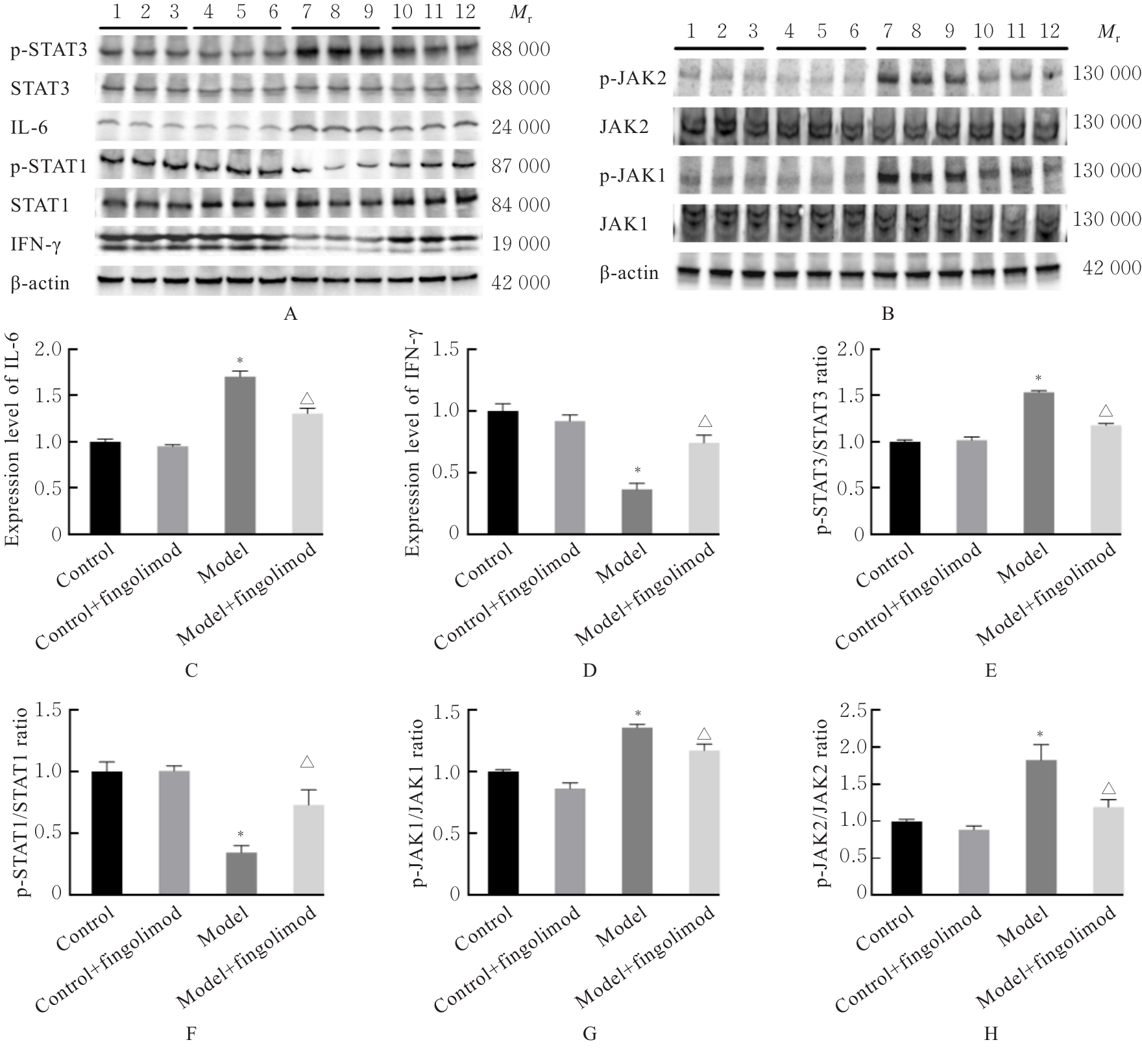
目的 探讨芬戈莫德对2型糖尿病(T2DM)小鼠肝纤维化的改善作用,并阐明其作用机制。 方法 60只雄性C57BL/6J小鼠随机分为对照组、对照+芬戈莫德组、模型组和模型+芬戈莫德组,每组15只。采用高脂饲料联合低剂量链脲佐菌素(STZ)腹腔注射诱导T2DM模型。模型建立后,模型+芬戈莫德组和对照+芬戈莫德组小鼠每日腹腔注射芬戈莫德(1.0 mg·kg?1)干预8周。检测各组小鼠肝脏系数和空腹血糖(FBG)水平。采用试剂盒检测各组小鼠血清中丙氨酸氨基转移酶(ALT)和天冬氨酸氨基转移酶(AST)活性及甘油三酯(TG)、总胆固醇(TC)、高密度脂蛋白胆固醇(HDL-C)和低密度脂蛋白胆固醇(LDL-C)水平,HE染色观察各组小鼠肝组织病理形态表现,Masson染色观察各组小鼠肝纤维化形态表现,油红O染色检查各组小鼠肝组织脂质沉积情况,实时荧光定量PCR(RT-qPCR)法检测各组小鼠肝组织中α平滑肌肌动蛋白(α-SMA)和Janus激酶(JAK)/信号转导与转录激活因子(STAT)通路相关分子JAK1JAK2STAT1STAT3、干扰素γ(IFN-γ)和白细胞介素6(IL-6) mRNA表达水平,Western blotting法检测各组小鼠肝组织中α-SMA、IFN-γ、IL-6、STAT1、磷酸化STAT1(p-STAT1)、STAT3、磷酸化STAT3(p-STAT3)、JAK1、磷酸化JAK1(p-JAK1)、JAK2及磷酸化JAK2(p-JAK2)蛋白表达水平。 结果 与对照组比较,模型组小鼠肝脏系数和FBG水平明显升高(P<0.001);肝细胞肿胀、肝血窦变窄,肝组织内出现大量脂滴和明显胶原积累,肝组织CVF和脂滴面积占比明显升高(P<0.001);血清中ALT和AST活性及TC、TG和LDL-C水平明显升高(P<0.001),HDL-C水平明显降低(P<0.001);肝组织中IL-6α-SMA mRNA和蛋白表达水平明显升高(P<0.001),IFN-γ mRNA和蛋白表达水平明显降低(P<0.001),p-STAT3/STAT3、p-JAK1/JAK1和p-JAK2/JAK2比值明显升高(P<0.001),p-STAT1/STAT1比值明显降低(P<0.001)。与模型组比较,模型+芬戈莫德组小鼠肝脏系数和FBG水平明显降低(P<0.01);肝细胞脂肪变性减轻,脂滴减少,纤维化程度减轻,肝组织CVF和脂滴面积占比明显降低(P<0.001);血清中ALT和AST活性及TC、TG和LDL-C水平明显降低(P<0.001),HDL-C水平明显升高(P<0.001);肝组织中IL-6α-SMA mRNA和蛋白表达水平明显降低(P<0.001),IFN-γ mRNA和蛋白表达水平明显升高(P<0.001),p-STAT3/STAT3、p-JAK1/JAK1和p-JAK2/JAK2比值明显降低(P<0.001),p-STAT1/STAT1比值明显升高(P<0.001)。 结论 芬戈莫德可改善T2DM小鼠糖脂代谢紊乱、肝功能损伤和肝组织内脂质沉积,减轻肝纤维化,其作用机制可能与上调IFN-γ和p-STAT1表达及下调IL-6和p-STAT3表达有关。

关键词: 芬戈莫德, 2型糖尿病, 代谢相关脂肪性肝病, 肝纤维化, 代谢紊乱

Abstract:

Objective To discuss the improvement effect of fingolimod on liver fibrosis in the mice with type 2 diabetes mellitus (T2DM), and to clarify its mechanism. Methods Sixty male C57BL/6J mice were randomly divided into control group, control+fingolimod group, model group, and model+fingolimod group, with 15 mice in each group. The T2DM model was induced by high-fat diet combined with low dose of streptozotocin (STZ) intraperitoneal injection. After the model was established, the mice in model+fingolimod group and control+fingolimod group were given daily intraperitoneal injection of fingolimod (1.0 mg·kg-1) for 8 weeks. The liver coefficients and fasting blood glucose (FBG) levels of the mice in various groups were detected. The kits were used to detect the activities of alanine aminotransferase (ALT) and aspartate aminotransferase (AST), and the levels of triglyceride (TG), total cholesterol (TC), high-density lipoprotein cholesterol (HDL-C), and low-density lipoprotein cholesterol (LDL-C) in serum of the mice in various groups; HE staining was used to observe the pathomorphology of liver tissue of the mice in various groups; Masson staining was used to observe the morphology of liver fibrosis of the mice in various groups; Oil red O staining was used to detect the lipid deposition in liver tissue of the mice in various groups; real-time fluorescence quantitative PCR(RT-qPCR) method was used to detect the expression levels of α-smooth muscle actin (α-SMA) and Janus kinase (JAK)/signal transducer and activator of transcription (STAT) pathway-related molecules JAK1JAK2STAT1STAT3, interferon-γ (IFN-γ), and interleukin-6 (IL-6) mRNA in liver tissue of the mice in various groups; Western blotting method was used to detect the expression levels of α-SMA, IFN-γ, IL-6,STAT1, phosphorylated STAT1 (p-STAT1), STAT3, phosphorylated STAT3(p-STAT3), JAK1, phosphorylated JAK1(p-JAK1), JAK2 and phosphorylated JAK2(p-JAK2) proteins in liver tissue of the mice in various groups. Results Compared with control group, the liver coefficient and FBG level of the mice in model group were significantly increased (P<0.001); the hepatocytes were swollen, the hepatic sinusoids were narrowed, a large number of lipid droplets and obvious collagen accumulation in liver tissue were observed, and the CVF and lipid droplet area proportion of liver tissue were significantly increased (P<0.001); the activities of ALT and AST and the levels of TC, TG, and LDL-C in serum were significantly increased (P<0.001), while the level of HDL-C was significantly decreased (P<0.001); the expression levels of IL-6 and α-SMA mRNA and protein in liver tissue were significantly increased (P<0.001), the expression levels of IFN-γ mRNA and protein were significantly decreased (P<0.001), the ratios of p-STAT3/STAT3, p-JAK1/JAK1, and p-JAK2/JAK2 were significantly increased (P<0.001), and the ratio of p-STAT1/STAT1 was significantly decreased (P<0.001). Compared with model group, the liver coefficient and FBG level of the mice in model+fingolimod group were significantly decreased (P<0.01); the hepatocyte steatosis, lipid droplets, and fibrosis degree were alleviated, and the CVF and lipid droplet area proportion of liver tissue were significantly decreased (P<0.001); the activities of ALT and AST and the levels of TC, TG, and LDL-C in serum were significantly decreased (P<0.001), while the level of HDL-C was significantly increased (P<0.001); the expression levels of IL-6 and α-SMA mRNA and protein in liver tissue were significantly decreased (P<0.001), the expression levels of IFN-γ mRNA and protein were significantly increased (P<0.001), the ratios of p-STAT3/STAT3, p-JAK1/JAK1, and p-JAK2/JAK2 were significantly decreased (P<0.001), and the ratio of p-STAT1/STAT1 was significantly increased (P<0.001). Conclusion Fingolimod can improve glucose and lipid metabolism disorders, liver function injury, and lipid deposition in liver tissue of T2DM mice, and alleviate liver fibrosis. Its mechanism may be related to up-regulating the expressions of IFN-γ and p-STAT1 and down-regulating the expressions of IL-6 and p-STAT3.

Key words: Fingolimod, Type 2 diabetes mellitus, Metabolism-associated fatty liver disease, Liver fibrosis, Metabolic disorder

中图分类号: 

  • R587.2