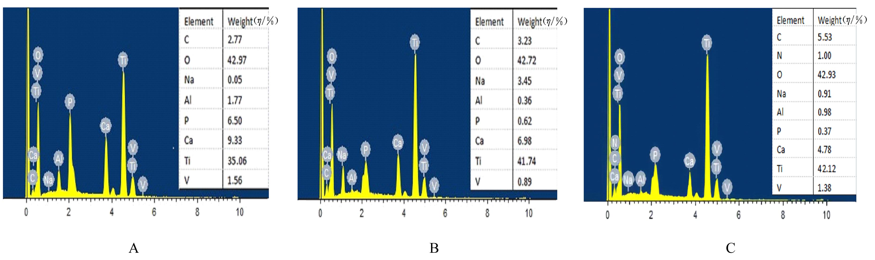
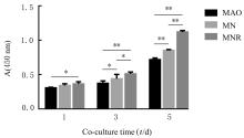
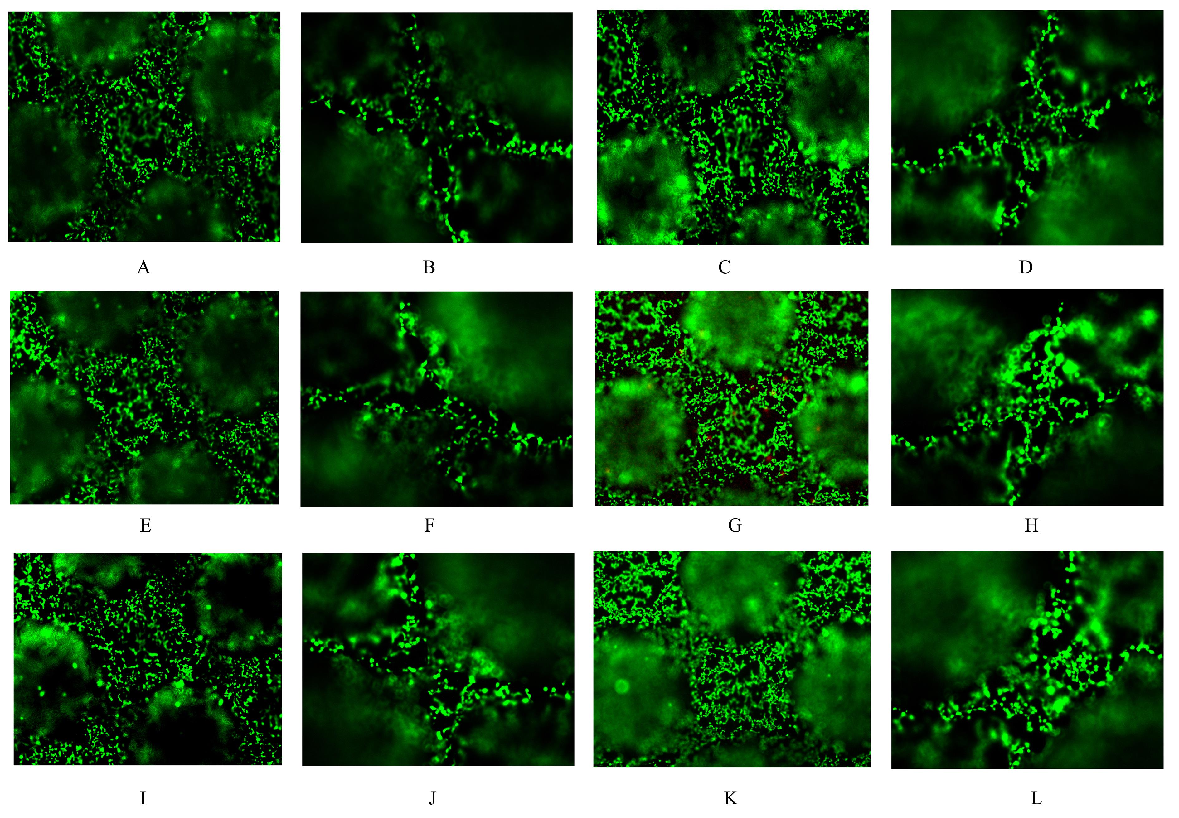
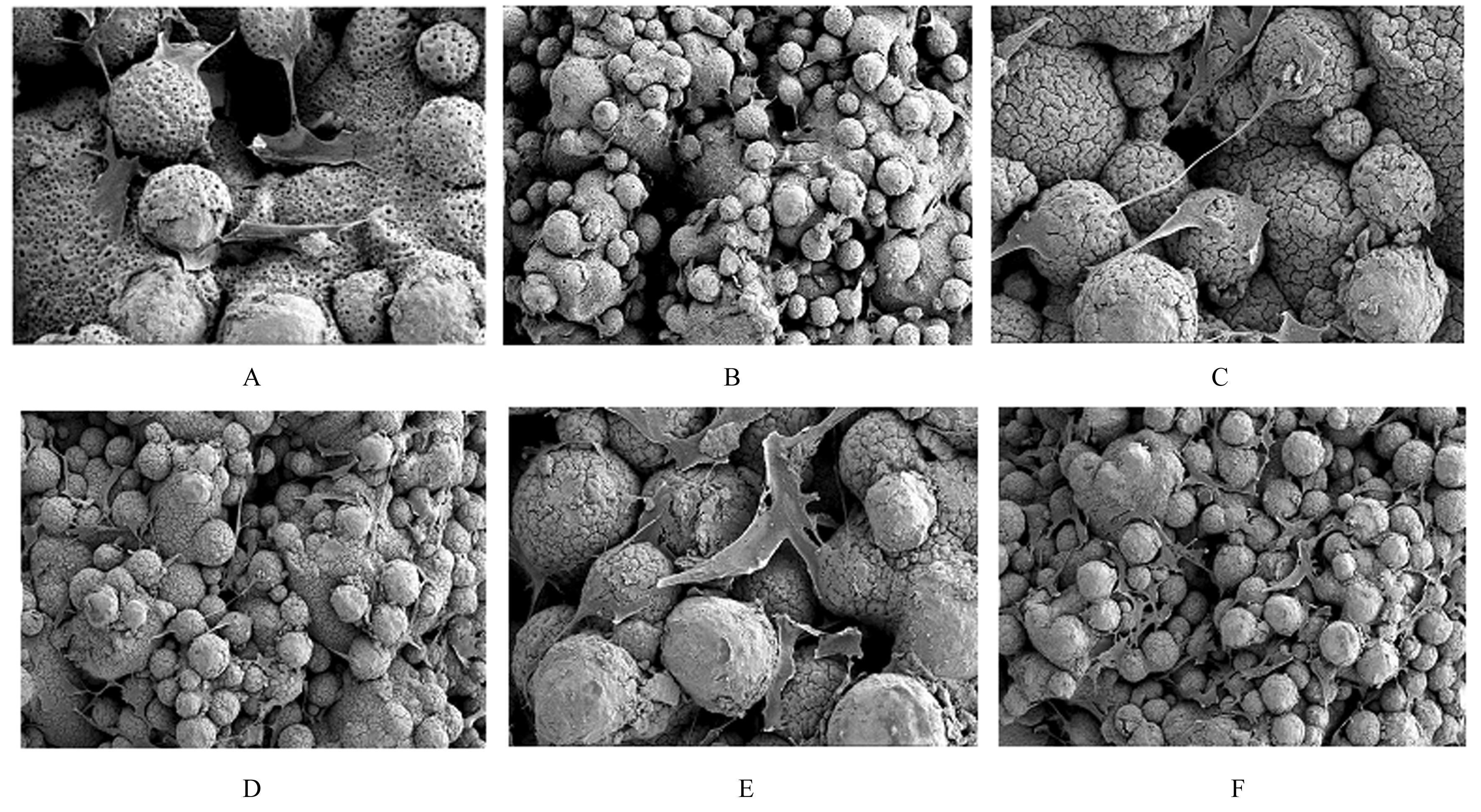
| 1 |
郭佳明, 梁精龙, 沈海涛, 等. 生物医用钛合金材料制备方法及应用进展[J]. 热加工工艺, 2021, 50(20): 30-34.
|
| 2 |
HARDT M, KLOCKE F, DÖBBELER B, et al. Experimental study on surface integrity of cryogenically machined Ti-6Al-4V alloy for biomedical devices[J]. Procedia CIRP, 2018, 71: 181-186.
|
| 3 |
LIU W W, YANG D, WEI X H, et al. Fabrication of piezoelectric porous BaTiO3 scaffold to repair large segmental bone defect in sheep[J]. J Biomater Appl, 2020, 35(4/5): 544-552.
|
| 4 |
LI J M, JANSEN J A, WALBOOMERS X F, et al. Mechanical aspects of dental implants and osseointegration: a narrative review[J]. J Mech Behav Biomed Mater, 2020, 103: 103574.
|
| 5 |
GINESTRA P, FERRARO R M, ZOHAR-HAUBER K,et al. Selective laser melting and electron beam melting of Ti6Al4V for orthopedic applications: a comparative study on the applied building direction[J]. Materials (Basel), 2020, 13(23): E5584.
|
| 6 |
梁鹏晨, 史俊峰, 孙苗苗, 等. 聚多巴胺辅助红景天苷改善微弧氧化纯钛的成骨性能[J]. 中国组织工程研究, 2022, 26(10): 1522-1529.
|
| 7 |
HUANG Y Z, HE S K, GUO Z J, et al. Nanostructured titanium surfaces fabricated by hydrothermal method: influence of alkali conditions on the osteogenic performance of implants[J]. Mater Sci Eng C Mater Biol Appl, 2019, 94: 1-10.
|
| 8 |
VERSTAPPEN J F M, JIN J F, KOÇER G, et al. RGD-functionalized supported lipid bilayers modulate pre-osteoblast adherence and promote osteogenic differentiation[J].J Biomed Mater Res A,2020,108(4): 923-937.
|
| 9 |
GUO X B, LIU Y, BAI J X, et al. Efficient inhibition of wear-debris-induced osteolysis by surface biomimetic engineering of titanium implant with a mussel-derived integrin-targeting peptide[J]. Adv Biosyst, 2019, 3(2): e1800253.
|
| 10 |
HAMDAN F, BIGDELI Z, ASGHARI S M, et al. Synthesis of modified RGD-based peptides and their in vitro activity[J].ChemMedChem,2019,14(2): 282-288.
|
| 11 |
TAMADDON M, SAMIZADEH S, WANG L, et al. Intrinsic osteoinductivity of porous titanium scaffold for bone tissue engineering[J]. Int J Biomater, 2017, 2017: 5093063.
|
| 12 |
LI G Y, WANG L, PAN W, et al. In vitro and in vivo study of additive manufactured porous Ti6Al4V scaffolds for repairing bone defects[J]. Sci Rep, 2016, 6: 34072.
|
| 13 |
ZHANG T, WEI Q G, ZHOU H, et al. Sustainable release of vancomycin from micro-arc oxidised 3D-printed porous Ti6Al4V for treating methicillin-resistant Staphylococcus aureus bone infection and enhancing osteogenesis in a rabbit tibia osteomyelitis model[J]. Biomater Sci, 2020, 8(11): 3106-3115.
|
| 14 |
YIN L H, CHANG Y R, YOU Y H, et al. Biological responses of human bone mesenchymal stem cells to Ti and TiZr implant materials[J]. Clin Implant Dent Relat Res, 2019, 21(4): 550-564.
|
| 15 |
WANG X H, CAO Y, YANG L, et al. Bioactivity study of the titanium plates treated with microarc oxidation and alkali[J]. J Nanosci Nanotechnol, 2011, 11(11): 9650-9655.
|
| 16 |
KARAMAN O, KELEBEK S, DEMIRCI E A, et al. Synergistic effect of cold plasma treatment and RGD peptide coating on cell proliferation over titanium surfaces[J]. Tissue Eng Regen Med, 2018, 15(1): 13-24.
|
| 17 |
张 辉, 王少华, 王 茜, 等. RGD多肽修饰多孔钽可激活MG63细胞整合素/黏着斑激酶信号通路[J]. 中国组织工程研究, 2021, 25(16): 2535-2540.
|
| 18 |
TAPIAL MARTÍNEZ P, LÓPEZ NAVAJAS P, LIETHA D. FAK structure and regulation by membrane interactions and force in focal adhesions[J]. Biomolecules, 2020, 10(2): 179.
|
| 19 |
LI Z C, LIU T Q, YANG J X, et al. Characterization of adhesion properties of the cardiomyocyte integrins and extracellular matrix proteins using atomic force microscopy[J]. J Mol Recognit, 2020, 33(4): e2823.
|
| 20 |
SUNDEN F, ALSADHAN I, LYUBIMOV A, et al. Differential catalytic promiscuity of the alkaline phosphatase superfamily bimetallo core reveals mechanistic features underlying enzyme evolution[J]. J Biol Chem, 2017, 292(51): 20960-20974.
|
| 21 |
GUO X B, BAI J X, GE G R, et al. Bioinspired peptide adhesion on Ti implants alleviates wear particle-induced inflammation and improves interfacial osteogenesis[J]. J Colloid Interface Sci, 2022, 605: 410-424.
|
| 22 |
WANG Z H, SUN J, LI Y L, et al. Experimental study of the synergistic effect and network regulation mechanisms of an applied combination of BMP-2, VEGF, and TGF-β1 on osteogenic differentiation[J]. J Cell Biochem, 2020, 121(3): 2394-2405.
|
| 23 |
KUNG F C. Injectable collagen/RGD systems for bone tissue engineering applications[J]. Biomed Mater Eng, 2018, 29(2): 241-251.
|
| 24 |
YANG X L, LIU X, WU Q Q, et al. Dopamine-assisted immobilization of peptide arginine-glycine-aspartic acid to enhance the cellular performances of MC3T3-E1 cells of carbon-carbon composites[J]. J Biomater Appl, 2019, 34(2): 284-296.
|
 )
)