Journal of Jilin University(Medicine Edition) ›› 2025, Vol. 51 ›› Issue (3): 567-575.doi: 10.13481/j.1671-587X.20250302

• Research in basic medicine • Previous Articles    

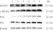
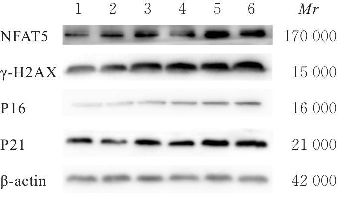
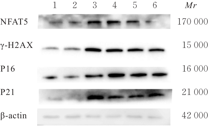
Effect of nuclear factor of activated T lymphocytes 5 on senescence of smooth muscle cells of mice induced by high-salt and its mechanism

Wei ZHONG1,Zhiyin DAI1,Xinggang CUI1,Bo LI1,Yu JIANG1,2()   

  1. 1.Department of Cardiology,Affiliated Hospital,Jiangsu University,Zhenjiang 212001,China
    2.Department of Cardiology,Maternal and Child Health Care Hospital,Changzhou City,Jiangsu Province,Changzhou 213003,China
  • Received:2024-08-29 Accepted:2024-10-20 Online:2025-05-28 Published:2025-07-18
  • Contact: Yu JIANG E-mail:jy950927@163.com

Abstract:

Objective To discuss the role of nuclear factor of activated T-cells 5 (NFAT5) inhibitor KRN5 in high salt-induced senescence of mouse vascular smooth muscle cells (VSMCs), and to clarify its mechanism. Methods Thirty 8-week-old male ApoE-/- mice were divided into normal group, senescence group and high-salt treatment senecence group, with 10 mice in each group; the mice in senescence group and high-salt treatment senecence group were used to establish natural senecence mouse models; the mouse VSMCs were isolated and cultured, and divided into normal group, senescence group, high-salt treatment senecence group and high-salt treatment senecence+KRN5 group. β-galactosidase (Sa-β-gal) staining was used to detect the senescence of aortic tissues and VSMCs in various groups; immunofluorescence method was used to detect the expressions of NFAT5 and phosphorylated histone H2A variant X (γ-H2AX) proteins in mouse aortic tissues and VSMCs in various groups; real-time fluorescence quantitative PCR (RT-qPCR) method was used to detect the mRNA expression levels of NFAT5, γ-H2AX, cyclin-dependent kinase inhibitor 2A (P16) and cyclin-dependent kinase inhibitor 1A (P21) in the cells in various groups; Western blotting method was used to detect the protein expression levels of NFAT5, γ-H2AX, P16 and P21 in VSMCs in various groups. Results The Sa-β-gal staining results showed that compared with normal group, the proportions of senescence-positive area in aortic tissues of the mice in senescence group and high-salt treatment senecence group were significantly increased (P<0.05), and the proportion of senescence-positive cells in the VSMCs of the mice was significantly increased (P<0.05); compared with senecence group, the proportion of senescence-positive cells in the VSMCs mice in high-salt treatment senecence group was significantly increased (P<0.05); compared with high-salt treatment senecence group, the proportion of senescence-positive cells in the VSMCs of the mice in high-salt treatment senecence+KRN5 group was significantly decreased (P<0.01). The immunofluorescence results showed that compared with normal group, the expression level of γ-H2AX protein in mouse VSMCs of the mice in senescence group was significantly increased (P<0.05); compared with senescence group, the expression levels of SA-β-gal staining and NFAT5 protein in aortic tissue of the mice in high-salt treatment senecence group were significantly increased (P<0.05); compared with normal group, the expression level of NFAT5 protein in the VSMCs of the mice in senecence group and high-salt treatment senecence group was significantly increased (P<0.05); compared with senecence group, the expression level of NFAT5 protein in the VSMCs of the mice in high-salt treatment senecence group was significantly increased (P<0.05). The RT-qPCR results showed that compared with normal group, the expression levels of NFAT5, γ-H2AX, P16, and P21 mRNA in the VSMCs of the mice in senescence group and high-salt treatment senecence group were significantly increased (P<0.05); compared with senecence group, the mRNA expression levels of NFAT5, γ-H2AX, P16, and P21 mRNA in the VSMCs of the mice in high-salt treatment senecence group were significantly increased (P<0.05); compared with senecence group, the expression levels of NFAT5, γ-H2AX, P16, and P21 mRNA in the VSMCs of the mice in high-salt treatment senecence group and high-salt treatment senecence+KRN5 group were significantly increased (P<0.05); compared with high-salt treatment senecence group, the mRNA expression levels of NFAT5, γ-H2AX, P16, and P21 in the VSMCs of the mice in high-salt treatment senecence+KRN5 group were significantly decreased (P<0.05). The Western blotting results showed that compared with normal group, the expression levels of NFAT5, γ-H2AX, P16, and P21 proteins in the VSMCs of the mice in senescence group and high-salt treatment senecence group were significantly increased (P<0.05); compared with senescence group, the expression levels of NFAT5, γ-H2AX, P16, and P21 proteins in the VSMCs of the mice in high-salt treatment senecence group were significantly increased (P<0.05); compared with senecence group, the expression levels of NFAT5, γ-H2AX, P16, and P21 proteins in the VSMCs of the mice in high-salt treatment senecence group and high-salt treatment senecence+KRN5 group were significantly increased (P<0.05); compared with high-salt treatment senecence group, the expression levels of NFAT5, γ-H2AX, P16, and P21 proteins in the VSMCs of the mice in high-salt treatment senecence+KRN5 group were significantly decreased (P<0.05). Conclusion NFAT5 may play a promoting role in high salt-induced senescence of the mouse VSMCs.

Key words: Vascular aging, Nuclear factor of activated T-cells 5, KRN5, Vascular smooth muscle cells, β-galactosidase

CLC Number: 

  • R339.38